Ventricular Septal Perforation Following Takotsubo Cardiomyopathy
Received: 03-Oct-2016 / Accepted Date: 03-Nov-2016 / Published Date: 10-Nov-2016
Abstract
Ventricular septal perforation is a well-known complication following acute myocardial infarction, but it rarely complicates takotsubo cardiomyopathy. A 76-year-old woman, who had dyspnea, had been admitted for takotsubo cardiomyopathy. Systolic murmur that was loudest at the apex was heard. Transthoracic echocardiography revealed shunt flow from the left ventricle to the right ventricle. Ventricular septal perforation following takotsubo cardiomyopathy was diagnosed. Intra-aortic balloon pumping was immediately started. We successfully performed patch repair. Takotsubo cardiomyopathy complicated by ventricular septal perforation is a rare and critical condition that requires optimal treatment and careful monitoring.
Keywords: Takotsubo Cardiomyopathy; Ventricular Septal Perforation; cardiacarrest; myocardialinfarction
78723Case Report
Ventricular septal perforation is a well-known complication following acute myocardial infarction, but it has rarely complicated takotsubo cardiomyopathy [1].
We report a rare case of ventricular septal perforation after takotsubo cardiomyopathy (TCM-VSP), which was successfully treated by surgery.
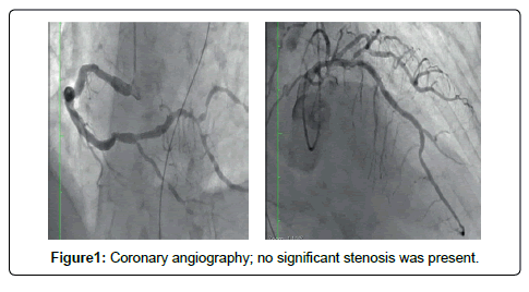
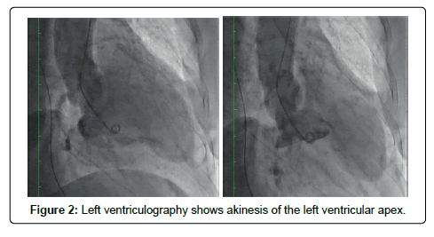
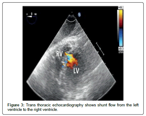
A 76-year-old woman was admitted for bowel obstruction caused by an ascending colon tumor. The patient was under treatment for hypertension and diabetes. Dyspnea occurred after three days. Electrocardiography showed ST elevation in leads V1-V6. The elevation suggested acute myocardial infarction and coronary arteriography was performed. No coronary lesion was present (Figure 1), but an absence of apical contraction and an excessive base contraction were noted on the left ventriculography (Figure 2). Takotsubo cardiomyopathy was diagnosed. The symptoms improved after inpatient treatment, but there was sudden onset of severe dyspnea after four days. Levine 4/6 pansystolic murmur that was loudest at the apex was heard. Transthoracic echocardiography revealed shunt flow from the left ventricle to the right ventricle (Figure 3). TCM-VSP was diagnosed. Intra-aortic balloon pumping was immediately started.
Results
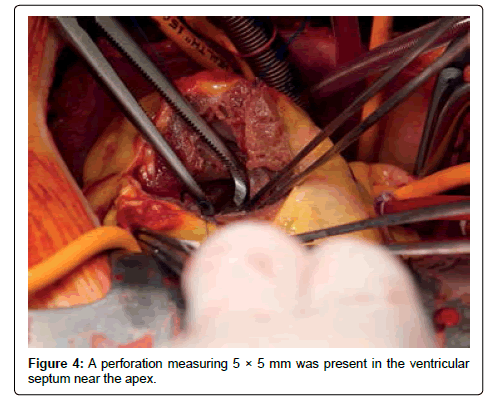
Median sternotomy was performed. The left ventricle was incised using cardiopulmonary bypass and induced cardiac arrest. A perforation measuring 5 x 5mm was present in the ventricular septum near the apex (Figure 4). The ventricular septum of the perforated region was not fragile, but relatively firm. Two pairs of hemashield patches were attached to the perforated region from the right and left ventricle. Teflon felt strip was applied to close the left ventricle incision. The postoperative course was uneventful.
Conclusion
Takotsubo cardiomyopathy has a good prognosis and is associated with a higher prevalence in neurologic or psychiatric disorder [1]. The mechanism of TCM-VSP is unclear, but a case of TCM-VSP on pathological examination showed myocardial necrosis in the ruptured region [2]. Ventricular septal perforation is surgically treated by the infarct exclusion technique [3,4]. P atch repair for TCM-VSP can be a viable treatment, because the ventricular septum of the perforated region is not fragile. TCM-VSP is a rare and critical condition that requires optimal treatment and careful monitoring.
Refrences
- Kenya S, Ochiai H, Katayama N, Nakamura K, Arataki K, et al. (2005) Ventricular Septal Perforation in a Patients With Takotsubo Cardiomyopathy. Circ J 169: 365-367.
- Kenta I, Tada S, Yamada T (2008) A case of Takotsubo Cardiomyopathy Complicated by Ventricular Septal Perforation. Circ J 72: 1540-1543.
- Giovanni M, Cattaneo P, Rossi A, Baravelli M, Piffaretti G, et al. (2010) Tako-tsubo cardiomyopathy complicated by ventricular septal perforation and septal dissection. Heart Vessel 25: 73-75.
- Tadao A, Sakakibara M, Takahashi M, Asakawa K, Dannoura Y, et al. (2015) Critical Takotsubo cardiomyopathy Complicated by Ventricular Septal Perforation.
Intern Med 54: 37-41.
Citation: Yamazaki S, Kato M, Toyama M (2016) Ventricular Septal Perforation Following Takotsubo Cardiomyopathy. Cardiovasc Ther 1: 113.
Copyright: © 2016 Yamazaki S, et al. This is an open-access article distributed under the terms of the Creative Commons Attribution License, which permits unrestricted use, distribution, and reproduction in any medium, provided the original author and source are credited.
Select your language of interest to view the total content in your interested language
Share This Article
Open Access Journals
Article Usage
- Total views: 3986
- [From(publication date): 0-2016 - Jul 12, 2025]
- Breakdown by view type
- HTML page views: 3059
- PDF downloads: 927


