Research Article Open Access
Varicella Pneumonia in an Immunocompetent Adult: A Case Report
Fatma Medhioub Kaaniche*, Anis Chaari, Rania Ammar, Mabrouk Bahloul and Mounir Bouaziz
Intensive care department, CHU Habib Bourguiba, Road El Ain
- *Corresponding Author:
- Kaaniche FM
Intensive care department, CHU Habib Bourguiba
Road El Ain Km 0.5, postal code 3029, Tunisia
Email: fatma_kaaniche@yahoo.fr
Received Date: April 22, 2015 Accepted Date: May 23, 2015 Published Date: May 25, 2015
Citation: Chaari A, Ammar R, Bahloul M, Bouaziz M (2015) Varicella Pneumonia in an Immunocompetent Adult: A Case Report. J Clin Diagn Res 3:116. doi:2376-0311.1000116
Copyright: © 2015 Kaaniche FM, et al. This is an open-access article distributed under the terms of the Creative Commons Attribution License, which permits unrestricted use, distribution, and reproduction in any medium, provided the original author and source are credited.
Visit for more related articles at JBR Journal of Clinical Diagnosis and Research
Abstract
The clinical presentation of varicella in adults is frequently associated with complications, particularly varicella pneumonia. Most cases occur in immunocompromised adults but immunocompetent can have serious pulmonary compromise. We report the case of an immunocompetent patient admitted to the intensive care unit with dyspnea and an exanthematous vesicular rash. We diagnosed varicella pneumonia based on the presence of a typical skin rash, pulmonary symptoms and contact with a child with chickenpox, confirmed by positive serology of varicellazoster virus (VZV). The patient made a full recovery with mechanical ventilation and anti-viral therapy.
Keywords
Varicella pneumonia; Immunocompetent adult; Risk factors; Antiviral therapy; Intensive care
Introduction
Varicella is a common benign childhood illness caused by varicellazoster virus (VZV), typically associated with fever and a characteristic exanthematous vesicular rash [1]. The clinical presentation in adults is more severe and more commonly associated with complications, particularly varicella pneumonia [1,2]. Varicella pneumonia has been reported in 5-50% of adults with chickenpox [1-4]. This complication was mainly reported in immuocompromised patients with markedly increased morbidity and mortality [1,2]. Varicella pneumonia was rarely reported in immunocompetent patients and risk factors for such condition are not fully established. We report the case of immunocompetent patient who developed severe varicella pneumonia with an acute respiratory failure leading to intensive care unit (ICU) admission. Clinical factors and therapeutic management are discussed.
Case presentation
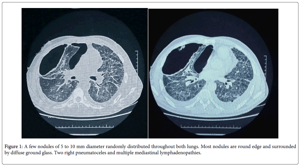
A 36-year old man presented to the intensive care unit with sudden onset of fever, dyspnea, myalgia, and an exanthemata’s vesicular rash. He had no remarkable medical history, including chickenpox in his childhood. He was a smoker and his child had suffered from chickenpox, two weeks earlier. On examination, he was febrile (39ºC) with signs of respiratory distress (respiratory rate was 24 breaths/min and oxygen saturation was 90% on room air). Heart rate was 110 beats/min. pulmonary auscultation revealed bilateral crackles. An extensive polymorphic pruritic rash with macular, vesicles, pustules and crusty lesions was noted. Laboratory studies showed lymphocytic leukocytosis and elevation of liver enzymes (Alanine transaminase (ALT); Aspartate transaminase (AST)). Arterial blood gases were consistent with hypoxemic respiratory failure (PaO2 of 71.8 mmHg). Human immunodeficiency virus (HIV) testing according to the enzyme-linked immunosorbent assay (ELISA) technique was negative. The serum titer of VZV IgM according to the ELISA technique was positive. Microbiological sputum examination did not show any evidence of bacterial infection. A chest roentgenogram revealed diffuse, poorly defined, bilateral alveolar-interstitial opacities. Highresolution computerized tomography (CT) showed ground glass appearance, nodules whose diameter is 5 to 10 millimeters (mm), two right pneumatoceles and multiple mediastinal lymphadenopathies (figure 1). We diagnosed varicella pneumonia based on the presence of a typical skin rash, pulmonary symptoms, and contact with a child with chickenpox, confirmed by positive serology of VZV.
The diagnosis of varicella pneumonia in an immunocompetent patient was considered. Nasal oxygen (8 liters/min), intravenous Acyclovir (10 mg/kg every 8 hours) and skin care (showers with lukewarm water and application of antiseptic and cicatrizing creams) were promptly initiated. The patient's condition deteriorated necessitating intubation and the use of mechanical ventilation. A distal tracheal sample taken on the fourth day of hospitalization identified Acinetobacter baumannii infection. The patient was treated with intravenous Imipenem (0.5 grams every 6 hours), Colistin (3 million international units every 8 hours) and Acyclovir. On day 8 of hospitalization, a tracheostomy was placed due to prolonged mechanical ventilation. The patient's respiratory function progressively improved, and he was subsequently decannulated on day 20 of the ICU hospitalization. He was discharged from our ICU after 22 days of hospitalization. The diagnosis of varicella pneumonia in an immunocompetent patient was considered. Nasal oxygen (8 liters/ min), intravenous Acyclovir (10 mg/kg every 8 hours) and skin care (showers with lukewarm water and application of antiseptic and cicatrizing creams) were promptly initiated. The patient's condition deteriorated necessitating intubation and the use of mechanical ventilation. A distal tracheal sample taken on the fourth day of hospitalization identified Acinetobacter baumannii infection.
The diagnosis of varicella pneumonia in an immunocompetent patient was considered. Nasal oxygen (8 liters/min), intravenous Acyclovir (10 mg/kg every 8 hours) and skin care (showers with lukewarm water and application of antiseptic and cicatrizing creams) were promptly initiated. The patient's condition deteriorated necessitating intubation and the use of mechanical ventilation. A distal tracheal sample taken on the fourth day of hospitalization identified Acinetobacter baumannii infection. The patient was treated with intravenous Imipenem (0.5 grams every 6 hours), Colistin (3 million international units every 8 hours) and Acyclovir. On day 8 of hospitalization, a tracheostomy was placed due to prolonged mechanical ventilation. The patient's respiratory function progressively improved, and he was subsequently decannulated on day 20 of the ICU hospitalization. He was discharged from our ICU after 22 days of hospitalization.
Discussion
Primary chickenpox involving the skin is a benign and self-limiting condition. It rapidly spreads among humans with close proximity [5]. Varicella pneumonitis is the most common complication of the virus and can be fatal, usually occurs 1 to 6 days after the onset of a varicella-zoster infection. It is more prevalent among adults [4]. The clinical manifestations include cough, dyspnea, tachypnea, fever, cyanosis, and sometimes pleuritic chest pain or hemoptysis [1,2]. However, chest symptoms may start before the appearance of the skin rash [6]. Physical findings are often minimal and chest radiographs typically reveal nodular or interstitial pneumonitis [6].
The risk factors for acute lung injury due to varicella infection are as the following: male sex, adult, smoking, greater than 100 skin lesions, pregnancy and close contact with an infected person or any immunosuppression [6]. Our patient had several of these risk factors (adult, male sex, smoking, Florida skin disease); moreover he was not previously exposed to chickenpox in childhood.
Histological features of pneumonia caused by the VZV consist of an interstitial mononuclear inflammatory infiltrate, intraalveolar proteinaceous exudate, hyaline membrane formation, and alveolar type II cells hyperplasia [4]. After recovery from the initial disease, spherical nodules are seen, randomly distributed in the lung parenchyma. The nodules are composed of an outer, often lamellated fibrous capsule and frequently enclosing areas of hyalinized collagen or necrotic tissue. The nodules frequently calcify [2].
The radiological characteristics consist of multiple bilateral 5-10 mm, ill-defined nodules, which may be confluent [1]. Occasionally, hilar lymphadenopathy, reticular opacities and pleural effusions may occur [1,2].
Kim et al. [2] presented high-resolution CT findings for three immunocompetent patients with varicella pneumonia. The abnormalities included 1-10 mm diameter well- and ill-defined nodules, diffusely dispersed throughout both lungs; these nodules had a surrounding halo of ground-glass attenuation, patchy ground-glass attenuation, and coalescence of lesions. The nodules were randomly distributed in the secondary pulmonary lobules and showed no predilection for any particular area of the lung. None of the patients had pleural effusion or lymphadenopathy. A few the high-resolution CT findings in our patient were similar to previous report [2], consisting of some 5-10 mm diameter nodules. Most of them were round edged and surrounded by diffuse ground glass. However, our case had two right pneumatoceles which were not previously reported in patients with varicella pneumonia. These features can be related to the varicella pneumonia itself, but may also be explained by the consequences of mechanical ventilation [7].
Acyclovir is an established therapy for treating varicella-related illnesses [6]. Moreover, timely initiation of Acyclovir therapy may avoid the further complications of the varicella infection [6]. Current consensus supports a 7-10 day course of intravenous Acyclovir for varicella pneumonitis. Early intervention may modify the natural course of this complication [8]. Pregnant women with signs of severe disease or risk of premature labor, and immunocompromised patients require close monitoring [8]. In patients who are immunocompromised, the disease may progress rapidly into acute respiratory distress syndrome and respiratory failure, with mortality reaching 50% despite aggressive treatment [8]. Most healthy adults have favorable outcomes with complete recovery [3].
In our patient, acute respiratory failure occured 5 days after the onset of the disease. Despite being immunocompetent, he had significant radiologic changes. The outcome was favorable in this case.
For this preventable illness, there is an effective vaccine that is highly protective against varicella and especially protective against severe disease [9]. The recommended adult immunization schedule in the USA suggests that all adults without evidence of immunity to varicella should receive two doses of varicella vaccine if not previously vaccinated, unless there is a contraindication [10]. In Tunisia, vaccination against varicella is for immunocompromised children [11]. The duration of immunity is estimated at 7-10 years [11]. Our patient who did not receive immunization shows that pneumonitis is a severe complication of varicella infection in adults. In a patient with known risk factors, the diagnosis of varicella infection and pneumonitis should be considered early on in the clinical course and treated aggressively.
Conclusion
Pneumonia caused by varicella infection is a serious and severe complication of the disease when it occurs in adults. Patients with varicella pneumonia are at high risk for acute respiratory failure leading to mechanical ventilation. Early antiviral treatment with Acyclovir is recommended in case of varicella pneumonia proven.
References
- Emerson L Gasparetto, Danny Warszawiak, Priscilla Tazoniero, Dante L Escuissato et. Al (2002) Severity of illness and outcome in adult patients with primary varicella pneumonia. Respiration 69: 330-4.
- Kim JS, Ryu CW, Lee SI, Sung DW, Park CK (1999) High-resolution CT findings of varicella-zoster pneumonia. AJR Am J Roentgenol 172: 113-116.
- Jones AM, Thomas N, Wilkins EG (2001) Outcome of varicella pneumonitis in immunocompetent adults requiring treatment in a high dependency unit. J Infect 43: 135-139.
- Gasparetto EL, Warszawiak D, Tazoniero P, Escuissato DL and Marchiori E (2005) Varicella pneumonia in immunocompetent adults: report of two cases, with emphasis on high-resolution computed. The Brazilian journal of infectious diseases : an official publication of the Brazilian Society of Infectious Diseases 9:3 Jun pg 262-5.
- Brisson M, Edmunds WJ (2003) Varicella vaccination in England and Wales: cost-utility analysis. Arch Dis Child 88: 862-869.
- Mohsen AH, Peck RJ, Mason Z, Mattock L, McKendrick MW (2001) Lung function tests and risk factors for pneumonia in adults with chickenpox. Thorax 56: 796-799.
- J C Chevrolet, D Tassaux, P Jolliet and J Pugin. Syndrome de détresserespiratoireaiguë. EMC-Pneumologie (2004) 143–186.
- Mohsen AH, McKendrick M (2003) Varicella pneumonia in adults. EurRespir J 21: 886-891.
- Gershon AA, Takahashi M and Seward J (2003) Varicella vaccine. In: Plotkin S, Orenstein W, eds Vaccines 4th edn. Philadelphia: WB Saunders, 783-823.
- Advisory Committee on Immunization Practices (2009) Recommended adult immunization schedule: United States, 2009. Ann Intern Med 150: 40-44.
- Tunisian Health Public Ministry. National vaccination program (January 2002).www.unicef.org.tn/medias/pnv.pdf
--
Relevant Topics
- Back Pain Diagnosis
- Cardiovascular Diagnosis
- Clinical Diagnosis
- Clinical Echocardiography
- COPD Diagnosis
- Diabetes Diagnosis
- Diagnosis Methods
- Diagnosis of cancer
- Diagnosis of CNS
- Diagnosis of Diabetes
- Diagnostic Products
- Diagnostics Market Analysis
- Heart diagnosis
- Immuno Diagnosis
- Infertility Diagnosis
- Medical Diagnostic Tools
- Preimplementation Genetic Diagnosis
- Prenatal Diagnostics
- Ultrasonography
Recommended Journals
Article Tools
Article Usage
- Total views: 16717
- [From(publication date):
December-2015 - Apr 02, 2025] - Breakdown by view type
- HTML page views : 11998
- PDF downloads : 4719

