Vagus Nerve Schwannoma: A Rare Origin of Dysphonia and Dysphagia
Received: 02-Jan-2024 / Manuscript No. roa-24-125072 / Editor assigned: 05-Jan-2024 / PreQC No. roa-24-125072 / Reviewed: 19-Jan-2024 / QC No. roa-24-125072 / Revised: 26-Jan-2024 / Manuscript No. roa-24-125072 / Published Date: 31-Jan-2024
Abstract
Cervical schwannoma is an uncommon benign tumor, posing diagnostic challenges that necessitate clinical and radiological assessments, ultimately confirmed through histological examination. The clinical presentation lacks pathognomonic features, often presenting as an isolated, asymptomatic lateral cervical mass that progressively enlarges. Imaging studies play a pivotal role in determining the tumor's size, para-pharyngeal location, and vascular relationships. The definitive diagnosis is established through histopathological examination of the surgical specimen. Complete excision of the tumor is the sole guarantee to prevent recurrence, offering an excellent prognosis with rare instances of relapse.
Keywords
Schwannoma; Vagus nerve; CT
Clinical History
72 years old man, chronic smoker (20 packs/year), high blood pression under medical treatment for 10 years, who present a dysphonia, dyspnea, and progressive dysphagia for 4 months. A physical examination without abnormality. A cervico-abdominopelvic CT is required.
Imaging Findings
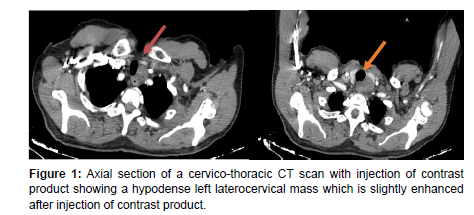
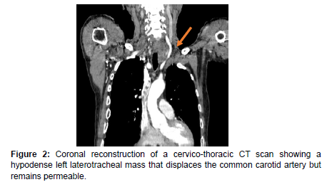
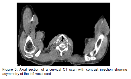
The CT shows a left tracheal mass with a very limited regular contour, hypodense, slightly enhanced after injection of contrast agent, measuring 22x21x45mm. This mass comes into contact with the esophagus without a fatty separation line behind it, and pushes the thyroid forward. The left common carotid artery remains permeable. Also note the asymmetry of the vocal cords (left vocal cord paralysis).
Our patient underwent an ultrasound-guided biopsy with a histological study which came back in favor of a morphological and immunohistochemical appearance of schwannoma (Figures 1-3).
Under general anesthesia, complete surgical resection of vagus nerve was performed by lateral cervical incision. Anatomical and pathological examination confirmed the diagnosis of vagus nerve schwannoma.
Discussion
Schwannoma is a rare neurogenic benign tumor developed at the expense of nerve sheath, and about one third of schwannomas occur in the head and neck region, vagus nerve schwannoma is an extremely rare tumor [1]. The diagnosis is difficult, it is clinical and radiological, its evocation is not of 1st attention.
The clinical presentation is not pathognomonic. Most often, it is an isolated, asymptomatic, later cervical mass that gradually increases in size.
The size of this mass may lead to pharyngeal compression and nonspecific signs such as pharyngeal discomfort or even odynophagia.
When cervical mass is associated with dysphonia of ipsilateral vocal cord paralysis, vagus nerve shwannoma is more likely to be caused [2]. Imaging plays an important role in determining the size and parapharyngeal location of tumors.
Diagnosis is made anatomopathologically on surgical specimens, in the presence of Schwann cells of different morphology and organization, Immunohistochemistry shows predominant S100 protein expression [3,4].
Complete excision of the tumour is the only way to prevent recurrence. Conservation of the original nerve is often possible, followed by rehabilitation to reduce complications, with an excellent prognosis and rare recurrence.
Final diagnosis
Vagus nerve schwannoma
Differential diagnosis list
Esophageal tumor
Adenopathy
Vagus nerve paraganglioma
References
- Chai CK, Tang IP, Prepageran N, FRCSEdin ORL, Jayalakshmi P, et al. (2012) An Extensive Cervical Vagal Nerve Schwannoma: A Case Report. Med J Malaysia 67: 343.
- Najib Benmansour, Yasine Elfadl, Amal Bennani, Mustapha Maaroufi, Leila Chbani, et al. (2013) Schwannome cervical du nerf vague: Stratégies diagnostique et thérapeutique. Pan African Medical Journal 14: 1.
- Behuria S, Rout TK, Pattanayak S (2015) Diagnosis and management of schwannomas originating from the cervical vagus nerve. Ann R Coll Surg Engl 97: 92-97.
- Kanatas A, Mücke T, Houghton D, Mitchell DA (2009) Schwannomas of the head and neck. Oncol Rev 3: 107-111.
Indexed at, Google Scholar, Crossref
Indexed at, Google Scholar, Crossref
Citation: Ez-zaky S, Imrani K, Jellal S, Nassar I, Billah NM (2024) Vagus NerveSchwannoma: A Rare Origin of Dysphonia and Dysphagia. OMICS J Radiol 13:526.
Copyright: © 2024 Ez-zaky S, et al. This is an open-access article distributed underthe terms of the Creative Commons Attribution License, which permits unrestricteduse, distribution, and reproduction in any medium, provided the original author andsource are credited.
Share This Article
Open Access Journals
Article Usage
- Total views: 218
- [From(publication date): 0-0 - Aug 16, 2024]
- Breakdown by view type
- HTML page views: 187
- PDF downloads: 31


