Research Article Open Access
Utility of Calretinin Stain in Work-up of Inadequate Biopies in Patient’s with Hirschsprung’s Disease.
Sikandar M*, Nagi AH and Naseem NDepartment of Morbid Anatomy and Histopathology, University of Health Sciences, Lahore, Pakistan
- Corresponding Author:
- Mishal Sikandar
Department of Morbid Anatomy and Histopathology
University of Health Sciences, 96-Z
Street 32, Phase 3, DHA, Punjab 042, Lahore, Pakistan
Tel: 9203346508547
E-mail: mishal_sikandar@yahoo.com
Received Date: June 08, 2017; Accepted Date: July 05, 2017; Published Date: July 12, 2017
Citation: Sikandar M, Nagi AH, Naseem N (2017) Utility of Calretinin Stain in Work-up of Inadequate Biopies in Patient’s with Hirschsprung’s Disease. J Gastrointest Dig Syst 7:515. doi:10.4172/2161-069X.1000515
Copyright: © 2017 Sikandar M, et al. This is an open-access article distributed under the terms of the Creative Commons Attribution License, which permits unrestricted use, distribution, and reproduction in any medium, provided the original author and source are credited.
Visit for more related articles at Journal of Gastrointestinal & Digestive System
Abstract
Background and Objective: A thorough study was designed with an objective of observing utility of calretinin as immune-histochemical marker for aganglionosis and for detection of ganglion cells in the inadequate biopies of affected areas for more accurate and better diagnosis of the disease.
Study Design: It was observational, descriptive study.
Setting: It was carried out at the Department of Morbid Anatomy and Histopathology in University of Health Sciences (UHS) Lahore.
Period: The study commenced in March 2016 after approval of the synopsis by the Advance Studies and Research Board of UHS and was successfully completed in December 2016.
Methodology: Biopsy specimens of colon which were considered for the study were collected from 73 patients from Mayo Hospital and Jinnah Hospital, Lahore with established histopathologically diagnosed HSCR on H&E staining.
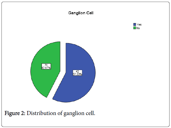
Results: The mean age was 12.52 ± 9.21 months. On the basis of Calretinin staining Ganglion cells were present in 42/73 (57.53%) and absent in 31/73 (42.47%) respectively.
Conclusion: It was consummated that Calretinin provides a reliable and very cost effective adjunctive test to be used routinely with H&E in diagnosing HSCR and consequently waiving off the need for unnecessary surgeries and repeated biopsies.
Keywords
Hirschsprung’s disease; Diagnosis; Rectal biopsy; Aganglionosis; Acetyl-cholinesterase; Calretinin
Introduction
Hirschsprung’s disease, a malformation of the hindgut, is characterized by congenital mega-colon due to the absence of ganglion cells in our enteric nervous system [1]. It is a very important colonic disease in children that causes life-threatening constipation [1]. A Danish pediatrician in 1988 first described it as a chronic severe constipation which leaded to a mega-colon [2,3]. Embryologically it is due to lack of migration of neural crest cells which results in the absence of parasympathetic ganglion cells in the meissner’s and the myenteric plexuses. Estimated incidence is 1 out of 5000 live births with a male to female preponderance and ratio of 4:1.1 [2].
The disease primarily presents in the period of infancy, in which some patients present with insistent, debiliatating and severe constipation later on in life. Symptoms in infancy include impaired peristalsis, vomiting, feeding poorly, insufficient gain of weight, poor milestones and progressive distention of the abdomen [4]. Gastrointestinal functional disorders predominantly constipation are common cause of morbidity in otherwise healthy persons and patients with various predisposing diseases [5].
Definitive curative treatment, i.e. resection surgically, depends on a definitive diagnosis of HD histopathologically which rests upon aganglionosis in the tissue biopsy. Absence of a particular histological feature can be a result of improper presentation of specimen or inadequacy of specimen, so, a suitable ancillary technique would be extremely useful in providing diagnostic accuracy [6]. So an early diagnosis is cardinal to overcome developing complications (e.g., enterocolitis, colonic rupture) [4]. Diagnosis of Hirschsprung’s disease (HSCR) rests upon histologic and/or histochemical staining of sections from rectal suction biopsies. Acetyl-cholinesterase histochemistry (AChE) aids diagnosis but has its pitfalls as it requires special handling of the tissue [7]. One study further added that a certain type of nerve cell bodies in submucosa and the myenteric ganglia of the gastrointestinal tract were seen to show immunopositivity for calretinin, a calcium binding protein playing an important role in the functioning and organisation of our central nervous system [8]. Recently, it is reported that calretinin immunohistochemical staining is found to be superior to conventional acetylcholinesterase staining to confirm absence of ganglions [9].
In this study, we observed calretinin’s expression as a marker for aganglionosis and hypertrophic nerve fibers in biopsies of patients with HD. The proposed study was an attempt to identify the role of calretinin in diagnosing Hirschsprung’s disease which can be practiced as reliable method in routine laboratory setups.
Materials and Methods
Study design and setting
It was an observational, descriptive study carried out at Department of Morbid Anatomy and Histopathology in University of Health Sciences (UHS) Lahore. The study commenced in March 2016 after approval of the synopsis by the Advance Studies and Research Board of UHS and was successfully completed in November 2016.
Sample size
It was calculated with confidence level of 95% and 5% margin of error and taking expected positivity of calretinin immunostaining for diagnosing the aganglionic HD intestinal specimens. The sample size was worked out as 73, out of which 2 were considered as control whereas the remaining 71 samples were used for detailed evaluation.
Sampling technique
Non-purposive convenient sampling technique was employed for the study.
Sample selection
Inclusion criteria
• 73 Cases of Hirschsprung's disease (HD) on histopathology were included in this study, irrespective of gender and age limits.
Exclusion criteria
• Blocks with insufficient/non diagnostic biopsies and autolysed specimens.
• Biopsies from anorectal transformation zone were excluded from the study.
Sample preparation
The samples of HD colorectal biopsies for the study were collected from Mayo and Jinnah Hospital, Lahore. For the purpose of data collection, the pediatric surgery wards of mentioned hospitals along with their associated Pathology departments/laboratories at associated medial colleges were contacted. Most of the cases for rectal biopsies were available at Mayo Hospital and the samples were initially processed at pathology laboratory of affiliated King Edward Medical College, Lahore. Throughout the research, close contact was maintained with the hospital and allied pathology lab for acquisition of the study samples including paraffin embedded blocks, histological reports of the diagnostic biopsies and diseased colon specimens for cases where surgeries were performed. The collected data/materials were then brought to the department of Morbid Anatomy and Histopathology UHS for further processing and utilization for the study.
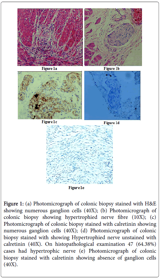
As stated earlier, the study samples were collected from children with HD without any consideration for gender and age. The approach for sampling was generally the same for all the patients however there was a very slight difference for children who were less than 4 months and for those older than 4 months. For children with less than 4 months of age diagnostic biopsies were taken to the associated pathology laboratory where they were processed, paraffin embedded blocks were made for cutting, slides with routine H&E staining procedure were prepared for diagnosis to be given to surgeon (Figure 1). The paraffin embedded blocks were brought to laboratory of Department of Morbid Anatomy and Histopathology which had already been undertaken for diagnostic purposes and HD had been established at the laboratory of pathology department of the respective hospital. Only 1 or 2 biopsies were available for each case, blocks where they were re-embedded, and new slides were made to be studied in detail at our laboratory and results were correlated with those of pathology laboratory of King Edward Medical College, Lahore.
Figure 1: (a) Photomicrograph of colonic biopsy stained with H&E showing numerous ganglion cells (40X); (b) Photomicrograph of colonic biopsy showing hypertrophied nerve fibre (10X); (c) Photomicrograph of colonic biopsy stained with calretinin showing numerous ganglion cells (40X); (d) Photomicrograph of colonic biopsy stained with showing Hypertrophied nerve unstained with calretinin (40X). On histopathological examination 47 (64.38%) cases had hypertrophic nerve (e) Photomicrograph of colonic biopsy stained with calretinin showing absence of ganglion cells (40X).
Likewise for children with ages greater than 4 months, undergoing colectomy, the biopsies had already been taken for diagnostic purposes and HD had been established at the pathology lab of the hospital. However, for such cases, fresh biopsies were undertaken during surgeries in the presence of the researcher. Several biopsies were taken from the proximal and distal colostomy margins and also from the middle aganglionic portion of the diseased colon segments removed during the colectomies. Biopsies were taken to the laboratory at the Department of Morbid Anatomy and Histopathology, UHS for processing and also to the associated pathological laboratory of the hospital and the slides were cut and processed for H&E staining. In both the cases, the acquired biopsy sections were used for diagnostic procedures including H&E staining and calretinin immunohistochemistry.
Data collection
Proformas were prepared to record the socio-demographic information (name, age, gender, family history) as well as clinical details of the patients including presenting complaints, age of weaning and associated syndrome. Data from biopsy specimens like morphologic features (in case of full thickness biopsy) and microscopic features of mucosa, submucosa, muscularispropria and serosa were also recorded in separate proformas (Table 1).
| IHC Positivity | Results |
| No. staining of ganglion | Positive for HD, if any specific staining (excluding mast cells) is present within the submucosal nerve plexus, muscularis mucosa or lamina propria |
| Staining of ganglion cells | Negative for HD |
Table 1: Quantification: Reporting technique [10].
Results
Demographics: The mean age of patients was 12.52 ± 9.21 days.
Sign and symptoms: Patients showed signs and symptoms of fever, constipation, vomiting, enterocolitis, failure to thrive and had palpable abdominal masses.
Histopathological examination: In histopathological examination 47/73 (64.38%) cases had hypertrophic nerves. A total of 57/73 (78%) cases had chronic inflammation. Ganglion cells were present in 42 (57.53%) and absent in 31 (42.47%) respectively. In patients with ganglion cell, 30/42 (71.4%) had hypertrophic nerve and 12/42 (28.6%) patients did not have hypertrophic nerve. Among those who did not have ganglion cells, 17/31 (54.8%) patients had hypertrophic nerve while 14/31 (45.2%) did not have hypertrophic nerve; there was no significant difference of hypertrophic nerve and ganglion cell, pvalue> 0.05 (Table 2).
| Symptoms | Ganglion Cell | p-value | ||
|---|---|---|---|---|
| Yes | No | |||
| Hypertrophic Nerve | Yes | 30(71.4%) | 17(54.8%) | 0.143 |
| No | 12(28.6%) | 14(45.2%) | ||
| Chronic Inflammation | Yes | 37(88.1%) | 20(64.5%) | 0.016 |
| No | 5(11.9%) | 11(35.5%) | ||
Table 2: Comparison of ganglion cell findings with hypertrophied nerves and chronic inflammation.
In patients with ganglion cell, 37/42 (88.1%) had chronic inflammation and 5/42 (11.9%) patients did not have chronic inflammation. Among those who did not have Ganglion cells, 20/31(64.5%) patients had chronic inflammation while 11/31 (35.5%) did not have chronic inflammation; there was no significant difference of chronic inflammation and ganglion cell, p-value>0.05 (Figure 2).
Discussion
Definitive diagnosis of the disease is basically in view of the proof of the aganglionosis in the pathological areas of the colon. The process is extremely troublesome and tedious, furthermore needs a few serial cut sections of various segments of colon. There are numerous proposed methods in this field, yet none of them has been agreed upon by pathologists to be accepted completely. The standard criterion to diagnose a case of HSCR is the absence of ganglion cells in affected portion of bowel wall.
Routinely used gold standard is H&E with acetyl-cholinestrase. Although acetyl-cholinesterase histochemistry can be a useful ancillary technique to help in the diagnosis and preoperative planning, some studies suggest that immunohistochemical (IHC) staining for calretinin might be more accurate than acetyl-cholinesterase staining in diagnosing congenital aganglionosis in suction biopsy specimens. Acetyl-cholinesterase just identifies hypertrophic nerves not the presence or absence of ganglion cells. Calretinin is the only non-toxic and cost effective marker to reliably detect ganglion cells. So, it should be used as an adjunct in the diagnosis of Hirschsprung’s disease. Calretinin immunohistochemistry (IHC) was presented as a diagnostic marker to beat the issues in analysis of this pathology around 5 years back [11]. The free calcium focus intracellularly subserves brain’s complex flagging part. Calcium (Ca2+) manages variety of neurons and neuronal survival [12]. Calretinin, calbindin D-28, and parvalbumin have a place with a group of Ca2+-binding proteins, which are more than 200 in man [12].
In current study with mean age of the patient, 12.52 ± 9.21 days, there were 48/73 (65.8%) cases who were ≤ 12 months old, 20/73 (27.4%) were 12.1-60 months old and 5/73 (6.8%) cases were 60.10-120 months old. The mean age of our patients was almost consistent with review of literature by Friedmacher et al. In another study conducted on 101 patients with Hirschsprung’s disease at a University Teaching Hospital in Northwestern Tanzania, maximum numbers of patients were less than 1 year of age.
We found that after calretinin staining ganglion cells were present and absent in 42/73 (57.53%) and 31/73 (42.47%) respectively. Amongst various markers used to diagnose HSCR, calretinin has been seen to have the most potential to be used as a robust ancillary test. The expression of Caretinin was not seen in HD in other studies. The ratio of calretinin expression is almost same in different studies as we found in our patients. The first study to demonstrate differences in immunohistochemical staining for calretinin between the ganglionic and aganglionic portions of bowel in HD was done by Barshack. He studied ten large bowels. Full thickness biopsy specimens from patients with a classic rectosigmoid HD were selected from the pathology repository. In total 54 paraffin wax blocks were processed, out of which 24 were from the ganglionic zone, 17 were from the aganglionic zone and 13 were from the transitional zone [8]. Other subsequent studies by Guinard-Samuel et al. they took 131 rectal biopsy among them 130 biopsies were correctly diagnosed on the basis of the positive and negative calretinin staining. Initially 12 more cases considered doubtful for HD, diagnosed using the standard method, were accurately diagnosed with calretinin immunohistochemistry. Calretinin immunohistochemistry overthrows most of the obstacles encountered using combination of the histology and acetylcholinesterase staining, and detects almost all cases of HD with confidence, with no false positives [13]. According to Gonzalo et al. all 12 of the patients without the Hirschsprung’s disease had Calretininpositive nerve fibers in lamina propria or the muscularis mucosae, and all 5 of patients with Hirschsprung disease had no staining of the nerves i-e Calretinin-negative [10]. One more study reported that there was great concordance between the final diagnosis of both pathologists and gold standard (k>0.9). Calretinin immunostaining showed 100% specificity and positive predictive value and more than 90% sensitivity and negative predictive value. High agreement was present between the two pathologists (k>0.9) [11]. One more study was done with similar objectives, they reported that in the normal rectal suction biopsies, stained with calretinin IHC, thin linear nerve fibrils were found in the lamina propria, the muscularis mucosae and the superficial submucosa, but did not show ganglion cells [14]. Another study reported that out of the 72 non-HD patients, three false positive results were obtained, which were due to diminished immunoreactivity of previously frozen biopsy specimens. In blinded slide review, 2 of the reviewers correctly reported 100/101 biopsies (from 99 patients) whereas 1 reviewer correctly reported 99/101 biopsies. All contradictory findings by the reviewers were due to examination of the sections at only low (40X and 100X) magnification and then misinterpreting positive calretinin staining as absent [6]. Our results are in consistence to these statistics. Moreover, we in current study found significant association of Ganglion cells with history of constipation, vomiting, enterocolitis and chronic inflammation, pvalue< 0.05. The use of calretinin may help the pathologists in making accurate and reliable diagnosis for HD and consequently eliminating the need for repeated biopsies and unnecessary surgeries. The same findings were observed and validated in the literature.
Conclusion
Through the findings of this study it can be concluded that Calretinin as immunohistochemical marker provides a reliable, accurate and a very cost effective adjunctive test to be used routinely with H&E for diagnosis of HSCR in inadequate Rectal Section Biopsies (RSBs) consequently eliminating the need of repeated biopsies with unnecessary surgeries.
Acknowledgements
The author acknowledges the encouragement extended by the Vice Chancellor of University of Health Sciences, Lahore. Also the colleagues and the laboratory staff of Department of Morbid Anatomy and Histopathology, UHS, Lahore, Pakistan.
Contribution of Authors
• Dr Mishal Sikandar carried out the whole research work and wrote the article.
• Prof. A.H. Nagi helped in providing material for research work and guided throughout the research.
• Dr Nadia Naseem helped in research work and writing the article.
References
- Chia ST, Chen SC, Lu CL, Sheu SM, Kuo HC (2016) Epidemiology of Hirschsprung'sdisease in Taiwanese children: A 13-year nationwide population-based study. PediatrNeonatol57: 201-206.
- Amiel J, Lyonnet S(2008) Hirschsprung disease, associated syndromes and genetics: A review. J Med Genet 45: 729-739.
- Skaba R (2007) Historic milestones of Hirschsprung'sdisease (commemorating the 90th anniversary of Professor HaraldHirschsprung's death). J PediatrSurg 42:249-251.
- Kessmann J (2006) Hirschsprung’s disease: Diagnosis and management. Am Family Physician 74:1319-1322.
- Peppas G, Alexiou VG, Mourtzoukou E, Falagas ME (2008) Epidemiology of constipation in Europe and Oceania: A systematic review. BMC Gastroenterology 8:5.
- Lim KH, Wan WK, Lim TKH, Loh AHL, Nah SA, et al. (2014) Primary diagnosis of Hirschsprungdisease-Calretininimmunohistochemistry in rectal suction biopsies, with emphasis on diagnostic pitfalls. World Journal of Pathology 3: 3.
- Kapur RP, Reed RC, Finn LS, Patterson K, Johanson J, et al. (2008) Calretinin immunohistochemistry versus acetyl-cholinesterase histochemistry in the evaluation of suction rectal biopsies for Hirschsprung disease. PediatrDevPathol 12:6-15.
- Barshack I, Fridman E, Goldberg I, Chowers Y, Kopolovic J (2004) The loss of calretinin expression indicates aganglionosis in Hirschsprung’s disease. J Clinic Pathol 57:712-716.
- Holland SK, Ramalingam P, Podolsky RH, Reid-Nicholson MD, Lee JR (2011) Calretininimmunostaining as an adjunct in the diagnosis of Hirschsprung disease. Ann Diagnostic Pathol15:323-328.
- Gonzalo DH, Plesec T (2013) Hirschsprung disease and use of calretinin in inadequate rectal suction biopsies. Arch Pathol Lab Med 137:1099-1102.
- AnbardarMH, Geramizadeh B, Foroutan HR (2015) Evaluation of calretinin as a new marker in the diagnosis ofHirschsprungdisease. Iran J Pediatr25: e367.
- Zündorf G, Reiser G (2011) Calcium dysregulation and homeostasis of neural calcium in the molecular mechanisms of neurodegenerative diseases provide multiple targets for neuroprotection. Antioxid Redox Signal 14:1275-1288.
- Guinard-Samuel V, Bonnard A, De Lagausie P, Philippe-Chomette P, Alberti C, et al. (2009) Calretinin immunohistochemistry: Asimple and efficient tool to diagnose Hirschsprung disease. Mod Pathol 22:1379-1384.
- Alexandrescu S, Rosenberg H,Tatevian N (2013) Role of calretininimmunohistochemical stain in evaluation of Hirschsprung disease: An institutional experience.Int J Clinic Experiment Pathol 6:2955-2961.
Relevant Topics
- Constipation
- Digestive Enzymes
- Endoscopy
- Epigastric Pain
- Gall Bladder
- Gastric Cancer
- Gastrointestinal Bleeding
- Gastrointestinal Hormones
- Gastrointestinal Infections
- Gastrointestinal Inflammation
- Gastrointestinal Pathology
- Gastrointestinal Pharmacology
- Gastrointestinal Radiology
- Gastrointestinal Surgery
- Gastrointestinal Tuberculosis
- GIST Sarcoma
- Intestinal Blockage
- Pancreas
- Salivary Glands
- Stomach Bloating
- Stomach Cramps
- Stomach Disorders
- Stomach Ulcer
Recommended Journals
Article Tools
Article Usage
- Total views: 2673
- [From(publication date):
August-2017 - Nov 21, 2024] - Breakdown by view type
- HTML page views : 2057
- PDF downloads : 616


