Uterine Procidentia: Prolapse in its Most Severe Form
Received: 04-Apr-2023 / Manuscript No. roa-23-94921 / Editor assigned: 06-Apr-2023 / PreQC No. roa-23-94921 (PQ) / Reviewed: 20-Apr-2023 / QC No. roa-23-94921 / Revised: 22-Apr-2023 / Manuscript No. roa-23-94921 (R) / Published Date: 29-Apr-2023 DOI: 10.4172/2167-7964.1000438
Image Article
Pelvic organ prolapse corresponds to an abnormal downward herniation of various pelvic organs whether through the perineum or into it. Uterine prolapse (UP) corresponds to its descent from normal anatomical borders to outside the vaginal introitus (or within it).
In its most severe form it is called procidentia. It results from pelvic muscles weakening and can be associated to different prolapse symptoms such as a sensation of fullness inside and a bulge outside the vagina as well as sexual, urinary, and bowel dysfunction. Major risk factors are pregnancy at an early age, multiparity, advanced age, delivery by unskilled attendants, obesity, chronic obstructive pulmonary diseases, and also other factors which may chronically cause an intraabdominal pressure raise [1].
Physical examination is essential for diagnosing pelvic organ prolapse in its mild form. However, in patients with severe symptoms, such as serious urinary incontinence, procidentia and fecal incontinence, clinical examination may be difficult, and imaging is the useful tool [2].
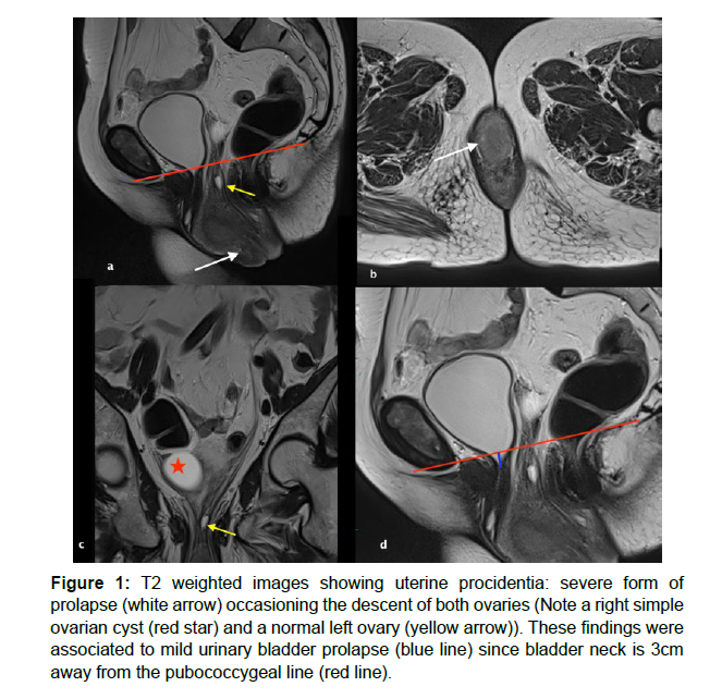
MRI has emerged as a an invaluable tool to assess severe forms of pelvic floor dysfunction, as it also helps in preoperative planning since it provides detailed anatomic information of all three compartments of the pelvic floor (anterior, middle and posterior) [1]. Dynamic MRI diagnoses can also help to visualize these abnormities, since images are obtained while the patient is contracting and relaxing the pelvic muscles. In this technique, we use the pubococcygeal line to demarcate the level of the pelvic floor on the mid sagittal images. It extends from the most inferior portion of the pubic symphysis to the last horizontal sacrococcygeal joint. Moreover, on the image showing the most severe degree of pelvic organ herniation, we may draw perpendicular lines from the PCL to the anterior aspect of the anorectal junction, the bladder neck, and the anterior cervical lip (or in the case of a hysterectomy: vaginal apex), to evaluate the degree of descent of pelvic compartments [2] (Figure 1).
Figure 1: T2 weighted images showing uterine procidentia: severe form of prolapse (white arrow) occasioning the descent of both ovaries (Note a right simple ovarian cyst (red star) and a normal left ovary (yellow arrow)). These findings were associated to mild urinary bladder prolapse (blue line) since bladder neck is 3cm away from the pubococcygeal line (red line).
References
- Badacho AS, Lelu MA, Gelan Z, Woltamo DD (2022) Uterine prolapse and associated factors among reproductive-age women in south-west Ethiopia: A community-based cross-sectional study. PLoS One 17: e0262077.
- Salvador JC, Coutinho MP,Venancio JM, Viamonte B. Dynamic magnetic resonance imaging of the female pelvic floor—a pictorial review. Insights Imaging 10: 4.
Indexed at, Google Scholar, Crossref
Citation: HARRAS Y, MANDOUR J, CHOAYB S, ALLALI N, CHAT L, et al. (2023)Detecting Calcified Liver Metastasis in Breast Cancer. OMICS J Radiol 12: 437. DOI: 10.4172/2167-7964.1000438
Copyright: © 2023 HARRAS Y, et al. This is an open-access article distributedunder the terms of the Creative Commons Attribution License, which permitsunrestricted use, distribution, and reproduction in any medium, provided theoriginal author and source are credited.
Share This Article
Open Access Journals
Article Tools
Article Usage
- Total views: 1572
- [From(publication date): 0-2023 - Mar 29, 2025]
- Breakdown by view type
- HTML page views: 1279
- PDF downloads: 293

