Update of Usefulness and Adverse Effects of Nanoparticles on Animals and Human Health
Received: 20-Mar-2018 / Accepted Date: 02-Apr-2018 / Published Date: 11-Apr-2018
Abstract
Nanotechnology has a potential application and a great biological role in veterinary diagnosis and in animal reproductive biotechnologies beside great impact on diagnosis in animal diseases. There is also a great interest from toxicologist for research on safety of different nanoparticles that used in cure of diseases. Nanoparticles reported to have various adverse effects in different animal tissue and also in human health. Various nanoparticles have toxic effect on liver, lung, skin, eye, reproductive disturbance and blood cells. The key mechanism of toxicity of nanoparticle is due to increase its concentration in non-target tissue of therapy through genotoxicity, oxidative stress or hemotoxic effects. On other word, nanoparticles could have adverse effects on healthy rather than diseased tissue.
Keywords: Nanoparticles; Animals; Toxicity
Introduction
Nanotechnology is defined as an application of scientific knowledge for manipulation and control in nanometric scale (1- 100 nm) with specific function at the cellular, atomic and molecular levels [1]. Nanostructures may be a new physical and chemical characteristic, demonstrating high solubility levels, reactivity and better stability than its original compound [2]. The application of the novel nanoparticles generated the new research field of Nano biotechnology, which plays a central role in disease diagnosis, drug design and delivery [3]. It holds a major promise for animal health, veterinary medicine and animal production and also has a key role in treatment of diseases by development of smart drug delivery systems which providing targeted time controlled self-regulated, pre-programmed and effective dosage of drug to site of disease. Moreover, Nano medicine includes the use of nanoparticles for diagnosis and treatment of a variety of diseases, as well as in regenerative medicine [4]. Currently, employing nanoparticles in medicine as a drug delivery, heat, light or other substances to a specific types of cells such as cancer cells [5]. Nanoparticles are also used in reduction of FMD which is controlling disease of cattle and other ruminants that had severe consequences in United Kingdom, as nanotechnology is the key enabling sensitive detection in a very small scale by a rapid sensitive test of virus prior to disease symptoms (nanomaterial are bound as hand-held detector) and its application poses little to risk and great social benefits [6].
Silver nanoparticles (Ag NPs) could be used as coating of the device. Nano-silver coating has been applied to several medical catheters, drains and wound dressings are the most prominent as it leading to reduce colonization, infection rate, and hospitalization days, wound healing and show economic benefits. The efficiency of silver to improve clinical outcome depend on application and device. Wound dressing used for treatment of burns and non-healing wound with silver sulfadiazine crème or with other silver compound or salts [7]. Key mechanisms of toxicity of Silver nanoparticles include oxidative stress and genotoxicity, disruption of actin cytoskeleton, activation of lysosomal AcP activity, and stimulation of phagocytosis in blood cells and increase of MXR transport activity and inhibition of Na-K-ATPase in gill cells of fish [8].
Silver nanoparticles had the ability to inhibit the activities of interferon gamma and tumor necrosis factor alpha which involved in inflammation. The study proved that silver nanoparticles had anti-inflammatory action which can be used in many therapies. Also Silver nanoparticles (AgNPs) are widely used because of their anti-bacterial and anti-inflammatory properties; however, the adverse health effects of these nanoparticles, especially to the lungs [9]. Silver nanoparticles are also shown toxic effect on male reproductive system as it could pass blood-testis barrier and deposited in testes and adversely affect the sperm [10].
Additionally, the aquatic animals affected by silver nanoparticles, as silver nanoparticles could interact with gills of fish and inhibit basolateral Na+-K+-ATPase activity which inhibits osmoregulation in the fish [11]. Additionally, a combination of a low level of the NPs in the chitosan matrix improved their antimicrobial benefit. However, aqueous exposure to these materials still had hazardous effects on fish health [12].
Prostate cancer in dog was considered a highly dangerous disease but scientists at university of Missouri developed a new formulation of gold nanoparticles for treatment of dog prostate cancer. This new treatment doses that thousands times smaller than used in chemotherapy and didn’t travel through body so not cause damage to the healthy tissues [13]. Notably, gum arabic-coated radioactive gold nanoparticles therapy had no toxicity in the treatment of natural occurring prostatic cancer in dog [14].
Mastitis is a disease of high milk yielding animals and causing high economic loss include decreased milk yield and increased use of drugs and veterinary services which caused by Staphylococcus, Streptococcus and E. coli could be treated by zinc oxide nanoparticles (ZnONPS), where they could reach the target organism generally inaccessible to antibiotics [15].
Titanium dioxide, due to its spherical shape with zero dimensionality had a large specific surface area, it used in a higher rate of photo catalytic decomposition of organic pollutants [16]. Although the unique physical and chemical properties of silver, an excellent anti-microbial and anti-inflammatory action it has adverse effect on environment and human. Titanium in many studies causes pulmonary toxicity of acute exposure to anatase form of titanium in terms of bronco-alveolar lavage (BAL) inflammation [17]. Also, mixtures of anatase and rutile particles of Tio2 were able to induce increase in BAL fluid inflammatory parameters and lung histopathological alteration by inhalation in rat’s sub acutely [18].
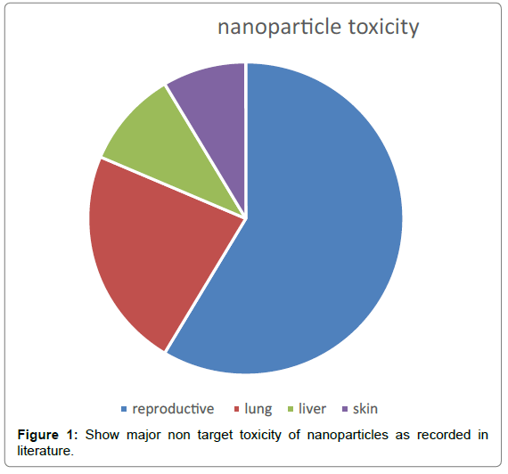
The trace mineral elements in brain were altered after the exposure to Tio2 NP which may responsible of the spatial recognition memory impairments reported in treated mice due to the disrupted homeostasis of trace elements, neurotransmitters and enzymes in the brain [19,20]. Also Titanium dioxide (TiO2) has been recorded to induce diverse pulmonary responses in exposed animals (Figure 1) [21].
In human exposed to free silver ions caused permanent bluish-grey discoloration of skin (argyria) or argyrosis due to silver ions in aqueous phase as a result from industrial wastes but the exposure to soluble silver caused toxic effect on liver and kidney and damage of eye, skin, respiratory tract and intestinal tract irritation and disturbances of blood cells [22].
Results
It was notice from analysis of previous literature that different nanoparticles used in veterinary fields for diagnosis, treatment of natural occurring disease especially cancer in valuable breeds of dog and cat. Also nanoparticles used in domestic animals for treatment of diseases as mastitis and used frequently in fish and treatment and safe of zoo or wild animals in captive protected area. The key mechanism of toxicity of nanoparticles in veterinary is due to increase in its leakage of nanoparticles in non-target tissue of therapy through genotoxicity, oxidative stress or hemotoxic effects. On other word, nanoparticles could have adverse effects on healthy rather than diseased tissue.
Discussion
Nanoparticles is promising tools in future medicine even there were toxic for reproductive organ [10,15], lung [9], liver and skin [22]. Toxicity of nanoparticles used in veterinary due to increase its accumulation of nanoparticles in non-target tissue of therapy [13,14] through genotoxicity, oxidative stress or hemotoxic effects [8]. On other word, nanoparticles could have adverse effects on healthy rather than diseased tissue. While adverse effect of nanoparticles includes liver, kidney and eye damage, skin, respiratory tract and intestinal tract irritation and disturbances of blood cells [22].
Finally, nanoparticles are hopeful agent in veterinary and human cure of diseases but still have adverse effects and need more investigation to reduce its toxicity.
References
- Teli MK, Mutalik S, Rajanikant GK (2010) Nanotechnology and Nanomedicine: going small means aiming big. Curr Pharm Des 16: 1882-92.
- Troncarelli MZ, Brandão HM, Gern JC, Guimarães AS, Langoni H (2013) Nanotechnology and Antimicrobials in Veterinary Medicine. Badajoz, Spain: FORMATEX.
- Ramos AP, Cruz MAE, Tovani CB, Ciancaglini P (2017) Biomedical applications of nanotechnology. Biophys Rev 9: 79-89.
- Chang EH, Harford JB, Eaton MA, Boisseau PM, Dube A, et al. (2015) Nanomedicine: Past, present and future - A global perspective. Biochem Biophys Res Commun 468: 511-17.
- Sahoo SK, Ma W, Labhasetwar V (2004) Efficacy of transferrin-conjugated paclitaxel-loaded nanoparticles in a murine model of prostate cancer. Int J Cancer 112: 335-40.
- Ward N, Donaldson A, Lowe P (2004) Policy framing and learning lessons from the UK's foot and mouth disease crisis. Environment and planning C: government and Policy. 22: 291-306
- Dowsett C (2004) The use of silver-based dressing in wound care. Nurs Stand 19: 56-60.
- Katsumiti A, Gilliland D, Arostegui I, Cajaraville MP (2015) Mechanisms of Toxicity of Ag Nanoparticles in Comparison to Bulk and Ionic Ag on Mussel Hemocytes and Gill Cells. PLoS One 10: e0129039.
- Fehaid A, Mohamed F, Hamed M, Abouelmagd, Akiyoshi T (2016) Time-dependent Toxic Effect and Distribution of Silver Nanoparticles Compared to Silver Nitrate after Intratracheal Instillation in Rats. Am J Nanomaterial 4: 12-19.
- Shin SH, Ye MK, Kim HS, Kang HS (2007) The effect of nano-silver on proliferation and cytokiene expression by peripheral blood mononuclear cell). Int Immune Pharmacol 7: 1813-1818.
- Erskine RJ, Wagner S, DeGraves FJ (2003) Mastitis therapy and pharmacology. Vet Clin North Am: Food Anim Pract 19: 109-138.
- Liu B, Nakata K, Sakai M, Saito H, Ochiai T, et al. (2011) Mesoporous TiO2 core shell spheres composed of nanocrystals with exposed high-energy facets: facile synthesis and formation mechanism. J Am Chemical Society 27: 8500-8508
- Panyala NR, Pena Mendes EM, Havel J (2008) Silver nanoparticles; a hazardous threat to the environment and human health? J Appt Biomed 6: 117-129.
- Axiak-Bechtel SM, Upendran A, Lattimer JC, Kelsey J, Cutler CS, et al. (2014) Gum arabic-coated radioactive gold nanoparticles cause no short-term local or systemic toxicity in the clinically relevant canine model of prostate cancer. Int J Nanomedicine 9: 5001-11.
- Brohi RD, Wang L, Talpur HS, Wu D, Khan FA, et al. (2017) Toxicity of Nanoparticles on the Reproductive System in Animal Models: A Review. Front Pharmacol 8: 606.
- Wood CM, Playle RC, Hogstrand C (1999) Physiology and modeling of mechanism of silver uptake and toxicity of fish. Environ Toxical Chem 18: 71-83.
- Abu-Elala NM, AbuBakr HO, Khattab MS, Mohamed SH, El-Hady MA, et al. (2018) Aquatic environmental risk assessment of chitosan/silver, copper and carbon nanotube Nano composites as antimicrobial agents. Int J Biol Macromol 12.
- Li J, Li Q, Xu J, Li J, Cai X, et al. (2007) Comparative study on the acute pulmonary toxicity induced by 3 and 20nm Tio2 primary particles in mice. Environmental Toxicology and Pharmacology 24: 239-244.
- Ravenzwaay B, Landsiedel R, Fabian E, Burkhardt S, Strauss V, et al. (2009) (Comparing fate and effect of three particles of different surface properties; nano-TiO2, pigmentary Tio2 and quartz) Toxicology Lettters. 186: 152-159.
- Hu R, Gong X, Duan Y, Li N, Che Y, et al. (2010) (Neurotoxicological effect and the impairment of spatial recognition memory in mice caused by exposure to TiO2 nanoparticles Biomaterial. 31: 8043-8050.
- Hu R, Gong X, Duan Y, Li N, Che Y, et al. (2010) Neurotoxicological effects and the impairment of spatial recognition memory in mice caused by exposure to TiO2 nanoparticles. Biomaterials. 31: 8043-50.
- Iavicoli I, Leso V, Bergamaschi A (2012) Toxicological Effects of Titanium Dioxide Nanoparticles: A Review of In Vivo Studies. J Nanomaterials 5: 1-36.
Citation: Elalfy MM, El-hadidy MG, Abouelmagd MM (2018) Update of Usefulness and Adverse Effects of Nanoparticles on Animals and Human Health. J Vet Med Health 2: 106.
Copyright: © 2018 Elalfy MM, et al. This is an open-access article distributed under the terms of the Creative Commons Attribution License, which permits unrestricted use, distribution, and reproduction in any medium, provided the original author and source are credited.
Share This Article
Recommended Journals
Open Access Journals
Article Usage
- Total views: 4583
- [From(publication date): 0-2018 - Mar 12, 2025]
- Breakdown by view type
- HTML page views: 3779
- PDF downloads: 804

