Make the best use of Scientific Research and information from our 700+ peer reviewed, Open Access Journals that operates with the help of 50,000+ Editorial Board Members and esteemed reviewers and 1000+ Scientific associations in Medical, Clinical, Pharmaceutical, Engineering, Technology and Management Fields.
Meet Inspiring Speakers and Experts at our 3000+ Global Conferenceseries Events with over 600+ Conferences, 1200+ Symposiums and 1200+ Workshops on Medical, Pharma, Engineering, Science, Technology and Business
Letter to Editor Open Access
Unusual Skin Toxicity after a Chemotherapic Combination
| Cantisani C1*, Paolino G1, Passarelli F2,Cigna E1, Rota L2, Pallotta S2, Bangrazi C3 and Calvieri S1 | |
| 1UOC of Dermatology, “Sapienza” University of Rome, Policlinico Umberto I | |
| 2IDI Rome, Italy | |
| 3Oncological Department “Sapienza” University of Rome, Policlinico Umberto I, Italy | |
| Corresponding Author : | Cantisani C MD UOC of Dermatology “Sapienza” University of Rome, Italy Tel: +393479385719 Fax: +390649976933 E-mail: carmencantisanister@ gmail.com |
| Received: July 15, 2015 Accepted: August 06, 2015 Published: August 13, 2015 | |
| Citation: Cantisani C, Paolino G, Passarelli F, Cigna E, Rota L, et al. (2015) Unusual Skin Toxicity after a Chemotherapic Combination. Clin Pharmacol Biopharm 4:141. doi:10.4172/2167-065X.1000141 | |
| Copyright: © 2015 Cantisani C et al. This is an open-access article distributed under the terms of the Creative Commons Attribution License, which permits unrestricted use, distribution, and reproduction in any medium, provided the original author and source are credited. | |
Visit for more related articles at Clinical Pharmacology & Biopharmaceutics
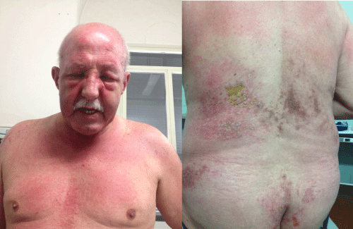
| A 66 year-old Caucasian patient presented to our attention complaining for an heliotrope rash and large diffuse erythematous itching with pustular-like lesions on his trunk, upper arms, involving the upper gluteus, with a yellowish necrotic like surface (Figure 1A,1B). Brown-to-purple lesions were also present on the right side of his back associated to muscle weakness. His personal medical history was positive for diabetes, hypertension and a 14 months history of squamous cell carcinoma of the lung. After 45 days of a sequential chemotherapic combination, with carboplatin/gemcitabine/taxotere/ bevacizumab regimen the patient referred the onset of the skin reaction. |
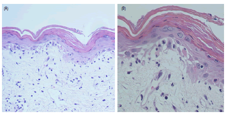
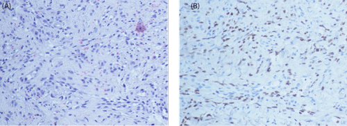
| Laboratory investigations showed red blood cells 3.6 X10³ uL (4.52-5.10 X 10³ uL), hemoglobin 11.6 g/dL(14-18 g/dL), haematocrit 34.4%( 42-50 %), MCV 16.2% femtolitre (80-96 femtolitre), white cells 10.9X10³ uL (4-8.500/mm3) with neutrophilia, ferritin 931 ng/ mL(20-300 ng/ml), LDH 480 U/L(122-222 U/L) and Potassium 5.4 g/dL (3.5-5.1 mEq/L). While the remaining laboratory investigations (including renal function, hepatic functions, autoantibody, calcium and phosphorous) were between normal ranges, as well as a chest X-Ray. The patient also showed an increased body temperature (maximum reached: 38°C), which disappeared after the administration of an intra-venous antibiotic therapy, with normalization of the laboratory tests. A cutaneous biopsy from the arm showed perivascular inflammatory cells in the dermis with increased dermal mucin, and atrophic epidermis with vacuolar basal changes and myofiber necrosis (Figure 2A,2B). According to the clinical and pathological findings, a final diagnosis of dermatomyositis with calciphylaxis aspect was made. The patient underwent to a steroidal immunosuppressive treatment (prednisolone 40 mg/day), with a fast response. However 6 weeks later, he developed a red-violaceous lesion on the lower-arms, histologically suggestive for Kaposi’s sarcoma (KS) (Figure 3A,3B); for which the patient underwent to clinical and instrumental evaluations. |
| As known calciphylaxis (CPX) is a rare condition involving subcutaneous vascular calcification and cutaneous necrosis, mostly observed in patients with renal failure. However CPX may also appear in patients affected by polymyositis, Sjogren syndrome, Lupus Erythematosus systemicus, Sarcoidosis and rheumatoid arthritis, especially in children. Clinically CPX can present itself as subcutaneous nodules, infiltrate plaques or purpuric-like and livedo-like plaques, while in the late stages necrotic ulcers (with a bizarre shape and severe pain) may be the main cutaneous manifestations. After the diagnosis, the percentage of death is around 50% (during the first year), often due to sepsis or organ failure. The reported frequency of malignancy in DM has varied from about 20% to 25%, and probably with more aggressive forms, in this view it can be considered a paraneoplastic manifestation, more frequent in gynecologic malignancy, nasopharyngeal, but also gastrointestinal and lung cancer, especially in patient more than 50 years old. In our patient the lung cancer was in remission and no metastasis was found. Up to date, dermatomyositis (DM) has rarely been described to present as a drug- induced CPX, although a case of CPX with harbinger’s myopathy was described in 1992 by Edelstein et al. [1]. In fact in a small percentage of patients the cutaneous lesions are due to or are exacerbated by drugs, but never reported with chemotherapic agents. Our report shows similarities with a recent published paper, where it was reported a gemcitabine-induced radiation myositis in a patient with dermatomyositis. At this point we are not able to state which drug induced skin toxicity. |
| To the best of our knowledge, in literature there are only 3 reports describing iatrogenic Kaposi’s sarcoma (KS) in patients with DM [2-6], after treatments with prednisolone, prednisone and azathioprine [2-5]. In all these cases KS developed after an immunosuppressive treatment, with a median time of 9 months (ranging between 1/2 months and 30 months). In our patient KS appeared 6 weeks after the immunosuppressive treatment beginning. In this regard KS should be seen as a result of the immunosuppressive treatment, concluding that KS, not usually associated with DM, can appear in this group of patients after an immunosuppressive treatment, with a faster onset time. The pathogenesis of the muscle disease is becoming better understood, but the cutaneous disease mechanisms remain enigmatic. Our patient was under carboplatin/gemcitabine/taxotere/bevacizumab regimen to treat the lung cancer, however, no similar skin toxicity has never been described in literature and is not possible to state which drug can be directly connected, therefore CPX could be explained as drug induced or as a paraneoplastic sign, which revealed in advance another tumour (i.d. KS). |
| This observation is important for rheumatologists, dermatologists and oncologists, due to the association between malignancy and dermatomyositis and the common use of gemcitabine as a chemotherapeutic agent, showing also how a steroid treatment can lead to an improvement of the cutaneous and muscular symptomatology, but it can induce immune disregulation [7]. Assuming that prognosis is poor in oncologic patients, a careful evaluation of each patient should be part of their initial and follow-up assessments. |
| Acknowledgements |
| we would like to thank Associazione Romana Ricerca Dermatologica. |
References
- Edelstein CL, Wickham MK, Kirby PA (1992) Systemic calciphylaxis presenting as a painful, proximal myopathy. Postgrad Med J 68:209-211.
- Simeoni S, Puccetti A, Moruzzi S, Tinazzi E, Peterlana D, et al (2007) Dermatomyositis complicated with Kaposi sarcoma: a case report. ClinRheumatol26: 440-442.
- Weiss VC, Serushan M (1982) Kaposi’s sarcoma in a patient with dermatomyositis receiving immunosuppressive therapy. Arch Dermatol118:183-185.
- Almong Y, Ben-Yehuda A, Ben-Chetrit E (1992). Dermatomyositisassociated with the recurrence of transitional cellcarcinomas and Kaposi’s sarcoma. ClinExpRheumatol92:285-288.
- Louthrenoo W, Kasitanon N, Mahanuphab P, Bhoopat L, Thongprasert S (2003) Kaposi’s sarcoma in rheumatic diseases. SeminArthritis Rheum 2003; 32:326-333.
- Weiss VC, Serushan M (1982) Kaposi’s sarcoma in a patient with dermatomyositis receiving immunosuppressive therapy. Arch Dermatol 118:183-185.
- Graf SW, Limaye VS, Cleland LG (2014) Gemcitabine-induced radiation recall myositis in a patient with dermatomyositis. Int J Rheum Dis. 7:696-697
Figures at a glance
 |
 |
 |
| Figure 1 | Figure 2 | Figure 3 |
Post your comment
Relevant Topics
- Applied Biopharmaceutics
- Biomarker Discovery
- Biopharmaceuticals Manufacturing and Industry
- Biopharmaceuticals Process Validation
- Biopharmaceutics and Drug Disposition
- Clinical Drug Trials
- Clinical Pharmacists
- Clinical Pharmacology
- Clinical Research Studies
- Clinical Trials Databases
- DMPK (Drug Metabolism and Pharmacokinetics)
- Medical Trails/ Drug Medical Trails
- Methods in Clinical Pharmacology
- Pharmacoeconomics
- Pharmacogenomics
- Pharmacokinetic-Pharmacodynamic (PK-PD) Modeling
- Precision Medicine
- Preclinical safety evaluation of biopharmaceuticals
- Psychopharmacology
Recommended Journals
Article Tools
Article Usage
- Total views: 13430
- [From(publication date):
August-2015 - Jul 12, 2025] - Breakdown by view type
- HTML page views : 8899
- PDF downloads : 4531
