Case Report Open Access
Unusual Cause of Gingival Ulcer
Daniel Fritz Zweifel*, Madrid C, Hoarau R, Zrounba H, Lanthemann E and Broome M
Division of Stomatology, University Hospital Lausanne CHUV, Oral and Maxillofacial Surgery, Rue du Bugnon 46, 1011 Lausanne, Switzerland
- *Corresponding Author:
- Daniel Fritz Zweifel
University Hospital Lausanne CHUV
Oral and Maxillofacial Surgery
Rue du Bugnon 46
1011 Lausanne, Switzerland
Tel: 0041 76 316 71 65
E-mail: daniel.zweifel@chuv.ch
Received Date: July 25, 2014; Accepted Date: August 21, 2014; Published Date: August 26, 2014
Citation: Zweifel DF, Madrid C, Hoarau R, Zrounba H, Lanthemann E, et al. (2014) Unusual Cause of Gingival Ulcer. J Oral Hyg Health 2:158. doi: 10.4172/2332-0702.1000158
Copyright: © 2014 Zweifel DF, et al. This is an open-access article distributed under the terms of the Creative Commons Attribution License, which permits unrestricted use, distribution, and reproduction in any medium, provided the original author and source are credited.
Visit for more related articles at Journal of Oral Hygiene & Health
Abstract
Background: The early diagnosis of malignant lesions of the oral cavity is one of the primary diagnostic aims of dental practitioners. Ulcerations are common primary lesions. We would like to demonstrate an uncommon differential diagnosis.
Case Description: An almost completely edentulous patient suffered from a growing, indolent gingival ulcer on his lower right mandible. He had avoided any kind of dental recall for many years due to a phobia. After radiological evaluation it was clear that his lower prosthesis had caused the frictional ulcer, ground through the bone to a completely retained horizontal second premolar and finally reached the pulp of the tooth itself.
Clinical Implications: This report underscores the importance of performing at least one complete radiological evaluation prior to patients receiving partial or complete dentures as well as checkups and oral mucosal examinations after adaptation of the prostheses.
Keywords
Oral ulcer; Malignancy; Dental prosthesis; Radiology; X-ray; Recall
Case Report
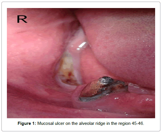
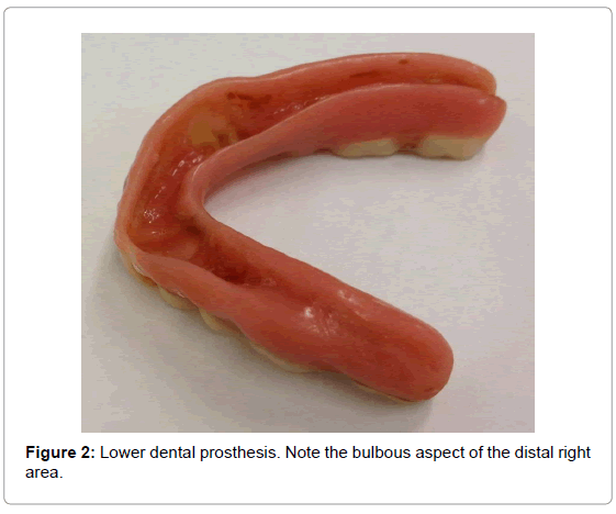
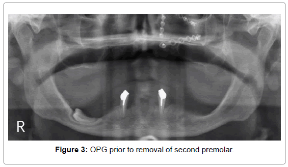
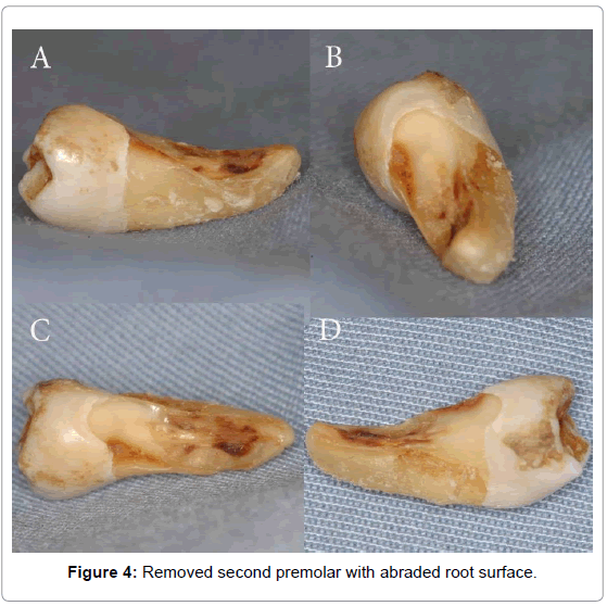
Oral ulcershave common occurrence, a multitude of causes and are prominent features in the practice of oral medicine [1]. On first presentation our 52 year old patient described a small, painless ulcer on the alveolar crest of the lower right jaw which he had first noted 2 years ago. He had sought no treatment and the ulcer slowly increased in size. His personal history was uneventful, suffering only from hypercholesterinemia treated with statins. There was no further regular medication and he had taken nothing in the days leading up to the consultation. 15 years ago he had received insufficient treatment for a left sided midface fracture in his home country. A revision osteotomy was performed a couple of years later and it was reported that the majority of his teeth were lost for reasons unrelated to the accident. He was able to retain the two lower canines, which were used as anchors for a lower dental prosthesis. During the interim period the patient had had various adaptations of the base of his prostheses which he rarely removed since he was embarrassed about being edentulous at such a young age. The lesion was situated on the alveolar crest in the region 45/46 and was 1.5x1cm in diameter; it did not show vascular injection but was slightly elevated at the border. On palpation the center was hard and completely indolent (Figure 1) and was considered to be denuded and discolored bone. It corresponded perfectly to the most convex part of his right lower dentures (Figure 2). There were no palpable lymph nodes in the neck and no other mucosal lesions. The OPG showed a distally inclined horizontal premolar with an indented crestal root surface (Figure 3). Perilesional biopsy demonstrated no malignancy. After removal of the tooth (Figure 4 A/B/C/D) and adaptation of the prosthesis the patient had no further issues and the ulcer did not recur.
Discussion
The term ulcer is commonly used for any lesion with central erosion of the skin or mucosal surface, usually accompanied by an inflammation and sloughing of the central/marginal areas [2]. Ulcers can be separated into the two large groups acute and chronic; the differentiation commonly accepted being that a chronic ulcer has been present for more than 2 weeks [3]. Acute ulcers are commonly of traumatic, allergic or infectious nature, while chronic ulcers can develop in cases of Lupus, pemphigoid, Lichen or oral squamous cell carcinoma (OSCC) to name but a few [1]. Mucosal lesions in denture wearers are sadly a frequent occurrence and pressure ulcers are one of the more common problems [4]. Since most ulcers are painful the fact that our patients was not had to raise concerns as to possible malignancy[5]. That the ulcer had been present for 2 whole years and grown only marginally during that time would however make the diagnosis less likely. The mobility the worn anchorage devices on the canines allowed was such that the round surface of the right lower prosthesis rubbed against the mucosa continually, and after wearing through that it ground through the overlying bone and one entire side of the root down to the pulp (Figures 2 and 4). Impacted teeth in edentulouspatients are less rare than one might think. While retained root fragments have been described between 15.3% and 9% completely retained and impacted teeth can be present in 4% to 6.2% [6,7]. Since the patient had had multiple occasions for radiographic evaluation the fact that the tooth had never been spotted or flagged up as a possible problem is astonishing. It has to be at least partially attributed to the fact that he was not amenable to regular recall. Problems with these retained teeth are however rarer and we were only able to find one publication describing paradental infection due to denture compression in the English speaking literature [8] and none illustrating actual dental abrasion. We would therefore like to emphasize the importance of performing at least one radiological evaluation of a patient receiving partial or complete dentures before beginning any kind of treatment [9] and the equal importance of checkups and oral mucosal exams after adaptation [10].
The patient gave written informed consent both for the removal of the tooth and for publication of anonymised data.
References
- Demko CA, Sawyer D, Slivka M, Smith D, Wotman S (2009) Prevalence of oral lesions in the dental office. Gen Dent 57: 504-509.
- Bhamrah G, Millwaters M (2008) Denture ulcerations. Br Dent J 205: 297.
- Muñoz-Corcuera M, Esparza-Gómez G, González-Moles MA, Bascones-Martínez A (2009) Oral ulcers: clinical aspects. A tool for dermatologists. Part II. Chronic ulcers. ClinExpDermatol 34: 456-461.
- Jainkittivong A, Aneksuk V, Langlais RP (2010) Oral mucosal lesions in denture wearers. Gerodontology 27: 26-32.
- Zygogianni AG, Kyrgias G, Karakitsos P, Psyrri A, Kouvaris J, et al. (2011) Oral squamous cell cancer: early detection and the role of alcohol and smoking. Head Neck Oncol 3: 2.
- Sumer AP, Sumer M, Güler AU, Biçer I (2007) Panoramic radiographic examination of edentulous mouths. Quintessence Int 38: e399-403.
- Soikkonen K, Ainamo A, Wolf J, Xie Q, Tilvis R, et al. (1994) Radiographic findings in the jaws of clinically edentulous old people living at home in Helsinki, Finland. ActaOdontolScand 52: 229-233.
- Albrecht D, Regina MS, Zix J (2010) Only a denture sore? Causes of a possible denture sore from a maxillary complete denture. SchweizMonatsschrZahnmed 120: 675-689.
- Angulo F (1989) Panoramic radiograph in edentulous and partially edentulous patients. ActaOdontolVenez 27: 60-67.
- Kivovics P, Jáhn M, Borbély J, Márton K (2007) Frequency and location of traumatic ulcerations following placement of complete dentures. Int J Prosthodont 20: 397-401.
Relevant Topics
- Advanced Bleeding Gums
- Advanced Receeding Gums
- Bleeding Gums
- Children’s Oral Health
- Coronal Fracture
- Dental Anestheia and Sedation
- Dental Plaque
- Dental Radiology
- Dentistry and Diabetes
- Fluoride Treatments
- Gum Cancer
- Gum Infection
- Occlusal Splint
- Oral and Maxillofacial Pathology
- Oral Hygiene
- Oral Hygiene Blogs
- Oral Hygiene Case Reports
- Oral Hygiene Practice
- Oral Leukoplakia
- Oral Microbiome
- Oral Rehydration
- Oral Surgery Special Issue
- Orthodontistry
- Periodontal Disease Management
- Periodontistry
- Root Canal Treatment
- Tele-Dentistry
Recommended Journals
Article Tools
Article Usage
- Total views: 16200
- [From(publication date):
November-2014 - Jul 15, 2025] - Breakdown by view type
- HTML page views : 11586
- PDF downloads : 4614


