Uncommon Location of a Hydatid Cyst: A Renal Parenchymal Encounter
Received: 03-Jun-2023 / Manuscript No. roa-23-101603 / Editor assigned: 05-Jun-2023 / PreQC No. roa-23-101603 (PQ) / Reviewed: 19-Jun-2023 / QC No. roa-23-101603 / Revised: 22-Jun-2023 / Manuscript No. roa-23-101603 (R) / Published Date: 29-Jun-2023 DOI: 10.4172/2167-7964.1000454
Clinical-Medical Image
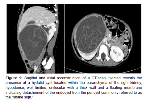
We encountered a 5-year-old girl with no previous medical history, who presented with pain in the right upper abdomen and mentioned contact with a dog. Upon conducting a control ultrasound, we discovered a large cystic mass in the right kidney. To further investigate, we ordered an abdominal CT scan which revealed a hydatid cyst located in the kidney, which is an uncommon occurrence. Within the cyst, we observed a floating membrane that resembled the shape of a snake, commonly known as the snake sign (Figure 1).
Figure 1: Sagittal and axial reconstruction of a CT-scan injected reveals the presence of a hydatid cyst located within the paranchyma of the right kidney, hypodense, well limited, unilocular with a thick wall and a floating membrane indicating detachement of the endocyst from the pericyst commonly referred to as the “snake sign.”
The snake sign is a distinctive radiological feature of hydatid cysts caused by Echinococcus infections. These structures represent the detached laminated membranes of the end cyst, floating within the cystic space, resembling the appearance of a serpent or a swirling motion [1]. A typical hydatid cyst consists of three layers: an outer fibrous capsule (pericyst) that provides protection, a laminated middle acellular membrane (ectocyst) that allows the passage of nutrients but can be easily ruptured, leading to infection, and an inner germ layer (endocyst) where the larval stage of the parasite resides. Detachment of the endocyst from the pericyst can occur due to various factors, including decreased intra-cystic pressure, degeneration, host response, trauma, medical treatment, or percutaneous drainage.
The “snake sign” can be detected on X-rays in cases of hydatid kidney disease. However, the presence of “serpentine membranes” within a hydatid cyst is classically described using abdominal ultrasound, computed tomography (CT), or MRI [2].
Conflict of Interest
The authors declared no potential conflicts of interest with respect to the research, authorship, and / or publication of this article.
References
- Giambelluca D, Cannella R, Caruana G, Picone D, Midiri M (2018) The “serpent sign” in hydatid disease. Abdom Radiol (NY) 43: 2523-2524.
- Pal RS, Gothi D, Singh B (2014) ‘‘Serpent’’ Sign, ‘‘Double Arch’’ sign and ‘‘Air-Bubble’’ sign in a case of ruptured hydatid cyst: A Case Report. Webmedcentral.
Indexed at, Google Scholar, Crossref
Citation: Chaimae ES, Zakaria A, Najlae L, Siham EH, Nazik A, Latifa C (2023) Uncommon Location of a Hydatid Cyst: A Renal Parenchymal Encounter. OMICS J Radiol 12: 454. DOI: 10.4172/2167-7964.1000454
Copyright: © 2023 Chaimae ES, et al. This is an open-access article distributed under the terms of the Creative Commons Attribution License, which permits unrestricted use, distribution, and reproduction in any medium, provided the original author and source are credited.
Share This Article
Open Access Journals
Article Tools
Article Usage
- Total views: 1424
- [From(publication date): 0-2023 - Mar 29, 2025]
- Breakdown by view type
- HTML page views: 1197
- PDF downloads: 227

