Ultrasound Imaging Technologies for Breast Cancer in Radiology
Received: 05-May-2022 / Manuscript No. roa-22-64948 / Editor assigned: 07-May-2022 / PreQC No. roa-22-64948 (PQ) / Reviewed: 21-May-2022 / QC No. roa-22-64948 / Revised: 23-May-2022 / Manuscript No. roa-22-64948 (R) / Published Date: 30-May-2022 DOI: 10.4172/2167-7964.1000384
Image Article
In medication Breast Imaging is a sub-speciality of symptomatic radiology that includes imaging of the breasts for screening or demonstrative purposes. There are different techniques for breast imaging involving an assortment of advancements as portrayed exhaustively beneath. Traditional screening and indicative mammography utilizes X-ray technology. Breast tomosynthesis is another computerized mammography procedure that produces 3D pictures of the breast using x-beams. Xeromammography and Galactography also use X-ray technology and are also used infrequently in the detection of breast cancer. Breast ultrasound is another technology employed in diagnosis and screening and specifically can help differentiate between fluid filled and solid lumps that can help determine if cancerous. Breast MRI is yet one more technology held for high-risk patients and can assist with deciding the degree of cancer whenever analyzed. Finally, scintimammography is utilized in a subgroup of patients who have abnormal mammograms or whose screening is not reliable on the basis of using traditional mammography or ultrasound [1].
Mammography (additionally called mastography) is the method of using low-energy X-rays (normally around 30 kVp) to analyze the human breast for analysis and screening. The objective of mammography is the early detection of breast cancer, regularly through detection of characteristic masses or micro calcifications [2].
Breast ultrasound is the utilization of clinical ultrasonography to perform imaging of the breast:
1. It tends to be considered either diagnostic or a screening procedure.
2. It may be used either with or without a mammogram.
3. It may be useful in younger women, where the denser stringy tissue of the breast might make mammograms more difficult to interpret.
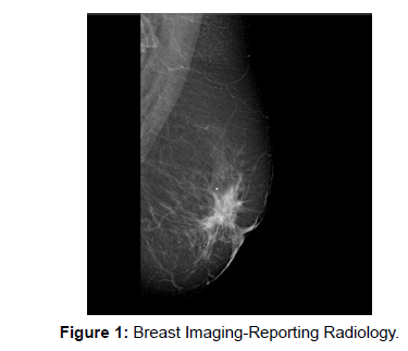
Automated whole-breast ultrasound (AWBU) is an ultrasound examination of the breast that is generally free of the operator skill and that permits the recreation of volumetric images of the breast (Figure 1).
Utilizing high-frequency ultrasound, an indicative assessment of the lactiferous ducts by means of ultrasound (duct sonography) can be performed. In this manner, dilated ducts and intraductal masses can be made visible. Another technique for visualizing the system of lactiferous ducts is galactography which allows a wider area of the lactiferous duct system to be visualized.
References
- Thomassin-Naggara I, Cornelis F, Kermarrec E (2019) Breast interventional imaging. Presse Med 48: 1169-1174.
- Whitman GJ (2021) Breast Ultrasound: An Essential Tool. Ultrasound Q 37: 1-2.
Indexed at, Google Scholar, Crossref
Citation: Albert P (2022) Ultrasound Imaging Technologies for Breast Cancer in Radiology. OMICS J Radiol 11: 384. DOI: 10.4172/2167-7964.1000384
Copyright: © 2022 Sharma P. This is an open-access article distributed under the terms of the Creative Commons Attribution License, which permits unrestricted use, distribution, and reproduction in any medium, provided the original author and source are credited.
Select your language of interest to view the total content in your interested language
Share This Article
Open Access Journals
Article Tools
Article Usage
- Total views: 1963
- [From(publication date): 0-2022 - Dec 01, 2025]
- Breakdown by view type
- HTML page views: 1432
- PDF downloads: 531

