Ultrasound Findings among Patients Who Have Chronic Abdominal Pain and Correlation between Site of Pain and Ultrasound Findings
Received: 22-Nov-2018 / Accepted Date: 22-Jan-2019 / Published Date: 29-Jan-2019 DOI: 10.4172/2167-7964.1000304
Abstract
Background: Upper abdominal pain is one of the most common complains by patients referred by GP. Abdominal ultrasound (USS) remains the primary imaging technique in majority of cases and considered safe, rapid and noninvasive method of abdominal examination.
Objectives: The aims of this study were to estimate the prevalence of relevant USS findings in patient with chronic abdominal pain and to determine its relation to the site of pain.
Methods: In all patients the site of pain was localized and abdominal USS was done. After collection and check of data, SPSS was used for data entry and analysis.
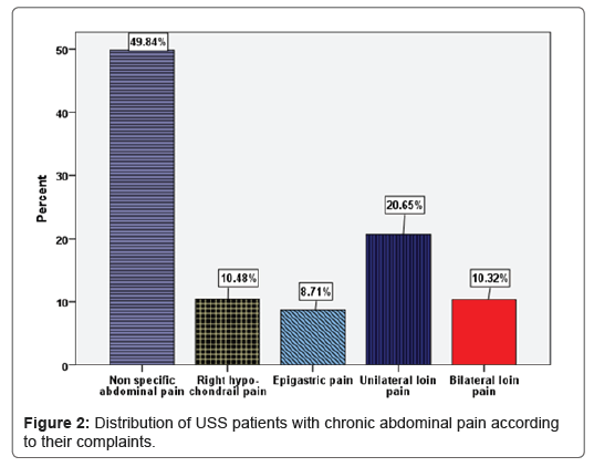
Results: A total of 620 patients were enrolled in the study; (10.48%) complained of RHC pain, (8.7%) complained of epigastric pain, (49.84%) complained of non-specific generalized pain, (20.65%) complained of unilateral loin pain and (10.3%) complained of bilateral loin pain. Less than half (44.8%) of cases had relevant findings such as; nonalcoholic fatty liver disease and cholelithiasis.
Conclusion: Abdominal ultrasound is an important modality in detection of relevant findings and can reduce the number of patients referred to specialist but in some cases can be of little value. Adequate clinical information and good examination are essential to enable the choice of appropriate modality and to reduce the number of unnecessary ultrasound examination.
Keywords: Abdominal ultrasound; Chronic abdominal pain; Site of pain; Ultrasound findings
Abbreviations
GP: General Practitioner; SPSS: Statistical Package for Social Sciences; RHC: Right Hypochondrial; CBD: Common Bile Duct; USS: Ultrasound Scan; SD: Standard Deviation
Introduction
Chronic abdominal pain can be defined as: continuous or intermittent abdominal discomfort lasting for at least six months [1]. Most patients have a benign and/or self-limiting etiology [2]. It may arise from gastrointestinal tract or adjacent organs, such as the hepatic, biliary tract, pancreas, genitourinary or gynecological organs [1]. In a minority of patients, the presence of negative findings could be attributed to presence of functional syndromes as; irritable bowel syndrome [1,3] Moreover, underlying malignancies as gastric, pancreatic, colonic and hepatic metastasis can cause chronic constant abdominal pain [3]. According to Clark and Silk, pain in certain abdominal regions may give a clue for certain diseases. They gave examples as; epigastric pain is very common and usually due to peptic ulcer disease, RHC pain usually is related with gall bladder or biliary tree and also its frequent complain in patient with functional bowel disorder [4]. Loin pain may be due to renal stone, renal tumors, infection or congenital abnormality [5].
Abdominal pain is among many indications for performing abdominal ultrasound, which is considered as an important tool for evaluation of abdominal organs and also it’s safe, noninvasive and lack of exposure to ionization radiation [2,6].
Subjects and Methods
Aims of the study
To estimate the prevalence of relevant ultrasound findings and the prevalence of each abnormality in patients with chronic abdominal pain; a further aim was to determine the relation between sites of pain and ultrasound findings.
Study design
Cross sectional descriptive study was applied in this research.
Study setting
Kish poly clinic in the Benghazi-Libya
Duration of study
June 2015 to May 2016.
Inclusion criteria of cases
Male and female patients aged 18 years and above who have chronic abdominal pain and were referred for abdominal ultrasound by GP
Exclusion criteria of cases
Male and female patients aged less than 18 years, patients presented with acute abdomen and patients with pelvic abnormality as cause of abdominal pain.
Tools: USS machine
High resolution gray scale B-mode trans-abdominal ultrasound (frequency 3.5 MHz, curvilinear transducer; GE LOGIQ F8).
Procedures
The ultrasound procedure was performed by the investigator herself and the examination performed after fasting for at least 8 hours unless the patient has a history of cholecystectomy
Questionnaire
The collected data included; age, gender, site of pain, and USS findings.
Approval
The approval of the director of the polyclinic was taken before starting collection of data.
Verbal consent of cases
Consent of each patient was taken after brief description about the research and its purpose.
Statistical analysis
After collection of data and checking for missing data, SPSS version 22 was used for data entry and analysis [7]. Descriptive statistics was applied as mean, standard deviation (SD) and presented in tabular and graphical forms.
Results
During the period of study, a total of 620 patients complained from chronic abdominal pain and USS was done for all of them. Patients’ complains were classified into five groups “non-specific generalized, epigastric, RHC, bilateral loin and unilateral loin pain”.
USS findings were classified into one of three groups: 1-no findings, when the findings were entirely normal, 2- non-relevant findings, when the findings were usually incidental and without any consequences and 3-clinically relevant findings, when the findings need referral to specialists for further managements.
The mean age of patients were 45.04 ± 14.11 years, the youngest patient aged 18 years and the eldest aged 86 years (Table 1).
| Age in years (N=620) | |
|---|---|
| Mean | 45.04 |
| Median | 45.00 |
| Mode | 50.00 |
| Std. Deviation | 14.11 |
| Minimum | 18.00 |
| Maximum | 86.00 |
Table 1: Descriptive statistics of age of patients with chronic abdominal pain
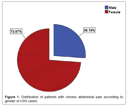
Figure 1 shows that majority of patients (73.87%) were females and 26.13% were males.
Figure 2 illustrates that; One tenth of patients; (10.48%) complained of RHC pain and their ultrasound findings were; 52.4% had gall bladder stones and 44.6% had fatty liver
Cases who complained of epigastric pain represented 8.7% out of the total 620 patients and the ultrasound findings were 90.7% had negative examination and 9.3% had gall bladder stones.
Nearly half (49.84%) of cases complained from non-specific generalized pain and the ultrasound findings were; 11.7%had gall bladder stones, and 36.2% had fatty liver.
One fifth (20.65%) of the cases complained of unilateral loin pain and the ultrasound findings were 39.9%had hydronephrosis, and 10 % had large renal cyst ≥ 5 cm.
One tenth (10.3%) of cases had complained from bilateral loin pain and their ultrasound findings were as follows: 81.2% had negative findings and 15.6 % had small renal cyst <5 cm.
Figure 3 shows distribution of patients with chronic abdominal pain with respect to the site of pain and commonest findings.
The Incidence of Relevant Abnormalities
Table 2 shows that Less than half (44.8%) of cases examined by abdominal USS; had abnormal findings. Among cases with abnormal findings; more than half (54.2%) had fatty liver, 22.3% had gall bladder stones, 1% had CBD stone, 0.3.% had chronic acalculus cholecystitis, 0.8% had gall bladder polyp, 0.5% adenomyomatosis, 2.6% had liver cirrhosis, 1.3% had liver metastasis, 0.3% had hydatid liver disease.
| Result of scan | % |
|---|---|
| Fatty liver | 54.2 |
| Gall bladder stones | 22.3 |
| Unilateral hydronephrosis | 18.7 |
| Liver cirrhosis | 2.6 |
| Small echogenic kidneys | 2.3 |
| Obstructing renal stones | 2 |
| Obstructing ureteric stone | 1.8 |
| Non- obstructed hernia | 1.5 |
| Liver metastasis | 1.3 |
| CBD stone | 1 |
| Large simple renal cyst | 1 |
| Non-obstructing renal stones | 1 |
| Gall bladder polyp | 0.8 |
| Adenomyomatosis | 0.5 |
| Complicated renal cyst | 0.5 |
| Bifid collecting system | 0.5 |
| Chronic acalculus cholycystitis | 0.3 |
| Hydatid liver disease | 0.3 |
| Bilateral hydronephrosis | 0.3 |
| Hydronephrosis with Fluid-Debris level | 0.3 |
| Nephrocalcinosis | 0.3 |
| Pelvic ureteric junction obstruction | 0.3 |
| Dilated ureter likely due to reflux | 0.3 |
| Peritoneal metastasis and ascites | 0.3 |
| Abdominal aorta aneurysm | 0.3 |
| Pancreatic pseudo-cyst | 0.3 |
| Lymphadenopathy | 0.3 |
| Dilated small bowel loops | 0.3 |
Table 2: Incidence of relevant abnormalities among patients with chronic abdominal pain.
Unilateral hydronephrosis was prevalent among 18.7%; equal proportions of cases (0.3%) had bilateral hydronephrosis and hydronephrosis with fluid-debris level. Regarding, obstructing and non-obstructing renal stones, represented (2% and 1% respectively). Obstructing ureteric stone was prevalent in 1.8%, Nephrocalcinosis (0.3%). Large simple renal cyst > or =5 cm (1%), complicated renal cyst (0.5%), small echogenic kidneys (2.3%), bifid collecting system (0.5%), pelvic ureteric junction obstruction (0.3%) and dilated ureter likely due to reflux (0.3%).
Concerning, other findings as; non- obstructed hernia (1.5%), (0.3%) peritoneal metastasis and ascites, equal proportions (0.3%) to abdominal aorta aneurysm, pancreatic pseudo-cyst, lymphadenopathy and dilated small bowel loops (0.3%).
The incidence of non-relevant findings (18.1%), these findings included hepatic cysts (2%), liver hemangioma (1.3%), and small simple renal cyst <5 cm (14.8%) (Table 3).
| Result of scan | % |
|---|---|
| Small simple renal cyst | 14.8 |
| Hepatic cysts | 2 |
| Liver hemangioma | 1.3 |
Table 3: Incidence of non- relevant abnormalities among patients with chronic abdominal pain.
The incidence of negative examination, in other words normal findings was 37.1%.
The most combination findings were gall bladder stone and fatty liver (3.9%) and fatty liver with hydronephrosis (1.9%).
Discussion
Many previous studies were done regarding the relevant findings in patients with chronic abdominal pain referred for abdominal ultrasound by GP. The relevant findings were ranging from 16%-30%, Colquhoun et al. [8], Mills et al. [9], Charlesworth et al. [10], Connor et al. [11], Speets et al. [12] and Speets et al. [13] reported the proportion of the clinically relevant findings as follows 30%, 27%, 25%, 28%, 29% and 16% respectively.
The prevalence of relevant findings in our study were 44.8% which is higher when compared to previous studies because in our study, we included the fatty liver as relevant finding as it is considered a significant finding in our population because it can lead to steatohepatitis which is associated with progressive liver damage and complications ranging from fibrosis, cirrhosis, and liver cancer [14]. In addition, it is also associated with metabolic syndrome and its components of obesity, hypertension, dyslipidemia, and insulin resistance and increased risk of atherosclerotic coronary heart disease and diabetes [15].
On the other hand, in other populations, where alcohol consumption is common, the fatty liver as an irrelevant findings in USS. Imran [2] reported that the incidence of positive findings were 50%; this percentage is in agreement with 44.8% significant abnormality found in our study. The prevalence of negative findings was 37.1 which is also important for disease exclusion and also decreases the number of referred patients to other specialists.
Highest proportion of patients (49.8%) complained from nonspecific generalized abdominal pain, the commonest finding among them was fatty liver. One tenth (10.48%) complained from RHC pain, the commonest finding among them was cholelithiasis. Cases who complained from epigastric pain represented 8.7% and most of their USS findings were negative. Yamamoto et al. [16] studied 489 patients complaining of abdominal pain, he reported that the commonest final diagnosis was intestinal disease in patient complained from whole (generalized) abdominal pain. They added that gastroduodenal disease was the final diagnosis in patients with epigastric pain. Regarding patients with right subcostal pain, hepatobiliary disease was the final diagnosis. The disagreement between our study and their study was due to lack of accessibility to do further investigations to reach to the final diagnosis which is considered one of the limitations of our study. The second limitation was that the patients with relevant findings were not followed up due to lack of tracking patients when referred to other specialists.
Conclusion
Abdominal ultrasound is an important modality in detection of relevant findings and can reduce the number of patients referred to specialists but in some cases can be of little value as in cases with epigastric pain.
Precise description of pain when referred from GP would reduce the number of unnecessary USS examination.
References
- Mendelson R (2015) Imaging for chronic abdominal pain in adults. Aust Prescr 38: 49-54.
- Imran S (2003) Accuracy of ultrasound in the diagnosis of upper abdominal pain. J Ayub Med Coll Abbottabad 15: 59-62.
- Palmar KR, Penman ID, Paterson BS, Boon NA, Walker BR, et al. (2006) Alimentary tract and pancreatic disease: Davidson's principles and practice of medicine. Elsevier, United Kingdom.
- Clark ML, Silk DB, Kumar P (2005) Gastrointestinal disease: Kumar Clark clinical medicine. Elsevier, USA.
- Goddard J, Boon NA, Colledge NR, Walker BR, et al. (2006) Kidney and urinary tract disease: Davidson's principles and practice of medicine. (20th edn) Elsevier science, London, United Kingdom.
- American College of Radiology (2001) ACR Standard for the performance of an ultrasound examination of the abdomen or retroperitoneum. pp: 1-5.
- IBM Corp. Released 2013. IBM SPSS Statistics for Windows, V.ersion 22.0.Armonk, NY: IBM Corp.
- Colquhoun IR, Saywell WR, Dewburry KC (1988) An analysis of referrals for primary diagnostic abdominal ultrasound at a general X-ray department. Br J Radiol 61: 297-300
- Mills P, Joseph AEA, Adam EJ (1989) Total abdominal and pelvic ultrasound: incidental findings and a comparison between outpatient and general practice referrals in 1000 cases. Br J Radiol 62: 974-976.
- Charlesworth CH, Sampsom MA (1994) How do general practitioners compare with the outpatient department when requesting upper abdominal examinations? Clin Radiol 49: 343-345.
- Connor SEJ, Banerjee AK (1998) General practitioner requests for upper abdominal ultrasound: their effect on clinical outcome. Br J Radiol 73: 1021-1025.
- Speets MA, Hoes AW, Graaf YV, Kalmijn S, Wit NJ, et al. (2006) Upper abdominal ultrasound in general practice: indications, diagnostic yield and consequences for patient management. Fam Pra 5-507: 511.
- Speets MA, Kalmijn S, Hoes AW, Graaf YV, Mali WP (2006) Yield of abdominal ultrasound in patients with abdominal pain referred by general practitioners. Eur J Gen Pract 12: 135-137.
- Neuschwander TBA, Caldwell SH (2003) Nonalcoholic steatohepatitis: summary of an AASLD single topic conference. Hepatol 37: 1202-1219.
- Targher G (2007) Non-alcoholic fatty liver disease, the metabolic syndrome and the risk of cardiovascular disease: the plot thickens. Diabet Med 24: 1-6.
- Yamamoto W, Kono H, Maekawa M, Fukui T (1996) The relationship between abdominal pain regions and specific diseases: an epidemiologic approach to clinical practice. J Epidemiol 7: 27-31
Citation: Abtehag AT, Azza SHG (2019) Ultrasound Findings among Patients Who Have Chronic Abdominal Pain and Correlation between Site of Pain and Ultrasound Findings. OMICS J Radiol 8:304. DOI: 10.4172/2167-7964.1000304
Copyright: © 2019 Abtehag AT, et al. This is an open-access article distributed under the terms of the Creative Commons Attribution License, which permits unrestricted use, distribution, and reproduction in any medium, provided the original author and source are credited.
Share This Article
Open Access Journals
Article Tools
Article Usage
- Total views: 4025
- [From(publication date): 0-2019 - Mar 29, 2025]
- Breakdown by view type
- HTML page views: 3205
- PDF downloads: 820


