Make the best use of Scientific Research and information from our 700+ peer reviewed, Open Access Journals that operates with the help of 50,000+ Editorial Board Members and esteemed reviewers and 1000+ Scientific associations in Medical, Clinical, Pharmaceutical, Engineering, Technology and Management Fields.
Meet Inspiring Speakers and Experts at our 3000+ Global Conferenceseries Events with over 600+ Conferences, 1200+ Symposiums and 1200+ Workshops on Medical, Pharma, Engineering, Science, Technology and Business
Editorial Open Access
Ultrasound Appearance of the Cutaneous Nerves of the Upper Limb: A Novel Description in Pain Management
| Rafael Blanco1*, Bárbara María Jiménez Gómez2 and José Manuel López González2 | |
| 1Complexo Hospitalario Universitario A Coruña, Spain | |
| 2Complexo Hospitalario Universitario de Vigo, Spain | |
| Corresponding Author : | Rafael Blanco Complexo Hospitalario Universitario A Coruña, Spain E-mail: rafablanco@mac.com |
| Received January 20, 2012; Accepted January 29, 2012; Published February 08, 2012 | |
| Citation: Blanco R, Jiménez Gómez BM, López González JM (2012) Ultrasound Appearance of the Cutaneous Nerves of the Upper Limb: A Novel Description in Pain Management. J Pain Relief 1:e109 doi: 10.4172/2167-0846.1000e109 | |
| Copyright: © 2012 Blanco R, et al. This is an open-access article distributed under the terms of the Creative Commons Attribution License, which permits unrestricted use, distribution, and reproduction in any medium, provided the original author and source are credited. | |
Visit for more related articles at Journal of Pain & Relief
| In recent years ultrasound has become the standard technique to locate peripheral nerves. These can be useful to block, so we can obtain pain relief during or after operative procedures. It can also be used in chronic pain management to treat long standing painful situations or as part of the armamentarium to neurolize small nerves or neuromas. Most of the nerves described up to now are commonly used by regional anesthetist in daily practice but the cutaneous sensory nerves have been left aside for two reasons: They are very small and superficial and classically they were difficult to locate because the technique used was nerve stimulation. Based on our experience with ultrasound of approximately 10 years we will describe in this article how to locate the sensory nerves of the upper limb [1]. These nerves play a vital role during common operations like Du Puy Trent, carpal tunnel release or many more reconstructive hand surgery techniques. A brief description of the brachial plexus is imperative. It’s originated by the anterior ramus of the spinal nerves from C5 to T1 and the main nerves derived from those spinal nerves are the musculocutaneous, median, radial,axillary, circumflex, ulnar, cutaneous nerve of the arm and cutaneous nerve of the forearm. The dermatomes are the part of the skin supplied by a specific spinal nerve (in this case C5-T1). From the sensitive point of view, any nerve injury will affect those areas and will cause different alterations from paresthesias to dysesthesia (Figure 1). In this paper and for the first time published we will describe the most common location of the sensitive nerves in the upper limb [2]. The main upper limb sensitive nerves are five: lateral cutaneous nerve of the forearm, median cutaneous nerve of the arm, median cutaneous nerve of the forearm, posterior cutaneous nerve of the arm and posterior cutaneous nerve of the forearm. |
| Lateral Cutaneous Nerve of the Forearm (C5-C7) |
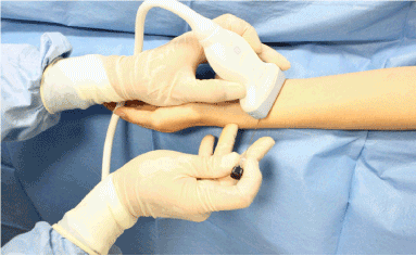
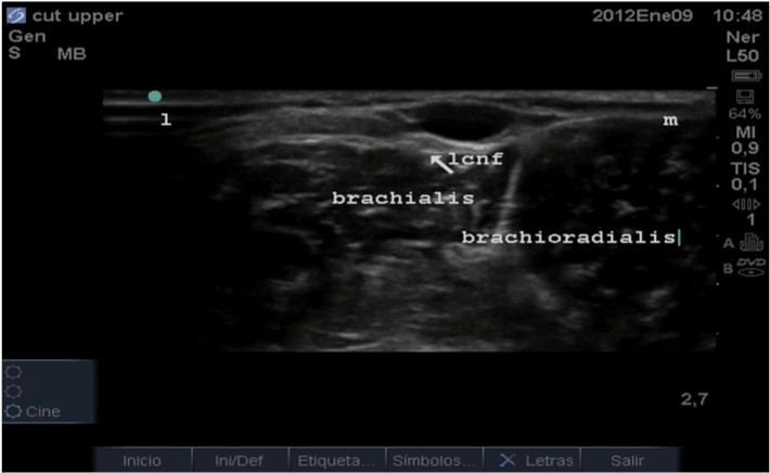
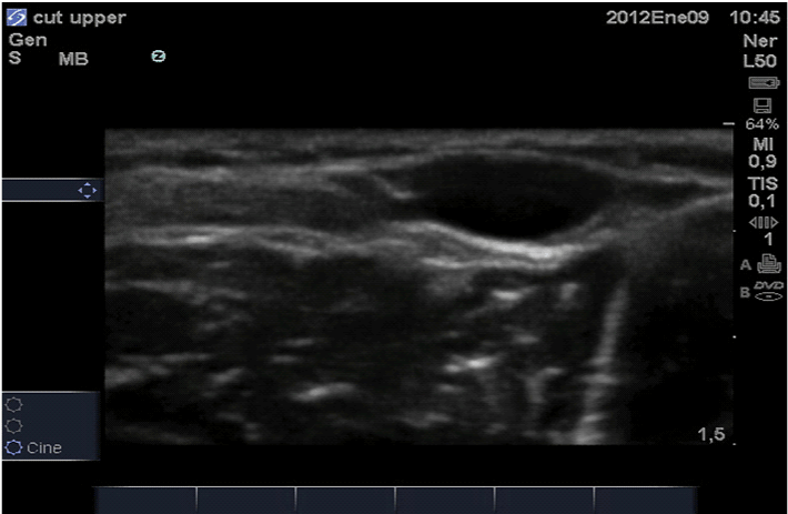
| It is the terminal branch of the musculocutaneous nerve. At the distal third of the forearm is located over the brachioradialis muscle. Under ultrasound guidance it can be track down from the musculocutaneous nerve when it becomes superficial after the biceps muscle fades away. An important sono landmark is the cephalic vein that runs in close proximity to this nerve in most of his path. This cutaneous nerve, like the others, runs in a fascial [3], subcutaneous compartment, between the skin and the muscle (Figure 2-4). |
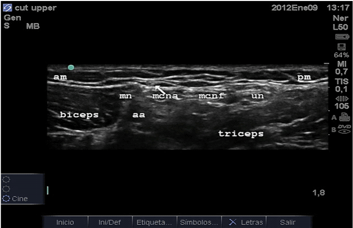
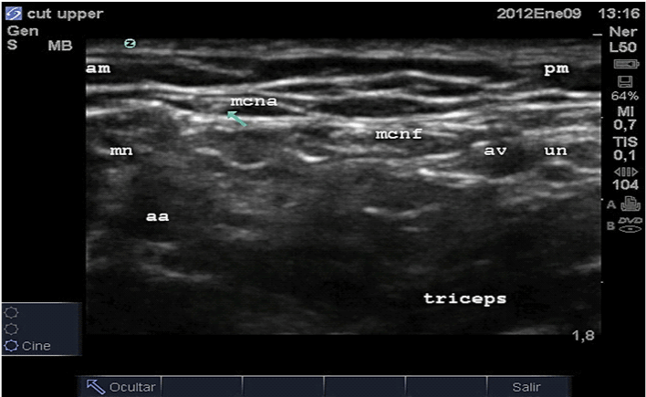
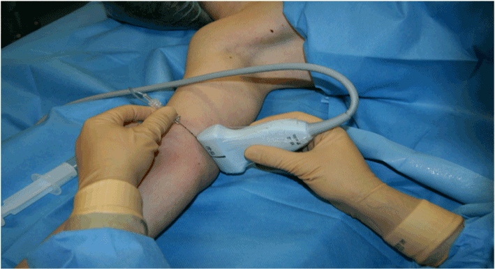
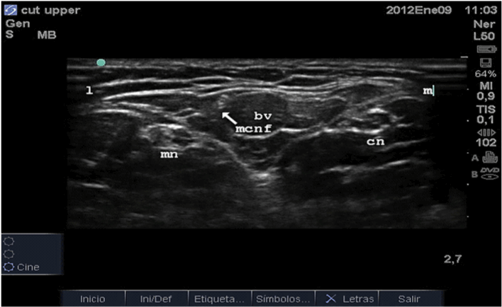
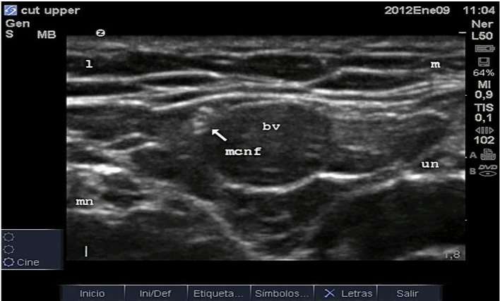
| It is the smallest nerve of the brachial plexus. It follows a path at subcutaneous level just underneath the fascia brachialis medial to the axillary artery and vein. It will communicate at a later stage with the medial cutaneous nerve of the forearm (Figure 5,6). |
| Medial Cutaneous Nerve of the Forearm (C8,T1) |
| It is found in the medial aspect of the forearm at subcutaneous level next to the basilic vein becoming more superficial as we go distal in the forearm. It will gives off anteriorly an intermediate branch running above pronator teres muscle (Figure 7-9). |
| Posterior Cutaneous Nerve of the Arm (C5-C8) |
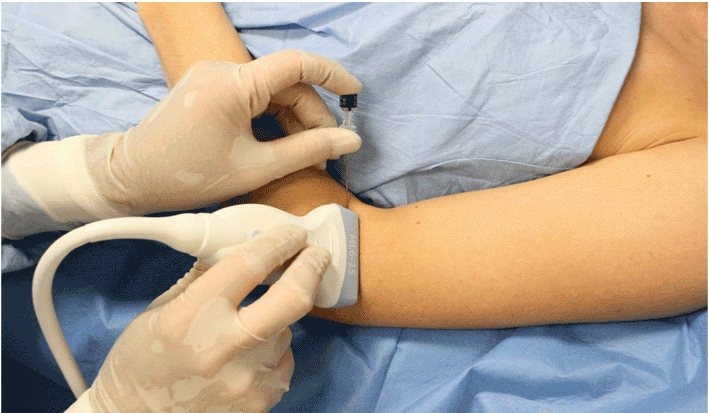
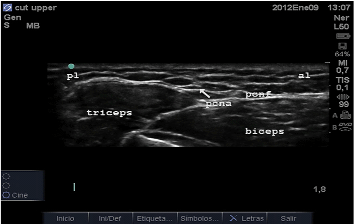
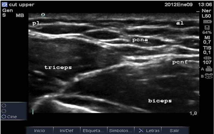
| It is one the posterior branches of the radial nerve. It is located in a fat pocket above the lateral head of the triceps muscle in the posterior side of the arm (Figure 10-12). |
| Posterior Cutaneous Nerve of the Forearm (C5-C8) |
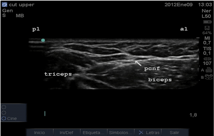
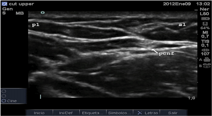
| It is another posterior branch of the radial nerve (Figure 13,14). It is located in a fat pocket above the long head of the triceps muscle in the posterior side of the arm, distal third. Along the forearm will be found above the extensor digitorum muscle. |
| Summary |
| The sensory nerves are very small and they are the ones involved when the surgeon or we perform a subcutaneous infiltration. With good anatomy knowledge we will be able to track them up into their main trunks so smaller and more precise amounts of local anesthetic can be delivered. In situations of lasting dysesthesias we will be able to perform successful chronic pain techniques very easily. |
References
- Keegan JJ, Garrett FD (1948) The segmental distribution of the cutaneous nerves in the limbs of man. Anat Rec 102: 409-437.
- Linell EA (1921) The distribution of nerves in the upper limb, with references to variabilities and their clinical significance. J Anat 55: 79-112.
- Nakajima H, Imanishi N, Fukuzumi S, Minabe T, Aiso S, et al. (1998) Accompanying Arteries of the Cutaneous Veins and Cutaneous Nerves in the Extremities: Anatomical Study and a Concept of the Venoadipofascial and/or Neuroadipofascial Pedicled Fasciocutaneous Flap. Plast Reconstr Surg 102: 779-791.
Figures at a glance
 |
 |
 |
 |
 |
| Figure 1 | Figure 2 | Figure 3 | Figure 4 | Figure 5 |
 |
 |
 |
 |
 |
| Figure 6 | Figure 7 | Figure 8 | Figure 9 | Figure 10 |
 |
 |
 |
 |
| Figure 11 | Figure 12 | Figure 13 | Figure 14 |
Post your comment
Relevant Topics
- Acupuncture
- Acute Pain
- Analgesics
- Anesthesia
- Arthroscopy
- Chronic Back Pain
- Chronic Pain
- Hypnosis
- Low Back Pain
- Meditation
- Musculoskeletal pain
- Natural Pain Relievers
- Nociceptive Pain
- Opioid
- Orthopedics
- Pain and Mental Health
- Pain killer drugs
- Pain Mechanisms and Pathophysiology
- Pain Medication
- Pain Medicine
- Pain Relief and Traditional Medicine
- Pain Sensation
- Pain Tolerance
- Post-Operative Pain
- Reaction to Pain
Recommended Journals
Article Tools
Article Usage
- Total views: 15564
- [From(publication date):
February-2012 - Apr 07, 2025] - Breakdown by view type
- HTML page views : 10872
- PDF downloads : 4692
