Make the best use of Scientific Research and information from our 700+ peer reviewed, Open Access Journals that operates with the help of 50,000+ Editorial Board Members and esteemed reviewers and 1000+ Scientific associations in Medical, Clinical, Pharmaceutical, Engineering, Technology and Management Fields.
Meet Inspiring Speakers and Experts at our 3000+ Global Conferenceseries Events with over 600+ Conferences, 1200+ Symposiums and 1200+ Workshops on Medical, Pharma, Engineering, Science, Technology and Business
Case Report Open Access
Ultrashort Common Bile Duct-A Novel Biliary Variant
| Prashant Naphade and Abhishek Keraliya* | |
| Dana Farber Cancer Institute, Boston, MA-02215, USA | |
| Corresponding Author : | Abhishek Keraliya Clinical Fellow Dana Farber Cancer Institute Imaging Boston, MA 02215, USA Tel: +16175436171 E-mail:abhishekkeraliya@gmail.com |
| Received November 25, 2014; Accepted December 22, 2014; Published December 26, 2014 | |
| Citation: Naphade P, Keraliya A (2015) Ultrashort Common Bile Duct – A Novel Biliary Variant. OMICS J Radiol 3:175. doi: 10.4172/2167-7964.1000175 | |
| Copyright: © 2015 Naphade P, Keraliya A. This is an open-access article distributed under the terms of the Creative Commons Attribution License, which permits unrestricted use, distribution, and reproduction in any medium, provided the original author and source are credited. | |
Visit for more related articles at Journal of Radiology
Abstract
Detection of variations in biliary ductal anatomy is of paramount importance as it may lead to diagnostic fallacies and ductal injury during therapeutic and surgical procedures. Authors report a unique case of ultrashort common bile duct (CBD), a novel biliary variant due to extreme low union of right and left hepatic ducts with cystic duct opening in right hepatic duct.
| Keywords |
| Biliary system; Magnetic resonance cholangiopancreatography; Anatomy |
| Introduction |
| A 65 year old woman presented to our hospital with complaints of right upper abdominal pain and vomiting. Clinical examination was unremarkable. Laboratory evaluation revealed mildly elevated total bilirubin levels (1.6 mg/dl). Ultrasound abdomen revealed dilated common bile duct (CBD) with mild intrahepatic biliary radical (IHBR) dilatation and was referred for magnetic resonance imaging (MRI) for further evaluation. |
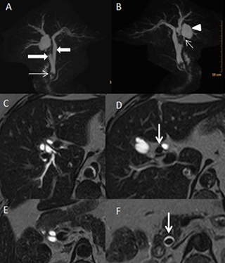
| Magnetic resonance cholangiopancreatography (MRCP) with heavily T2 weighted images confirmed mild IHBR dilatation. Right and left hepatic ducts were dilated and had separate extrahepatic and intrapancreatic course. Right hepatic duct passed posterior to the left hepatic duct in the intrapancreatic course and united with the left hepatic duct along the left lateral aspect just proximal to ampulla to form ultrashort CBD (1.6 cm length). T2 hypointense calculi were seen in the proximal right hepatic duct at the porta and in the terminal CBD just before its opening the ampulla, suggestive of choledocholithiasis (Figure 1). Cystic duct was seen joining the right hepatic duct. Pancreatic duct was normal in caliber and was opening normally at the major duodenal papilla. Gall bladder was unremarkable. |
| The patient underwent endoscopic retrograde cholangiopancreaticography (ERCP) which confirmed the MRCP findings and endoscopic sphincterotomy and removal of stones were performed. |
| Discussion |
| Detection of variations in biliary ductal anatomy is of paramount importance as it may lead to diagnostic fallacies and ductal injury during therapeutic and surgical procedures. MRCP allows noninvasive accurate multiplanar evaluation of biliary system using heavily T2 weighted 3D sequences. Right hepatic duct is normally formed by anterior and posterior branches of right lobe while left hepatic duct is formed by medial and lateral branches of left lobe. Common hepatic duct (CHD) is formed by union of right and left hepatic ducts at the porta. Cystic duct opens in the common hepatic duct below the hepatic ductal confluence to form CBD. This normal anatomy is seen in 60 to 65 % of patients [1,2]. The length of CBD varies from 5 to 15 cm [3]. Common variations include right posterior duct opening in left hepatic duct or right lateral aspect of right anterior duct and triple ductal confluence with CHD formation by union of right posterior, right anterior and left hepatic ducts [1,2,4,5]. |
| Conclusion |
| Detection of variations in biliary ductal anatomy is of paramount importance as it may lead to diagnostic fallacies and ductal injury during therapeutic and surgical procedures. This case demonstrates the extreme low union of right and left hepatic ducts just prior to the ampulla with formation of ultrashort CBD. Further cystic duct opening in right hepatic duct is very rare. To the best of authors’ knowledge, ultrashort CBD is a novel finding and has not been described in literature. |
References |
|
Figures at a glance
 |
| Figure 1 |
Post your comment
Relevant Topics
- Abdominal Radiology
- AI in Radiology
- Breast Imaging
- Cardiovascular Radiology
- Chest Radiology
- Clinical Radiology
- CT Imaging
- Diagnostic Radiology
- Emergency Radiology
- Fluoroscopy Radiology
- General Radiology
- Genitourinary Radiology
- Interventional Radiology Techniques
- Mammography
- Minimal Invasive surgery
- Musculoskeletal Radiology
- Neuroradiology
- Neuroradiology Advances
- Oral and Maxillofacial Radiology
- Radiography
- Radiology Imaging
- Surgical Radiology
- Tele Radiology
- Therapeutic Radiology
Recommended Journals
Article Tools
Article Usage
- Total views: 13483
- [From(publication date):
February-2015 - Jul 02, 2025] - Breakdown by view type
- HTML page views : 8929
- PDF downloads : 4554
