Case Report Open Access
Twin Reversed Arterial Perfusion Sequence in Dichorionic Triamniotic Triplet Pregnancy: Case Report
Abdelrahman RM*Department of Obstetrics and Gynecology, Ain Shams University, Cairo, Egypt
- Corresponding Author:
- Abdelrahman RM
Department of Obstetrics and Gynecology
Ain Shams University, Cairo, Egypt
Tel: 201005231096
E-mail: mohammedelsokkary1@yahoo.com
Received date: April 18, 2017; Accepted date: May 27, 2017; Published date: May 31, 2017
Citation: Abdelrahman RM (2017) Twin Reversed Arterial Perfusion Sequence in Dichorionic Triamniotic Triplet Pregnancy: Case Report. J Preg Child Health 4:327. doi:10.4172/2376-127X.1000327
Copyright: © 2017 Abdelrahman RM. This is an open-access article distributed under the terms of the Creative Commons Attribution License, which permits unrestricted use, distribution and reproduction in any medium, provided the original author and source are credited.
Visit for more related articles at Journal of Pregnancy and Child Health
Abstract
Twin reversed arterial perfusion sequence (TRAP) is a rare complication of spontaneous Dichorionic triamniotic triplet pregnancy and only few cases are documented. We report a case of twin reversed arterial perfusion (TRAP) sequence in a dichorionic triamniotic triplet spontaneous pregnancy, which was diagnosed at 27 weeks of gestation by four-dimensional ultrasound and revealed. The acardiac twin with omphalocele and the pump twin with cleft lip and palate. It highlights the risk of monochorionicity-associated morbidity in dichorionic triamniotic triplet pregnancies and role of four-dimensional ultrasound for diagnosis of trap and congenital anomalies.
Keywords
Triplet; Acardiac twin; Pump twin; Four dimensional ultrasound; Cleft lip; Omphalocele
Introduction
Metabolic syndrome (MS) and cardiovascular diseases (CVD) are common in patients with PCOS due to the occurrence of hypertension, dyslipidemia and insulin resistance. These abnormalities start in the adolescent period, and continue all through the patient life. It is well known that few adolescent females complaining of PCOS, have MS but other hormonal and metabolic aberrations are common with presence of at leaset two criteria of MS which is called pre-MS. There is increased potentiality of developing MS, DM, ischemic heart disease in patients with pre MS. Early detection and management of pre-MS would be beneficial to avoid the undesired fate of the disease.
Case Report
A 36 years old lady, PG, spontaneous Dichorionic triamniotic triplet pregnancy. One sac is Monochorionic Diamniotic; both are males and the other sac is monochrionic monamniotic; which is female. She had previous myomectomy from 2 years ago.
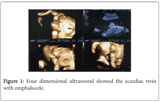
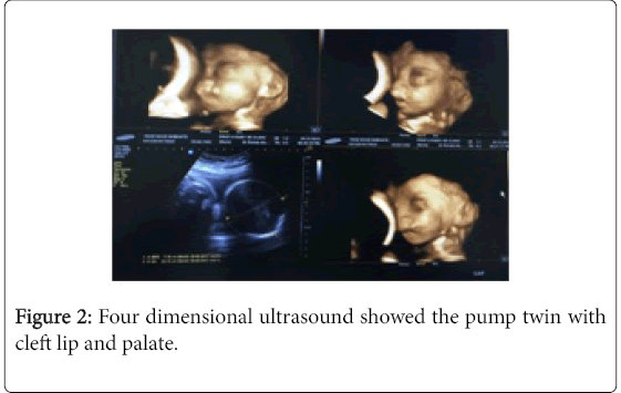
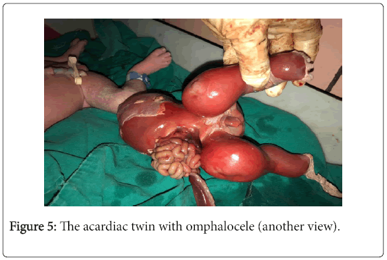
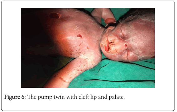
At 27 weeks of pregnancy, four-dimensional ultrasound was done and revealed twin reversed arterial perfusion sequence in monochorionic diamniotic sac. The Acardiac twin with omphalocele (Figure 1) and the pump twin with cleft lip and palate (Figure 2).
Hospital, Cairo, Egypt. Informed consent was obtained from the participant.
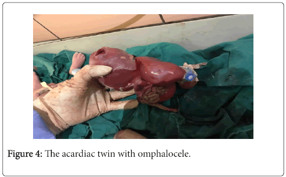
Cesarean section was performed and delivered living female 2500 g and the others died immediately after delivery (Figures 4-6).
Discussion
Twin pregnancy is associated with higher rates of almost every potential complication of pregnancy. Monochorionic twins are at risk for complications unique to these pregnancies, such as twin-twin transfusion syndrome (TTTS), which can be lethal or associated with serious morbidity. Twin reversed arterial perfusion sequence (TRAP) is a rare complication of monochorionic twins in which a living twin perfuses a non-living (acardiac) twin through patent vascular channels. The incidence of congenital anomalies is three to five folds higher in monozygotic twins than in singletons or dizygotic twins. Monochorionic twins have a higher rate of congenital anomalies than dichorionic twins and singletons [1].
Four-dimensional sonography refers to three-dimensional images that can be viewed in real-time. It is also called dynamic three dimensional sonography. It has been used to study the fetal heart, fetal movement and fetal behavioral states [2]. Fetal evaluation using this mode can be very helpful for example in the clear display of spinal and orbital anomalies, and fetal profile. Recently published studies have verified the utility of 4D-ultrasound in various diagnostic areas, including fetal biometry, skeletal dysplasia, facial anomaly and fetal echocardiography [3].
In this case, sonographic examination by four dimensional ultrasound revealed twin reversed arterial perfusion sequence in the monochorionic diamniotic sac and the associated anomalies as omphalocele in the acardiac twin and cleft lip and palate in the pump twin.
Conclusion
In conclusion, detailed sonographic examination by Four Dimensional Ultrasound should be considered for women with multiple pregnancies for complications as TRAP and congenital anomalies. Early diagnosis may allow earlier intervention that decreases maternal morbidity and mortality. So, clear evidence guiding the screening, diagnosis and management of this condition is needed [4].
References
- Chasen ST, Chervenak FA (2017) Twin pregnancy: Prenatal issues.
- Thomas DS (2015) Ultrasound examination in obstetrics and gynecology
- Paladini D, Sglavo G, Greco E, Nappi C (2008) Cardiac screening by STIC: Can sonologists performing the 20 week anomaly scan pick up outflow tract abnormalities by scrolling the A-plane of STIC volumes. Ultrasound Obstet Gynecol 32: 865-870.
- Hur H, Kim YH, Cho HY, Park YW, Won H, et al. (2015) Feasibility of three-dimensional reconstruction and automated measurement of fetal long bones using 5D long bone. Obstet Gynecol Sci 58: 268-276.
Relevant Topics
Recommended Journals
Article Tools
Article Usage
- Total views: 5754
- [From(publication date):
June-2017 - Dec 18, 2024] - Breakdown by view type
- HTML page views : 5011
- PDF downloads : 743

