Case Report Open Access
Treatment of a Young Permanent Maxillary First Molar with Two Palatal Roots
Tao N, Mei Y*, Zhou H and Yang YDepartment of Pathology, Affiliated Hospital of Stomatology Nanjing Medical University, Nanjing, Jiangsu, China
- *Corresponding Author:
- Yufeng Mei
Department of pathology
Affiliated Hospital of Stomatology Nanjing Medical University
Nanjing, Jiangsu
China
E-mail: yfmei@njmu.edu.cn
Received date: Aug 30, 2016; Accepted date: Sept 19, 2016; Published date: Sept 24, 2016
Citation: Tao N, Mei Y, Zhou H and Yang Y (2016) Treatment of a Young Permanent Maxillary First Molar with Two Palatal Roots. J Clin Diagn Res 4: 129. doi:10.4172/2376-0311.1000129
Copyright: © 2016 Tao N, et al. This is an open-access article distributed under the terms of the Creative Commons Attribution License, which permits unrestricted use, distribution, and reproduction in any medium, provided the original author and source are credited.
Visit for more related articles at JBR Journal of Clinical Diagnosis and Research
Abstract
The maxillary first molar usually has single palatal root. Incidence rate of maxillary first molar with two palatal roots is especially rare. We reported a case of young maxillary first molar with two palatal roots in this article. Anatomic characteristic of permanent maxillary molar is generally described as a group of teeth with three roots, one palatal and two buccalbut the root canal system is complexed. The second mesiobuccal root was common finding of the teeth studied, the second distobuccal and palatal root was rare. We reported the unusual variation in root and canal morphology of one four-rooted maxillary first molar with two palatal roots.
Keywords
Maxillary first molar; Two palatal roots; Carious lesion; Apexification
Case Report
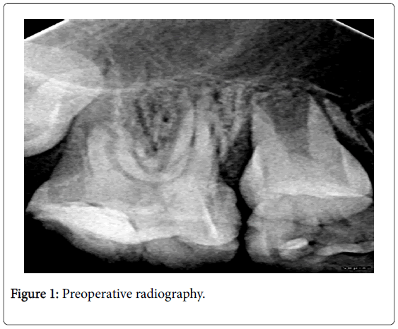
A ten-year old girl was referred to stomatological hospital of Jiangsu province with a history of toothache in the right maxillary region in March 16th, 2014. The patientâÂ?Â?s medical history was non-contributory. Clinical examination revealed an extensive distal obturation with secondary carious lesion in the right maxillary first molar and tenderness to percussion, with negative response to cold testing. The preoperative radiographic views revealed four roots with open apical foramen (Figure 1). The tooth was diagnosed with symptomatic apical peridontitis.
Figure 1: HPLC chromatogram of the nine reference compounds in 50% aqueous methanol, measured at 370nm. Retention times for rutin, sutherlandin A, sutherlandin B, kaempferol-3-O-rutinoside, sutherlandin C, sutherlandin D, quercitrin, quercetin and kaempferol were 11.9, 12.7, 13.8, 15.3, 16.2, 17.0, 18.0, 26.2 and 28.1 minutes, respectively.
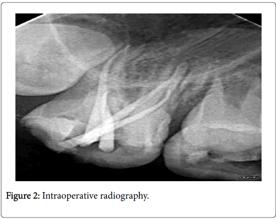
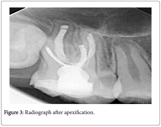
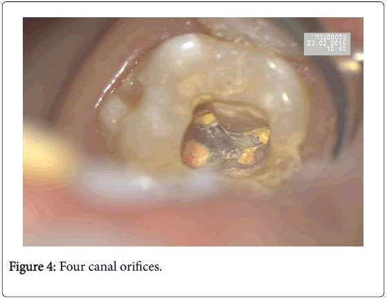
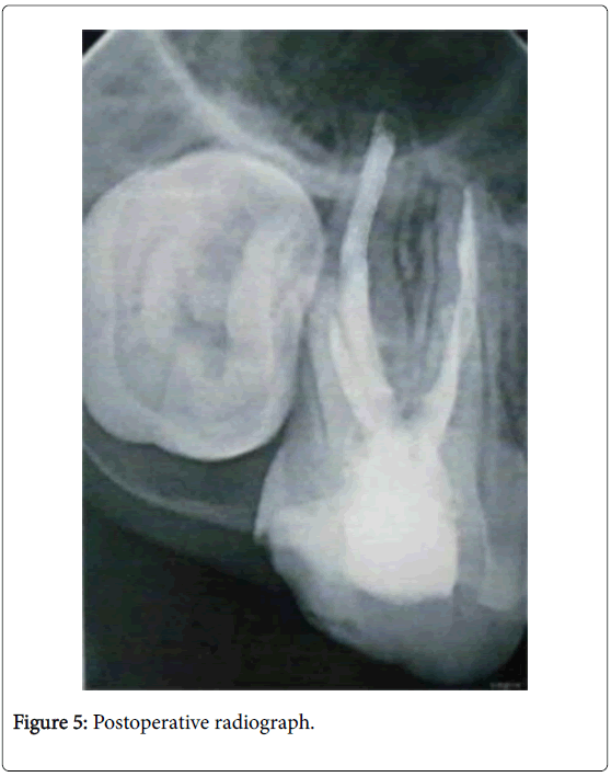
The patient was prepared for apexification. The access cavity of the maxillary first molar is usually triangular in shape. In the current case, the pulp chamber of this tooth was broader in the palatal areathe access cavity was prepared in trapezoidal shape. Apexification was performed and the root canals were cleaned and shaped under the copious irrigation with 3% H2O2 solution and normal saline. In the intraoperative radiographic views, the gutta-percha was put in the root to reveal the length of the roots (Figure 2). Then calcium hydroxide paste was placed in the root and zinc oxide ointment enveloped the tooth for a week. Then roots were filled with vitapex cataplasm MORITA, Japan and the pulp chamber was filled with zinc phosphate cement and glass ionomer. The patient reviewed once three months in regular. The periodic radiographic views (Figure 3) in February 16th, 2016 revealed apical barriers had sealed the apical foramen. Thus the patient was ready for endodontic treatment. The glass ionomer and zinc phosphate cement was removed and the root canals were cleaned and shaped using #15 and #20 stainless steel hand K-files, and Pro Taper nickel-titanium rotary instrumentation (Dentsply Maillefer, Switzerland) with crown-down technique up to X2 under the copious irrigation with 1% sodium hypochlorite. After a final ultrasonic rinse, the root canals were dried and the canals were obturated with guttapercha and AH-plus sealer (Dentsply, De Trey, Konstanz, Germany) using the lateral compaction technique and orifices were sealed (Figure 4). The postoperative radiographic views (Figure 5) revealed the root canals were densely covered with fillings. Finally, the patient was referred to the restorative department for final restoration.
Discussion
The maxillary first molar usually has only one palatal root. The present manuscript reported a maxillary first molar with two separate palatal roots and two buccal roots. The knowledge of root canal anatomy is the most important aspect for the successful root canal therapy. Finding all of the canals is essential for the success of the endodontic case, if clinicians fail to identify their presence, especially when the prevalence of the root is rare, nontreatment of the roots may lead to failure of root canal therapy. A large number of reports [2,3] showed the morphological variations of the root canal system of the maxillary molars.
Maxillary first molar has complexed root canal anatomy. Zheng et al. found that the mesial buccal root of maxillary first molar has higher incidence of double root: 52.24%, distal buccal and palatal root has lower incidence of double root, respectively, 1.12% and 1.76% [4]. And Yang found that the maxillary molar has [0.72% (14/1957)] incidence of palatal double root, palatal double root of the maxillary second molar incidence [1.12% (11/979)] is higher than the first molar [ 0.31% (3/978)] [5].
In clinical work, to detect presence of additional root, the clinician should not only be familiar with variations of root canal system, but also take care of the anatomic characteristic of the teeth, such as extra cusp and pulp chamber, additionally use radiographic examination and CT scan.
Conclusion
The variations in the anatomy of root canals have an important role in endodontic therapy. Clinicians ought to be aware of the possibilities, as nontreatment of one canal could lead to endodontic failure.
References
- Cleghorn BM, Christie WH, Dong CC (2006) Root and root canal morphology of the human permanent maxillary first molar: a literature review. J Endod32: 813-321.
- Caliskan MK, Pehlivan Y, Sepetçioglu F, Türkün M, Tuncer SS (1995) Root canal morphology of human permanent teeth in a Turkish population. J Endod21: 200-204.
- Taylor C, Roudsari RV, Jawad S, Darcey J, Qualtrough A (2016) Modern Endodontic Principles. Part 6: Managing Complex Situations. Dent Update 43: 218-220, 223-6, 229-32.
- ZhengQH,Wang Y,Zhou XD (2010) A cone-beam computed tomography study of maxillary first permanent molar root and canal morphology in a Chinese population. J Endod 36: 1480-1784.
- Yang B, Lu Q, Bai QX, Zhang Y, Liu XJ, et al. (2013) Evaluation of the prevalence of the maxillary molars with two palatal roots by cone-beam CT. Chinese J Stomatology 48: 359-62.
Relevant Topics
- Back Pain Diagnosis
- Cardiovascular Diagnosis
- Clinical Diagnosis
- Clinical Echocardiography
- COPD Diagnosis
- Diabetes Diagnosis
- Diagnosis Methods
- Diagnosis of cancer
- Diagnosis of CNS
- Diagnosis of Diabetes
- Diagnostic Products
- Diagnostics Market Analysis
- Heart diagnosis
- Immuno Diagnosis
- Infertility Diagnosis
- Medical Diagnostic Tools
- Preimplementation Genetic Diagnosis
- Prenatal Diagnostics
- Ultrasonography
Recommended Journals
Article Tools
Article Usage
- Total views: 4226
- [From(publication date):
December-2016 - Apr 02, 2025] - Breakdown by view type
- HTML page views : 3339
- PDF downloads : 887

