Case Report Open Access
Transient Sarcoid Reaction to a Thoracic Schwannoma
www.omicsonline.org/searchresult.phpCaillaud D1*, Horo K1, Kemeny JL2, Michel JL3 and Toloba Y1
1Pulmonology and Allergology Department, CHU G Montpied, Auvergne University, Clermont- Ferrand, France
2Pathology Department, CHU G Montpied, Auvergne University, Clermont-Ferrand, France
3Radiology Department, CHU G Montpied, Auvergne University, Clermont-Ferrand, France
- *Corresponding Author:
- Caillaud D
Pulmonology and Allergology Department
Hopital Gabriel Montpied
Montalembert Str Auvergne University
Clermont- Ferrand, 63003, France
Tel: 33-4-73751653
Fax: 33-4-73751656
E-mail: dcaillaud@chu-clermontferrand.fr
Received date: May 10, 2016; Accepted date: May 28, 2016; Published date: May 31, 2016
Citation: Caillaud D, Horo K, Kemeny JL, Michel JL, Toloba Y (2016) Transient Sarcoid Reaction to a Thoracic Schwannoma. Occup Med Health Aff 4: 236. doi:10.4172/2329-6879.1000236
Copyright: © 2016 Caillaud D, et al. This is an open-access article distributed under the terms of the Creative Commons Attribution License, which permits unrestricted use, distribution, and reproduction in any medium, provided the original author and source are credited.
Visit for more related articles at Occupational Medicine & Health Affairs
Abstract
Background: Sarcoid reactions to benign tumors are an exception. Only two cases have been reported where these reactions were associated with a benign teratoma. We report a new case where a sarcoid reaction is secondary to a thoracic schwannoma.
Observation: During a uveitis assessment performed on a 53 year-old man who was not displaying any other symptoms, a thoracic schwannoma and mediastino-hilar lymph nodes were fortuitously discovered. Surgical resection of the schwannoma leads to a regression of the mediastinal and hilar lymph nodes without any other medical treatment.
Conclusion: In case of thoracic schwannoma, a sarcoid reaction must be considered if mediastinal lymph nodes are present. Other similar cases are necessary to confirm the validity of this case report.
Keywords
Sarcoidosis; Schwannoma; Tumor; Benign
Introduction
Sarcoidosis is a multisystemic granulomatous disorder characterized by non caseating granulomas, of undetermined etiology, with bilateral mediastinal lymphadenopathy, pulmonary infiltration, ocular and skin lesions or some combination of these findings [1].
Malignant tumor-related tissue reactions resulting in the formation of epithelioid-cell granulomas have been termed sarcoid reactions. Sarcoid reactions are not exceptional and occur in 4.4% of carcinomas, 13.8% of patients with Hodgkin diseases, and in 7.3% of cases of non- Hodgkin lymphomas [2].
However, cases of sarcoid reactions associated with benign tumors are exceptional. Only two cases associated with thoracic mature teratoma have been reported in the literature [3,4]. We report a new case of sarcoid reaction in association with a benign thoracic schwannoma.
Case Report
A 53 year-old man with unilateral posterior uveitis was referred to the pulmonary department, for a small retrocardiac mass found on a routine chest radiograph performed for potential sarcoidosis. He had no previous respiratory problems and never smoked.
In the clinic, a physical examination revealed a heart rate of 72 beats/min, a BP of 130/80 mmHg, and a room oxygen saturation of 94%. Pulmonary examination was normal.
Chest X-ray showed the presence of large hilar opacities with polycyclic contours without parenchymal abnormalities.
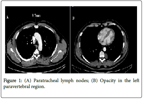
A computed tomography scan (Figure 1A) confirmed the presence of supra-centimeter lymph nodes in right paratracheal region, and bilateral hilar lymph nodes.
The scan also revealed the presence of a tissue opacity with a width of 3.5 cm located in the left paravertebral region, extending from the dorsal (D) vertebra D8 to D10, which suggested the presence of a neurinoma (Figure 1B).
Bronchoscopy and systematic bronchial biopsies were normal. A pulmonary function test revealed an FEV1 of 4.27 L (120% predicted), and FVC of 5.10 L (107% predicted), and a normal diffusing capacity.
Capillary blood gases found PaO2 at 70 mmHg, PaCO2 36 mmHg, pH 7.44 and an oxygen saturation at 94%. Laboratory results were all within normal limits (blood count, platelets, serum electrolytes, transaminases, alkaline phosphatases, calcemia, calciuria, as well as angiotensin converting enzyme).
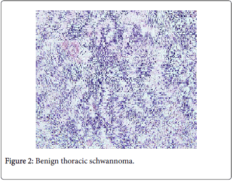
Costovertebral opacity was resected by left thoracoscopy, and histological examination revealed a schwannoma without evidence of malignancy (Figure 2).
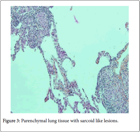
Due to the presence of micro-granules, a lung biopsy was performed on the lower left lobe; specifically on the lower left visceral and mediastinal pleura next to the ascending aorta and the posterior costal pleura.
Intraoperative histological examination of these micro-granules (Figure 3) revealed a sarcoid reaction (giant cell epithelioid granulomas without necrosis).
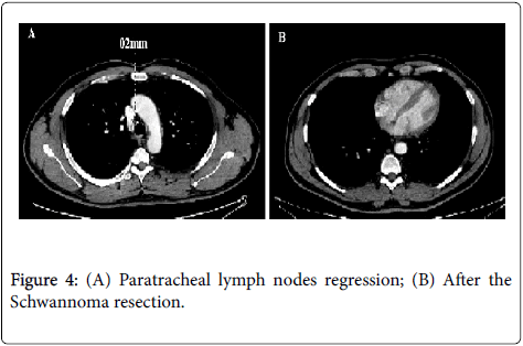
Regular monitoring was performed. A CT chest scan performed 6 months after surgical resection showed partial disappearance of lymph nodes. The latest CT scan, performed in April 2012 (4 years after the resection (Figure 4) showed complete disappearance of hilar and mediastinal lymph nodes, but calcification of the left basal pyramid was observed.
Discussion
This is a fortuitous discovery of a hilar-mediastinal sarcoid reaction to benign schwannoma during a uveitis assessment.
Sarcoid reaction incidence to cancers has been reported in 2.4 to 3% of cases. They may be interpreted as a defense reaction against a tumor’s antigens, and may have a more favorable prognosis than classical malignant nodes. The appearance of lymph nodes in a patient with a cancer history must first suggest a tumor progression. However, a sarcoid reaction possibility must be kept in mind by clinicians and oncologists because the therapeutic strategy and prognosis are radically different.
Sarcoid reactions to benign tumors, in contrast, are very rare and only two cases have been reported. The first case described in the literature [3], was a young man who exhibited signs and symptoms suggesting an acute myocardial infarction, and was thrombolyzed, but subsequently died. Post-mortem examination revealed a massive pericardial effusion and necrosis involving a mature teratoma of the mediastinum associated with sarcoid reaction. Gary-Rustom et al. [4] reported a second case of mature intra thymic teratoma associated with granulomatous sarcoid-like reaction. Mediastinal mature teratomas are benign and usually asymptomatic.
Benign schwannoma are the most common benign tumors of the peripheral nerves [5]. They are encapsulated lesions rarely causing neurological deficits, mainly in posterior mediastinal locations. Our case is the first to report a sarcoid reaction to this benign tumor. The regression of hilar and mediastinal lymph nodes without medical treatment after Schwannoma resection appears to establish a causal link.
Conclusion
In case of a thoracic schwannoma with mediastinal and hilar lymph nodes, the possibility of a sarcoid reaction must be considered. However, other cases are needed to confirm this first case report.
References
- Iannuzzi MC, Rybicki BA, Teirstein AS (2007)Sarcoidosis. N Engl J Med 357: 2153-2165.
- Brincker H (1986)Sarcoid reactions in malignant tumours. Cancer Treat Rev 13: 147-156.
- Madan V, Cottrell B, Sharpstone D (2007)Sarcoidalpericardial effusion masquerading as acute myocardial infarction. Internet J Intern Med 7: 1.
- Gary-Rustom L, Declercq PL, Veresezan L, Muir JF, Cuvelier A (2012) Mature mediastinal teratoma and sarcoid-like granulomatosis. Rev Mal Respir 29: 898-902.
- Das Gupta TK, Brasfield RD (1970) Tumors of peripheral nerve origin: benign and malignant solitary schwannomas. CA Cancer J Clin20: 228-233.
--
Relevant Topics
- Child Health Education
- Construction Safety
- Dental Health Education
- Holistic Health Education
- Industrial Hygiene
- Nursing Health Education
- Occupational and Environmental Medicine
- Occupational Dermatitis
- Occupational Disorders
- Occupational Exposures
- Occupational Medicine
- Occupational Physical Therapy
- Occupational Rehabilitation
- Occupational Standards
- Occupational Therapist Practice
- Occupational Therapy
- Occupational Therapy Devices & Market Analysis
- Occupational Toxicology
- Oral Health Education
- Paediatric Occupational Therapy
- Perinatal Mental Health
- Pleural Mesothelioma
- Recreation Therapy
- Sensory Integration Therapy
- Workplace Safety & Stress
- Workplace Safety Culture
Recommended Journals
Article Tools
Article Usage
- Total views: 11602
- [From(publication date):
June-2016 - Apr 16, 2025] - Breakdown by view type
- HTML page views : 10745
- PDF downloads : 857


