Case Report Open Access
Transcuneiform Crush Injury: A Case Report
| Laksha Dutt*, Susan Fisher, David J. Wilson and Paul Ryan | ||
| Orthopaedic Surgery, Madigan Army Medical Center (MAMC), Tacoma, Washington, USA | ||
| Corresponding Author : | Laksha Dutt Madigan Army Medical Center Orthopaedic Surgery, 9040 Fitzsimmons Ave tacoma, wa 98431, United States Tel: 510-557-5943 E-mail: laksha.dutt.mil@mail.mil |
|
| Received December 15, 2014; Accepted December 27, 2014; Published January 02, 2015 | ||
| Citation: Dutt L, Fisher S, Wilson DJ, Ryan P (2015) Transcuneiform Crush Injury: A Case Report. Clin Res Foot Ankle 3:162. doi:10.4172/2329-910X.1000162 | ||
| Copyright: ©2015 Dutt L, et al. This is an open-access article distributed under the terms of the Creative Commons Attribution License, which permits unrestricted use, distribution, and reproduction in any medium, provided the original author and source are credited. | ||
Related article at Pubmed Pubmed  Scholar Google Scholar Google |
||
Visit for more related articles at Clinical Research on Foot & Ankle
Abstract
Introduction: Transcuneiform fracture patterns without dislocations are uncommon. In a review of the literature, numerous case reports describe cuneiform fracture-dislocations involving either the transmetatarsal or midtarsal joint complexes due to direct trauma; however, an isolated transcuneiform fracture pattern without dislocation has not been reported.
Methods: We present a rare injury of the foot: an isolated transcuneiform fracture without associated dislocation of the tarsometatarsal joint or midtarsal joint complex.
Case: A 29 year old active duty Army male sustained direct blunt trauma to his foot when a 20 ton trailer fell onto it. His initial presentation prompted protected weight bearing after a clinical exam revealed soft tissue injury and tenderness to palpation about the dorsum of his midfoot. Due to persistent symptoms, he was referred to a musculoskeletal specialist. Radiographs and computed tomography (CT) images demonstrated a minimally displaced comminuted transcuneiform fracture involving the medial and intermediate cuneiforms without evidence of dislocation or subluxation. Treatment consisted of non-weight bearing cast immobilization with crutches for ten weeks before being transitioned into a weight bearing control ankle motion (CAM) boot. Radiographic evidence of healing was documented at 4 weeks by callus formation at the first metatarsal and medial cuneiform. The patient returned to low impact activity by 10 month follow-up without surgical intervention.
Discussion and Conclusions: Blunt trauma to the midfoot can result in isolated or complex fracture patterns without the typical fracture-dislocation following high mechanism trauma. Appropriate clinical suspicion and careful physical examination can facilitate timely diagnosis and treatment. Minimally displaced transcuneifom fracture patterns without associated midfoot dislocation injuries can be treated nonoperatively with protected weight bearing, close radiographic and clinical follow up, and gradual advancement to activity with acceptable clinical outcomes.
| Introduction | |
| The tarsometatarsal joints consist of articulations between the metatarsal bases, cuneiforms and the cuboid. This joint complex is primarily stabilized by the position of the second metatarsal base into the intercuneiform recess. The second metatarsal is considered the keystone in maintaining the tarsometatarsal joint. Cuneiform fractures are rare and account for approximately 4.2% of all tarsal bone fractures [1-3]. Transcuneiform fracture patterns involve more than one cuneiform and are more commonly associated with compression injuries. In a review of the literature, numerous case reports describe cuneiform fracture-dislocations involving either the transmetatarsal or midtarsal joint complexes due to direct trauma. There is, however, a paucity of literature describing isolated transcuneiform fracture in the absence of an associated dislocation. | |
| Isolated cuneiform fractures are uncommon and considered highenergy injuries [2-4]. Although the medial cuneiform is the most injured cuneiform, dislocations are very rare with the high-energy injuries due to the strong ligamentous attachment. Medial cuneiform fracture patterns are most commonly characterized as small, avulsion injuries [4]. Fracture displacement occurs through the pull of the tibialis anterior tendon. [Less commonly described crush injuries are the result of a direct blow to the dorsum of the foot] [1]. | |
| A thorough foot examination should be performed to rule out any bony or ligamentous injury. A complete plain radiographic work up can identify fractures and/or dislocations. On weight bearing radiographs, a gap sign between the first and second digits may represent intercuneiform instability [3]. | |
| Case | |
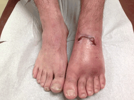
| A 29-year-old man sustained a direct blunt injury to his left foot when a large trailer landed on it. The patient presented to the emergency department where a clinical examination revealed a 5cm dorsal midfoot laceration, surrounding swelling, ecchymosis and tenderness. He was given crutches and sent home (Figure 1). | |
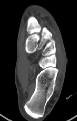
| Over the ensuing 48 hours, he continued to complain of pain isolated to the dorsal midfoot. He re-presented to an urgent clinic where non weight bearing radiographs (anterior-posterior, lateral, medial oblique) revealed a medial cuneiform fracture with no displacement of the tarsometatarsal joint and tarsals. A CT scan revealed minimally displaced comminuted transcuneiform fractures, involving the medial and intermediate cuneiforms and fractures of the first and fourth metatarsal without evidence of dislocation or subluxation (Figure 2). | |
| Treatment consisted of non-weight bearing cast immobilization with crutches for ten weeks. He then transitioned into a CAM boot and was made weight bearing as tolerated with crutch assistance. At six month follow-up, the patient continued to have minimal pain localized to the distal aspect of the second and third metatarsals. At ten month follow-up, the patient had already transitioned into a regular shoe with the use of custom made orthotics. He returned to low impact activity at 10 months. | |
| Discussion | |
| Medial cuneiform fractures are extremely rare, and the diagnosis is usually delayed or missed completely. It can be difficult to diagnose subtle isolated cuneiform and trans-cuneiform fracture patterns with non-weight-bearing standard views of the foot due to the overlap of the tarsal and metatarsal bones [3,4]. Isolated and trans-cuneiform fractures may also be missed due to the higher incidence of Lisfranc’s fracture-dislocation and/or Chopart joint injuries and the greater emphasis on trying to diagnosis and treat those foot pathologies. As an adjunctive diagnostic test, a bone scan using technetium has been a valuable tool in evaluation fractures especially in cases where overlapping of tarsal bones can impede the ability to interpret radiographs.2 Although displaced medial cuneiform fractures are rare, when present, anatomical reduction is indicated when there is disruption of the joint due to dislocation or fracture. | |
| Conclusion | |
| High-energy crush injuries can result in a transcuneiform fracture pattern without dislocation. This injury pattern may be difficult to see on plain radiographs due to the multiple overlapping shadows on nonweight bearing views and either CT or bone scan may be necessary to evaluate the extent of injury. As demonstrated in this case report, a stable transcunieform fracture pattern can be effectively treated nonoperatively. Although the fractures may heal in a predictable timeframe, the soft tissue component of the injury may delay return to activity over an extended period of time. | |
| References | |
| style="padding-left:35px;"> | |
References
- value="1" id="Reference_Titile_Link">Grivas T, Vasiliadis E. Koufopoulos G, Polyzois V, Polyzois D (2003) Midfoot Fractures: Clinical Podiatry Medical Surgery 23: 323-341.
- value="2" id="Reference_Titile_Link">Mandracchia VJ, Mandracchia DM, Pelsang DJ (1994) Isolated fracture of the lateral cuneiform. A rare tarsal injury. J Am Podiatr Med Assoc 84: 189-191.
- value="3" id="Reference_Titile_Link">Davies MS, Saxby TS (1999) Intercuneiform instability and the "gap" sign. Foot Ankle Int 20: 606-609.
- value="4" id="Reference_Titile_Link">Patterson RH, Petersen D, Cunningham R (1993) Isolated fracture of the medial cuneiform. J Orthop Trauma 7: 94-95.
Figures at a glance
 |
 |
|
| Figure 1 | Figure 2 |
Relevant Topics
Recommended Journals
Article Tools
Article Usage
- Total views: 16020
- [From(publication date):
May-2015 - Apr 07, 2025] - Breakdown by view type
- HTML page views : 11464
- PDF downloads : 4556
