Case Report Open Access
Total Ankle Replacement with a Staged Correction of a 20 Degree Post Traumatic Ankle Valgus and Medial Ankle Instability
| Lawrence A Di Domenico* and Danielle N Butto | |
| Ankle and Foot Care Centers, Lawrence Di Domenico, Ankle and Foot Care Centers, 8175 Market St Youngstown, OH 44512, USA | |
| Corresponding Author : | Lawrence Di Domenico Ankle and Foot Care Centers 8175 Market St Youngstown OH 44512, USA Tel: 330-629-8800 Fax: 330-758-4914 E-mail: Ld5353@aol.com |
| Received date: Sep 11, 2015; Accepted date: Jan 27, 2016; Published date: Jan 31, 2016 | |
| Citation: Domenico LAD, Butto DN (2016) Staged Correction of a 20 Degree Post Traumatic Ankle Valgus with Medial Ankle Instability. Clin Res Foot Ankle 4:179. doi:10.4172/2329-910X.1000179 | |
| Copyright: © 2016 Domenico LAD, et al. This is an open-access article distributed under the terms of the Creative Commons Attribution License, which permits unrestricted use, distribution, and reproduction in any medium, provided the original author and source are credited. | |
| Related article at Pubmed, Scholar Google | |
Visit for more related articles at Clinical Research on Foot & Ankle
Abstract
Total ankle replacement can be a challenging surgery especially when pre-operative deformity exists. Most authors advocate the indication for total ankle replacement should be narrowed to patients with less than 10 to 15 degrees of pre-operative varus or valgus. When greater than 10-15 degrees of coronal plane malalignment is found, ancillary procedures must be performed. We report a case of a 56 year old male with 20 degrees of pre-operative ankle valgus after a pronation-external rotation injury that was malreduced at an outside institution. In addition to the valgus, the patient presented with medial ankle instability, distal lateral tibial osteonecrosis and a shortened, posteriorly rotated fibula. Staged procedures were employed to successfully realign the patient’s ankle joint. The patient was first brought to the operating room and stressed under c-arm fluoroscopy. He was found to have instability of the deltoid complex. He subsequently underwent an ankle arthrotomy, synevectomy and deltoid imbrication to re-establish medial ankle stability. Second, the patient underwent a fibular lengthening and derotation, syndesmotic fusion and medial opening wedge tibial osteotomy. Once consolidation was confirmed by CAT scan, the patient had a gastrocnemius lengthening total ankle arthroplasty with a Zimmer trabecular metal implant (Zimmer Inc®, Warsaw, IN) and debridement and grafting of the distal lateral tibial osteonecrosis. The patient is now greater than 24 months post operatively and able to ambulate pain free without assistive devices.
| Keywords |
| Total ankle replacement; Ankle valgus; Tibial osteonecrosis; Post-traumatic arthritis |
| Introduction |
| Total ankle replacement and implant devices have evolved greatly. Nonetheless, the surgery can be challenging especially when preoperative deformity exists. Some authors have suggested the indication for total ankle replacement should be narrowed to patients with less than 10 to 15 degrees of pre-operative varus or valgus [1-4]. However, it was found that between 33% to 44% of patients with end-stage osteoarthritis present with malalignment of greater than 10 degrees in the coronal plane [1,3,5]. Depending on the degree of pre-operative malalignment, staged procedures may need to be considered before discounting the possibility of total ankle replacement. |
| Case Report |
| A 56 year old male presented for initial evaluation with complaint of right ankle pain. The patient underwent an open reduction internal fixation of a pronation-external rotation injury one year prior at an outside institution. The patient’s original fixation consisted of two, 4.0 cancellous medial malleolar screws and two, tightropes to fixate the syndesmosis. The surgeon of record felt that the fibula was noted to have good reduction and therefore was not fixated. At his first follow up he was informed his reduction was not satisfactory. Six weeks later he subsequently underwent a second surgery consisting of removal of hardware and revision surgery for open reduction internal fixation. The surgeon identified a butterfly fragment of the fibula fracture. At this time, the fibula was fixated with a 3.5 inter fragmentary screws and a semi-tubular plate. The medial malleolar fracture was fixated with circlage wire in a figure of eight pattern. The syndesmosis was fixated with a two, cortical screw engaging four cortices each. Post-operatively he was noted to have continued pain. He underwent a third surgery three months later with his original surgeon which included removal of the syndesmotic screws inserted during his second surgery. After his three surgeries, his ankle remained malaligned and painful. |
| The patient presented to our institution 13 months after his initial surgery with a complaint of persistent right ankle pain. His past medical history was significant for only hypertension. On physical examination, the patient ambulated in an apropulsive manner with the assistance of a cane. His vascular status, protective, and epicritic sensation were intact. Incisions sites were all well healed with no hypertrophic scar formation. Muscle strength was 4/5 to all muscles of the lower extremity. Range of motion of the ankle joint was severely limited with pain and crepitus noted. There was pain on palpation to both the lateral and medial ankle ligamentous structures. |
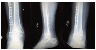
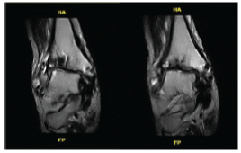
| Radiographic evaluation revealed a 20 degree ankle valgus, a malunion of the medial malleolus fracture, a 1cm increase in tibiafibular clear space, a shortened, posteriorly rotated fibula and lucency in the distal lateral tibia. A MRI revealed decreased signal uptake at the distal lateral tibia confirming the lucency was osteonecrosis of the distal lateral tibia (Figures 1 and 2). |
| The patient previously failed ankle bracing and physical therapy and was interested in surgical correction of his deformity. Both ankle fusion and replacement were discussed in detail. The patient desired a total ankle arthroplasty and was not interested in an ankle arthrodesis. The patient’s age, co-morbidities and physical demands were amendable to ankle replacement. The major concerns regarding his case included the medial deltoid instability, severe valgus rotation and tibial osteonecrosis. Given the patient’s complex deformity and history of multiple previous surgeries, a staged approach was employed. |
| The patient was brought to the operating room and the ankle joint was stressed under C-Arm fluoroscopy. The patient was noted to have significant instability in a valgus rotation with complete loss of his medial deltoid ligament function. His initial surgery consisted of an ankle joint arthrotomy with synevectomy and deltoid ligament imbrication. An oblique incision was made over the medial deltoid complex. The deltoid ligament was identified, incised and retracted. A medial capsular incision was made for the arthrotomy. The ankle joint had a tremendous amount of hypertrophic, chronic synovitis which was debrided. There were distal malleolus fracture fragments noted. The major fragments were identified and removed at this time. Removing the remaining fragments could compromise the repair medially and were left at this time. The ankle capsule was reapproximated and attention was drawn back to the deltoid ligaments. A wedge section was removed from the deltoid ligament complex and the ligament was primarily repaired with 0 vicryl. Prior to embarking on further reconstructive surgery, the surgeons wanted to evaluate if the medial deltoid reconstruction would stabilize the medial ankle. If the medial ankle could not be stabilized, an ankle arthrodesis would need to be performed instead of an ankle arthroplasty. Post-operatively it was felt that stability was re-established to the medial ankle. The decision to proceed with an ankle replacement was made with the thought process that bony realignment would be needed in order to support the medial deltoid ligament reconstruction. A fibular and tibial osteotomy would be necessary to realign the ankle joint appropriately in order for an ankle arthroplasty to be well maintained in the ankle joint. |
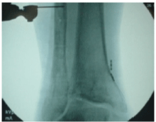
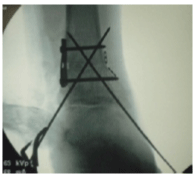
| A second surgery was planned 7 weeks after the first to address the patient’s shortened, posteriorly rotated fibula, syndesmotic rupture and valgus malalignment. All retained hardware from the previous open reduction internal fixation surgeries was removed. A 2-cm piece of fibula was resected from the middle - distal fibula to use for bone graft. Attention was then directed to the syndesmosis which was debrided and taken down and the bony surfaces were extensively debrided in preparation for syndesmotic arthrodesis. Attention was then directed to the tibia where an opening wedge osteotomy was performed superior to the area of osteonecrosis to address the 20 degrees of ankle valgus. The osteotomy was made with the base laterally and apex medially. The osteotomy site was packed with the autogenous fibula graft and allogenic bone. The tibial osteotomy was stabilized with a 1/3 tubular plate and 2, 3.5 bicortical screws. Once fully debrided, the fibula was able to be lengthened and anteriorly rotated into the incisura. The fibula was then brought to length and anteriorly rotated and held in a corrected position with a locking plate over the fibula. The plate extended to the proximal fibula to assist in maintaining anatomic alignment with the distal fibula. The syndesmosis was stabilized with two, 4.0 syndesmotic screws inserted across the syndesmosis through the fibula plate to fuse the syndesmosis (Figure 3). |
| Note the valgus ankle, the increase in the tibia-fibular syndesomsis, the mal union of the medial malleolus (Figure 4). |
| A distal tibial osteotomy was performed with the base being lateral and the apex medial. The osteotomy was bone grafted with autogenous fibular bone and back filled with allogenic cancellous bone. The fixation consisted of a two hole plate resulting in anatomic alignment. The weber clamp was used to pull the distal fibula inferiorly and anteriorly rotated. |
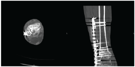
| A CT scan was ordered to evaluate the tibial osteotomy and the tibia-fibular syndesmotic arthrodesis. There was confirmed bony consolidation at the distal tibia-fibular syndesmotic fusion, and union of the graft at the tibial opening wedge osteotomy. The patient’s total ankle replacement surgery was then planned (Figures 5 and 6). |
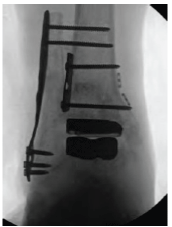
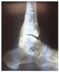
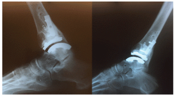
| Five months post operatively from the patients second staged procedure, the patient underwent removal of the large fibular plate, a gastrocnemius recession and total ankle arthroplasty with a Zimmer trabecular total ankle implant through a lateral approach. The lower extremity was aligned appropriately within the external fixator and a fibular osteotomy was made below the syndesmosis arthrodesis and proximal to the ankle joint. The tibial and talar bone surface preparation was performed as described by Zimmer technique utilizing a side cutting burr [6]. With this technique, the osteonecrosis was resected and inspected. Any suggesting there was a void was then packed with the autogenous bone graft. The implant was inserted and the fibular osteotomy was reduced and fixated with a plate and screws. A Zimmer trabecular total ankle implant (Zimmer Inc.®, Warsaw, IN) was utilized because the implant surfaces and bone resections are curved to help reduce the risk of implant subsidence. This also increases the effective contact area between the prostheses and the bones, allowing joint loads to be more broadly distributed. The design also places the implants in a region of greater bone density (Figures 7-9). |
| Movie (Supplementary file): This is a clinical movie demonstrating the range of motion the patient is experiencing following the total ankle replacement surgery. |
| The patient is now greater than 24 months status post total ankle arthroplasty. He reports an improvement in pain, function and range of motion. He is able to ambulate independently with no assistive devices. |
| Discussion |
| Complex pre-operative alignment presents as a challenging case for a surgeon to realign. The patient’s clinical picture and imaging studies should be carefully assessed for other areas in need of correction. One should refer back to the list of indications and contraindications and address deformities accordingly. This may require a series of procedures before the total ankle device can be inserted. |
| Medial ankle stability is crucial in a pre-operative valgus deformity and the survivorship of the ankle implant. The deltoid complex is the primary restraint against valgus tilting of the talus. In addition, the deep deltoid ligament is the secondary restraint against both lateral and anterior talar excursion [7]. The deltoid complex is often compromised in ankle injuries, with pronation-external rotation fractures being the most common injury associated with deltoid ligament rupture [8]. In this case the patient suffered an injury to the medial deltoid ligament, but additionally had deforming forces from malalignment. |
| Fibular position should also be carefully assessed, especially in posttraumatic arthritis. Shortening and lateral rotation of the fibula is a complication after open or closed treatment of malleolar fractures. A shortened and rotated fibula will interfere with the normal function of the ankle. Displacement of as little as 1 mm will change the mobility of the talus and fibula, as well as, the distribution of load in the talofibular and talotibial articulations [9]. Cadaveric studies showed that 30° of lateral rotation deformity decreases the tibiotalar contact area by 30% to 50% [9-11]. |
| There is debate in the literature regarding total ankle arthroplasty and pre-operative coronal plane deformity. Some authors argue malalignment of greater than 10 degrees in any plane in the supramalleolar or distal tibial region requires a corrective osteotomy at the level of the deformity before a TAR can be considered [3,12]. However, some authors have found deformity of up to 30 degrees can be corrected without a statistically significant increase in failure when compared to those with deformity less than 30 degrees. The primary mode of failure in pre-operative deformities greater than 20 degrees is instability [13]. In addition, the syndesmosis should be evaluated. After a malreduced ankle fracture, chronic instability can ensue. Syndesmotic instability is defined as chronic when present for greater than 6 months [14]. Syndesmotic fusion is the recommended treatment for chronic instability [14,15]. |
| Lastly, this patient had distal tibial osteonecrosis. Distal tibial osteonecrosis is not a common occurrence with only 11 cases published in the literature. It is thought to occur secondary to trauma and resultant intra-osseous compartment syndrome. The clinical entity has been a result of ankle fracture in all reports published including this case. Tibial osteonecrosis is typically treated with drilling, grafting, and realignment [16,17]. |
| While osteonecrosis and pre-operative coronal malalignment greater than 10-15 degrees are listed as contraindications, we were able to successfully insert a Zimmer total ankle implant with staged procedures to address the osteonecrosis, 20 degree ankle valgus, deltoid insufficiency and a shortened, rotated fibula with a good clinical outcome. We believe close attention to existing deformities and appropriate staging of procedures is key to a successful result. |
| References |
References
- value="1" id="Reference_Titile_Link">Trnka HJ, Muhlbauer M, Zettl R, Myerson MS, Ritschl P (1999) Comparison of the results of the Weil and Helal osteotomies for the treatment of metatarsalgia secondary to dislocation of the lesser metatarsophalangeal joints. Foot & Ankle Int 20: 72-79.
- value="2" id="Reference_Titile_Link">Scranton PE Jr (1981) Metatarsalgia: a clinical review of diagnosis and management. Foot Ankle 1: 229-234.
- value="3" id="Reference_Titile_Link">García-Bordes L, Jiménez-Potrero M, Collado-Sáenz F, Yunta-Gallo A (2010) Proximal metatarsal resection. Foot Ankle Surg 16: 65-69.
- value="4" id="Reference_Titile_Link">Sgarlato TE (1971) A compendium of podiatric biomechanics. San Francisco: California College of Podiatric Medicine.
- value="5" id="Reference_Titile_Link">Canadell J JE, Villas C (1979) Reseccioncuneiforme de las bases de los metatarsianoscomotratamiento de la metatarsalgiamecanica. Chirurgia del piede 3: 39.
- value="6" id="Reference_Titile_Link">Meisenbach R (1916) Painful anterior arch of the foot: an operation for its relief by means of raising the arch. Am J OrthopSurg 14: 206-211.
- value="7" id="Reference_Titile_Link">Lelievre J (1974)Tratamiento medico de lasmetatarsalgiascausadasportrastornosestaticos del antepie. Rev ChirOrthopReparatriceAppar Mot 60:174-192.
- value="8" id="Reference_Titile_Link">Helal B (1975) Metatarsal osteotomy for metatarsalgia. J Bone Joint Surg Br 57: 187-192.
- value="9" id="Reference_Titile_Link">Barouk LS (1996) [Weil's metatarsal osteotomy in the treatment of metatarsalgia]. Orthopade 25: 338-344.
- value="10" id="Reference_Titile_Link">Spence KF, O'Connell SJ, Kenzora JE (1990) Proximal metatarsal segmental resection: a treatment for intractable plantar keratoses. Orthopedics 13: 741-747.
- value="11" id="Reference_Titile_Link">Davidson M (1971) Non-stabilization metatarsal head osteotomies; a simple method for correcting second, third, fourth, and fifth metatarsal head pathology. J Foot Surg 10: 121-124.
- value="12" id="Reference_Titile_Link">Sullivan JD (1975) The dorsal displacement floating metatarsal subcapital osteotomy. J Foot Surg 14: 62.
- value="13" id="Reference_Titile_Link">Young DE, Hugar DW (1980) Evaluation of the V-osteotomy as a procedure to alleviate the intractable plantar keratoma. J Foot Surg 19: 187-189.
- value="14" id="Reference_Titile_Link">Lynch JR, Taitsman LA, Barei DP, Nork SE (2008) Femoral nonunion: risk factors and treatment options. J Am AcadOrthopSurg 16: 88-97.
Figures at a glance
 |
 |
 |
 |
 |
| Figure 1 | Figure 2 | Figure 3 | Figure 4 | Figure 5 |
 |
 |
 |
 |
|
| Figure 6 | Figure 7 | Figure 8 | Figure 9 |
Relevant Topics
Recommended Journals
Article Tools
Article Usage
- Total views: 11357
- [From(publication date):
March-2016 - Apr 06, 2025] - Breakdown by view type
- HTML page views : 10432
- PDF downloads : 925
