Research Article Open Access
Tinea Capitis at Charles Nicolle Hospital of Tunis (Tunisia)
| Kalthoum Dridi, Bouchekoua Myriam*, Sonia Trabelsi, Dorsaf Aloui and Samira Khaled | ||
| Laboratory of Parasitology-Mycology, Charles Nicolle hospital of Tunis, Tunisia | ||
| Corresponding Author : | Bouchekoua Myriam Laboratory of Parasitology-Mycology Charles Nicolle hospital of Tunis Rue 9 Avril 1938, 1006 Tunis, Tunisia Tel: 0021625842006 E-mail: myriambouchekoua@gmail.com |
|
| Received March 11, 2014; Accepted January 02, 2015; Published January 07, 2015 | ||
| Citation: Dridi K, Myriam B, Trabelsi S, Aloui D, Khaled S (2015) Tinea Capitis at Charles Nicolle Hospital of Tunis (Tunisia). J Infect Dis Ther 3:194. doi:10.4172/2332-0877.1000194 | ||
| Copyright: © 2015 Dridi K, et al. This is an open-access article distributed under the terms of the Creative Commons Attribution License, which permits unrestricted use,distribution, and reproduction in any medium, provided the original author and source are credited. | ||
Related article at Pubmed Pubmed  Scholar Google Scholar Google |
||
Visit for more related articles at Journal of Infectious Diseases & Therapy
Abstract
Introduction: Tinea capitis is an infection caused by dermatophytes which have a particular affinity for keratin. Its prevalence decreased significantly in developed countries due to the improvement of sanitary conditions and socioeconomic level. However, they are still common in developing countries including Tunisia. The aim of our study was to evaluate the epidemiological, clinical and mycological profile of tinea capitis diagnosed at Charles Nicolle Hospital of Tunis.
Methods: This is a cross-sectional study of 167 mycological samples scalp performed during four years (2009-2012).
Results: Tinea capitis was diagnosed in 88 patients (52.69%). Their mean age was 7.62 years. The sex ratio was 2.82. The main dermatophytes isolated were Microsporum canis and Trichophyton violaceum. The annual distribution of the dermatophytes isolated showed a decrease of the frequency of tinea capitis caused by Microsporum canis (36.36% in 2009 vs. 60.71% in 2012).
Conclusion: The study of etiological profile of tinea capitis encountered in Tunis showed an increase in the frequency of microsporic tinea that exceeds in recent years trichophytic tinea and emergence of zoophilic species such as Trichophyton verrucosum and Trichophyton mentagrophytes.
| Keywords | |
| Tinea capitis; Children; Microsporum canis; Trichophyton violaceum; Tunisia | |
| Introduction | |
| Tinea Capitis (TC) are a contagious infection that affects mainly children and teenagers. It is a serious public health problem especially in developing countries including Tunisia. However, its epidemiology differs from one country to another over the years and even from one region to another within the same country. Therefore, the study of epidemiological and clinical features of these infections is important. | |
| The aim of our study was to evaluate the epidemiological, clinical and mycological profile of TC encountered in the region of Tunis. | |
| Methods | |
| A cross-sectional study was realized at the Laboratory of Parasitology-Mycology at Charles Nicolle hospital of Tunis during four years (January 2009-December 2012). | |
| Study population | |
| The patients referred to the laboratory were from different regions of Tunis and consulting in dermatology for suspicion of ringworm of the scalp and having one or more plate’s alopecia with or without inflammatory signs. | |
| Data collection | |
| The age and gender of patients, the area where they live (urban/ rural), the history of contact with animals, the presence of similar cases in family and the clinical data were all collected on a questionnaire. | |
| Mycological study | |
| The hair sample was carried out using tweezers or by scraping scales. All samples were tested by direct microscopic examination with 30% potassium hydroxide solution to determine the type of parasitism hair and culture on Sabouraud media agar. Cultures were incubated at 27°C and examinated every week. They are considered negative after four weeks of incubation. The diagnosis of TC was confirmed when the direct microscopic examination and/or culture were positive. The identification of dermatophytes was based on macroscopic and microscopic appearance of colonies. In case of positive cultures with sterile hyphes on microscopy, we conducted subcultures on poor environments (water agar 2%) to stimulate fruiting. | |
| Statistical study | |
| The data has been collected and processed using Epi-Info version 3.3.2. Chi2 test was used to compare qualitative variables. The level of significance was 0.05. | |
| Results | |
| Epidemiologically | |
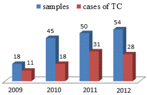
| During the study period, 167 hair samples were performed in our laboratory. Eighty-eight cases of TC (52.69% of samples received) were diagnosed. The average annual incidence was 19 cases/year (Figure 1). | |
| The average age of infected patients was 7.6 years (range one year 3 months -50 years). The sex ratio (65 men/23 women) was 2.82. | |
| The frequency of TC was higher among children younger than 10 years (82.9% of cases) mainly among boys (64.36% of cases) without statistically significant difference (Table 1). Adult’s tinea was encountered in 5.68% of cases. | |
| Concerning the origin of the patients, 46 were from a rural area and 42 patients were of urban origin. | |
| Sixteen patients (18.18%) had a family member suffering from ringworm of the scalp. Contact with animals (cats, dogs, hedgehogs, cattle) was found in 51 patients (57.95% of cases). | |
| Clinically | |
| The lesions were small and large plate’s alopecia, scaly and dry in 62 cases (70.5%). These moths were inflammatory with suppuration in 26 cases (29.5%). | |
| Biologically | |
| Direct microscopic examination was negative in 9 cases (10.22%). The culture was negative in 7 cases (7.95%) although a positive direct microscopic examination. The microsporic tinea with endo-ectothirx parasitism hair were found in 50 cases (54.54%) mainly due to Microsporum (M.) canis , while trichophytic tinea with endothrix parasitism hair in 22 cases (23.86%) due to Trichophyton (T.) violaceum (66.66%). Inflammatory TC were found in 5 patients and they were due to T. verrucosum (3 cases) and T. mentagrophytes (2 cases). No case of tinea favosa was dignosed. | |
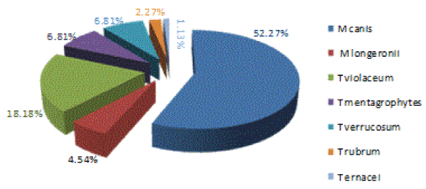
| The main dermatophytes isolated were M.canis and T. violaceum found respectively in 52.27 and 18.18% of cases (Figure 2). | |
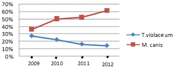
| The distribution of isolated species was different over the years. In fact, we noted a downward trend in the frequency of T. violaceum (27.27% in 2009 vs. 14.28% in 2012) and an increasing of the frequency of M. canis (36.36% vs. 60.71% in 2009 to 2012) (Table 2) (Figure 3). | |
| Discussion | |
| Tinea capitis remain a topical issue in many countries [1-3]. It affects children in preschool and school age [2-6]. In our study, children younger than 10 years old accounted for more 82% of the cases. Besides, adult’s tinea were relatively rare. In fact, TC is considered to be almost exclusive to children and rarely occurs after puberty, probably due to changes in the pH of the scalp and an increase in fatty acids serving a protective role [7]. | |
| Men were more affected than women (73.86% vs. 26.13%) without statistically significant difference. Men are usually reported in epidemiological studies [3,5,8] despite the women reported in some studies [2,9]. The high rate of tinea capitis in male may be attributed to the easy implantation of spores because of short hair and frequency of sharing comb, brushes and cups. | |
| Low standard of living and health education, overcrowding, poor hygiene, close personal and animal contacts are all favorable factors of TC [1]. In our series, the concept of animal contact was found in 51 patients, 16 patients had a family history of ringworm. In a Lybian study [10], 33.7% of children had at least another member in the family contaminated; in this reason, practician must examinate all members of family including children. | |
| Symptomatic tinea capitis has three main clinical forms: superficial tinea capitis with bald patches dry, inflammatory form and favosa tinea capitis [1]. In our study, lesions were dominated by bald patches dry in 70.5%, whereas inflammatory moths were found in 29.5%. | |
| The distribution of dermatophytes as the causative agent changes in recent decades in many countries [11]. In our study, the two main dermatophytes found were M. canis and T. violaceum. | |
| In Tunisia, although trichophytic moths caused by T. violaceum are frequent, there is a gradual decline in their incidence, which in our study increased from 27.27% in 2009 to 14.28% in 2012. Our results were similar to other Tunisian studies [8,9] and also Moroccan ones [3,12]. The regular health monitoring in schools, the improving hygiene and shaving habits of boys led to a decrease in trichophytic tinea caused by T. violaceum , which are anthropophilic and contagious mycoses. In contrast, the incidence of microsporic tinea due to M. canis showed an increase from 1% in 1950 when the first cases were reported [13] to 34.15% in 1997 [2]. This frequency has exceeded that of T. violaceum in a study reported by Belhadj [9] and Saghrouni [14]; this was found in our study where M. canis was the predominant species and its frequency is increased from 36.36% in 2009 to 60.71% in 2012 (Table 3). This increase of M. canis , also reported in other Mediterranean countries [15], can be explained by the frequent cohabitation with pets, particularly cats which are the main transmitter of M. canis [1]. | |
| Trichophyton verrucosum, zoophililc species present in the middle of breeding and T. mentagrophytes were present sporadically in previous studies [6,9,14]. In our study, these two species were found in 5 patients. All patients came from rural area and they were in close contact with animals. Moreover, T. erinacei was responsible for inflammatory ringworm in a child of 7 years living in a rural area in contact with animals, especially hedgehogs, the main reservoir of this species [16]. | |
| Trichophyton schoenleinii , agent of favus was very common in 1950 [13], has seen a spectacular decline reaching 0.24% in Tunisia [9] and 0.1% in Sfax [6]. In our study, no case of tinea favosa was found. | |
| Trichophyton rubrum , the most commonly isolated dermatophyte worldwide is responsible for tinea capitis in exceptional cases. In fact, in our study, it was diagnosed in two patients suffering from a dermatophytic disease. | |
| Changes dermatophytic flora in recent years is due to a change in the behavior of the Tunisian population even urban low socioeconomic level by a complementary breeding animals (rabbits, cattle) as well the company of pets at home (cats, dogs). | |
| Conclusion | |
| Tinea capitis remains a public health problem in Tunisia especially in children. The current etiologic profile study of TC confirmed the almost complete eradication of favus, the increasing frequency of microsporic tinea due to M. canis since recent years, which exceeds trichophytic tinea due to T. violaceum and the emergence of other zoophylic species: T. verrucosum and T. mentagrophytes . | |
References
- Contet-Audonneau N (2003) Teignes du cuirchevelu. Encyclopédiepratique de Médecine 8-0926: 1-5.
- Belhadj S, Kallel K, Boussen N, Chakroun R, Chaker E (1998) Evolution du profil des teignes du cuircheveludans la région de Tunis (1992-1997). Maghreb Medical 330: 25-26.
- Boumhil L, Hjira N, Naoui H, Zerrour A, Bhirich N, et al. (2010) Tineacapitis in the military hospital Mohammed V (Morocco). J Mycol Med 20: 97-100.
- Abu Shaqra QM, Al Momani W (2011) Cases of tineacapitis as encountered in a private practice laboratory from Jordan. See comment in PubMed Commons below J Mycol Med 21: 24-27.
- Aktas E, Karakusu A, Yigit N (2009) Etiological agents of tineacapitis in Erzurum, Turkey. J Mycol Med 19: 248-252.
- Makni F, Neji S, Sellami A, Cheikhrouhou F, Sellami H, et al. (2008) Tineacapitis in the area of Sfax. J Mycol Med 18: 162-165.
- Rebollo N, López-Barcenas AP, Arenas R (2008) Tineacapitis. ActasDermosifiliogr 99: 91-100.
- Mebazaa A, Fathallah A, El Aouamri K, GaiedMeksi S, Ghariania N, et al. (2010) Clinical and epidemiological profile of tineacapitis in central Tunisia. A 16- year retrospective study (1990-2005). J Mycol Med 20: 91-6.
- Belhadj S, Jeguirim H, Anane S, Kaouech E, Kallel K, et al. (2007) Evolution of Tineacapitis due to Microsporumcanis and Trichophytonviolaceum in Tunis. J Mycol Med 17: 54-57.
- Gargoom AM, Elyazachi MB, Al-Ani SM, Duweb GA (2000) Tineacapitis in Benghazi, Libya. Int J Dermatol 39: 263-265.
- Ginter-Hanselmayer G, Weger W, Ilkit M, Smolle J (2007) Epidemiology of tineacapitis in Europe: current state and changing patterns. Mycoses 50 Suppl 2: 6-13.
- Oudaina W, Biougnach H, Riane S, El Yaagoubil I, Tangi R, et al. (2011) Epidemiology of tineacapitis in outpatients at the Children's Hospital in Rabat (Morocco). See comment in PubMed Commons below J Mycol Med 21: 1-5.
- BIGUET J, COCHET G, COUTELEN F, DEBLOCK S, DOBY-DUBOIS M, et al. (1956) Epidemiological and mycological knowledge of tinea in children of Tunisia; research conducted principally in Moslem students. Ann Parasitol Hum Comp 31: 449-469.
- Saghrouni F1, Bougmiza I, Gheith S, Yaakoub A, Gaïed-Meksi S, et al. (2011) Mycological and epidemiological aspects of tineacapitis in the Sousse region of Tunisia. Ann DermatolVenereol 138: 557-563.
- Romano C (1999) Tineacapitis in Siena, Italy. An 18-year survey. Mycoses 42: 559-562.
- Quaife RA (1966) Human infection due to the hedgehog fungus, Trichophytonmentagrophytes var. erinacei. J ClinPathol 19: 177-178.
Tables and Figures at a glance
| Table 1 | Table 2 | Table 3 |
Figures at a glance
 |
 |
 |
||
| Figure 1 | Figure 2 | Figure 3 |
Relevant Topics
- Advanced Therapies
- Chicken Pox
- Ciprofloxacin
- Colon Infection
- Conjunctivitis
- Herpes Virus
- HIV and AIDS Research
- Human Papilloma Virus
- Infection
- Infection in Blood
- Infections Prevention
- Infectious Diseases in Children
- Influenza
- Liver Diseases
- Respiratory Tract Infections
- T Cell Lymphomatic Virus
- Treatment for Infectious Diseases
- Viral Encephalitis
- Yeast Infection
Recommended Journals
Article Tools
Article Usage
- Total views: 15215
- [From(publication date):
February-2015 - Apr 04, 2025] - Breakdown by view type
- HTML page views : 10559
- PDF downloads : 4656
