Theranostics Nanoparticles: Revolutionizing Cancer and Imaging
Received: 01-Oct-2022 / Manuscript No. omha-22-76757 / Editor assigned: 04-Oct-2022 / PreQC No. omha-22-76757 / Reviewed: 18-Oct-2022 / QC No. omha-22-76757 / Revised: 22-Oct-2022 / Manuscript No. omha-22-76757 / Published Date: 28-Oct-2022 DOI: 10.4172/2329-6879.1000435
Abstract
This literature review attempted to describe theranostic nanoparticles and their use in the treatment and diagnosis of cancer. Furthermore, this paper addressed nanoparticles and their potential ability to lower radioactive dose and improvement of quality in computed tomography examinations. This review also attempted to show the positive and negative effects implementing these nanoparticles have had within the modern healthcare system. Eighteen scholarly sources were selected and explored to acquire the relevant information for this review. Nanoparticles are an emerging technological trend being explored by many researchers and healthcare workers today. These technologies are revolutionizing the healthcare industry [1]. Newer prototypes and developments of nanotechnology is being introduced, created, and analyzed every day. There has been documenting about positive aspects of implementing nanoparticles into cancer treatment; their implementation as an aid to CT dose reduction and improved image quality is also to be noted. In contrast, there are many critics still opposed to the idea of utilizing this technology and view it as nothing more than a fantasy and/or novelty. Studies of the effects these technologies have on the healthcare industry, in particular cancer, show this technology has the potential to completely revolutionize the health care world. When analyzing all these factors, it would appear this technology is being adopted more frequently than ever before. Fear of change, negative perceptions, lack of specific work, cost, toxicity, and synthesis did not accurately satisfy this claim.
Keywords
Nanoparticle theranostic imaging; Radiography; Cellular theory; Oncogenes; CT scans; Modern sciences; Comparison with dyed imaging
Nanoparticle application in modern science
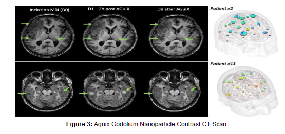
Nanoparticles are frequently suggested as diagnostic agents. However, except for iron oxide nanoparticles, diagnostic nanoparticles have been barely incorporated into clinical use so far. This is predominantly due to difficulties in achieving acceptable pharmacokinetic properties and reproducible particle uniformity as well as to concerns about toxicity, biodegradation, and elimination. Reasonable indications for the clinical utilization of nanoparticles should consider their biologic behavior. For example, many nanoparticles are taken up by macrophages and accumulate in macrophage-rich tissues. Thus, they can be used to provide contrast in liver, spleen, lymph nodes, and inflammatory lesions (eg, atherosclerotic plaques). The potential of using nanoparticles for molecular imaging is compromised because their pharmacokinetic properties are difficult to control. Ideal targets for nanoparticles are localized on the endothelial luminal surface, whereas targeted nanoparticle delivery to extravascular structures is often limited and difficult to separate from an underlying enhanced permeability and retention (EPR) effect. The majority of clinically used nanoparticle-based drug delivery systems are based on the EPR effect, and, for their more personalized use, imaging markers can be incorporated to monitor biodistribution, target site accumulation, drug release, and treatment efficacy. In conclusion, although nanoparticles are not always the right choice for molecular imaging (because smaller or larger molecules might provide more specific information), there are other diagnostic and theranostic applications for which nanoparticles hold substantial clinical potential.
Introduction
Article History
Since 2010s usage of nanoparticle imaging is emerging 18 relevant articles came forward referenced that gives the impactful idea about futuristic usage of this imaging technique, of course more mind word and resources needs to be invested to reach the conclusion of the pros and cons in comparison to dye imaging. Longer half-life on nanoparticle can cause renal consequences that are yet to be determined. Colloid structured particles stick better to ligands gives better imaging.
Theranostic Nanoparticles: Revolutionizing Cancer and Imaging
Nanotechnology, a technology once believed to be a figment of the imagination and part of the fictional world, is now revolutionizing cancer and diagnostic medical imaging. Featuring in many movies such as Avengers: Infinity War, Spiderman: Far from Home, and even children’s movies such as Big Hero 6, and Ben 10: Alien Swarm, nanotechnology is now a reality. It has gone from mere fantasy to making a revolutionary breakthrough in diagnosing and treating cancer. Cancer has been known to mankind for centuries. Although computed tomography is the most common imaging modality used for diagnosis and treatment of cancer, its high dosage is a pressing issue. Moreover, conventional drug treatment’s non-specific bio-distribution and ineffectiveness are concerning. With continued innovation, and human ingenuity, nanoparticles have the potential to be the long-awaited aid and tool to reversing cancer’s seemingly unending destructive path.
Methodology
A review of the literature was conducted by an extensive search of multiple scholarly databases. The search for articles was limited to a date range of no earlier than 2010. After extensively reading and critiquing many articles, 18 were selected. These 18 articles were selected in the pasis of nano particle application, The benefits of folic acid-modified gold nanoparticles showed quite resemblance to the application and how their duration was measured and accepted as satisfactory for this literature review. The keywords used to locate the correct articles were cancer Nanoparticles for improving cancer diagnosis. Materials Science & Engineering nanoparticles, theranostics, nanotechnology, nanoparticles in computed tomography, the future of nanotechnology in medicine. The article showed the modern application in medical sciences world Nanoparticles lowering radiographic dose, nanoparticles improving image quality, and limitations of nanoparticles. The exclusion criteria for the article that they didn’t share much resemblance with our systemic cause. Ambiguity was a major factor that becomes a big criterion for exclusion of the articles irrelevant to our systemic review [2]. Although the articles contains various knowledge but it didn’t settled without introduction and topic selection which is the most important part for beginning a study.
Discussion
This systemic review will attempt to prove the role of multifunctional nanoparticles as contrast agents and therapeutic drug carriers for diagnosing and treating cancer. This paper aims to provide basic information on how nanoparticles optimize computed tomography images by enhancing image contrast and lowering radiation dosage. Additionally, this paper will elaborate on the process in which nanoparticles deliver therapeutic drugs to specific targeted sites. This systemic review paper will conclude with the positives and limitations of this technology and briefly touch upon its future applications.
Results of Nanoparticles Technology Revolutionizing the Modern World and Imaging
Cancer
Cancer is a disease in which abnormal cells divide uncontrollably and may spread to surrounding tissues. A human body is made up of trillions of cells. These cells grow and reproduce to form new cells. As these cells grow old or become damaged, they undergo cellular apoptosis, and new cells take their place. However, this is not the case with abnormal cancerous cells. A cancer cell differs from its nonspecific function and overrides the signal of apoptosis. Cancer cells continue to divide, and form growths called tumors. Tumors may be non-cancerous (benign) or cancerous (malignant). There are various types of cancers; the difference among them is where cancer occurs. Similarly, the National Cancer Institute (2015) states there are more than a hundred types of cancer known; they are named after the location where the cancer forms or the type of cell forming them.
Cancer is one of the main causes of millions of deaths worldwide annually. If a patient is experiencing symptoms suggesting it may be cancer, a doctor may order a lab test such as an imaging test [3]. The physician may also order a biopsy to confirm his or her diagnosis. Imaging tests provide physicians with images of the inside of a patient’s body thus allowing a physician to see whether a tumor is present. There are various imaging tests such as x-rays, magnetic resonance imaging (MRI), nuclear scan, bone scan, and positron emission tomography scan (PET). However, the most commonly used imaging modality for the diagnosis of cancer is computed tomography (CT).
Computed Tomography
CT scans provide high-resolution images with fast scan time and low cost. Medical imaging plays a crucial role in the diagnosis, treatment, and monitoring of diseases. CT incorporates contrast agents to enhance the contrast of soft tissues, revealing anatomic information. Cormode, Naha, and Fayad (2014) stated small, iodinated molecules and barium suspensions are currently approved contrast agents for CT [4]. Although iodine contrast agents provide excellent vascular imaging, its short blood half-life and non-targeted imaging applications are a pressing issue. Moreover, iodinated based contrast agents are lethal for patients with compromised renal function and hypersensitivity towards iodine. Deviating from the previous assertions, the drawbacks associated with CT scans such as its increased ionizing radiation and patient dose cannot be ignored, although it provides superior spatial resolution. Reported 70% of the radiation dose received by patients undergoing imaging tests is acquired from CT scans. In the process of attaining optimal images with increased spatial resolution and density in computed tomography, the patient dose is compromised. Correspondingly, declared CT as one of the most important sources of ionizing radiation in diagnostic medical imaging.
ALARA is a principle proposed and enforced by the International Commission on Radiological Protection. ALARA stands for As Low as Reasonably Achievable. Radiologists must use ionizing radiation only when its use is justified; the imaging test must be optimized specifically using low dosage consistent with the diagnostic task. CT scans provide physicians with images essential for the diagnosis of tumors. However, patients’ exposure to ionizing radiation must be taken into account [5]. Many researchers have been working on a solution to eliminate the shortcomings associated with CT imaging.
Nanoparticles
Nanoparticles are colloidal particles, ranging in size between 10- 100nm. Due to the unique properties’ nanoparticles possess, they have become a subject for current biomedical applications specifically for imaging and therapy of various forms of cancer described how several biological mechanisms in the body occur at the nanoparticle scale, giving nanoparticles an advantageous edge to pass through biological barriers and interact with biomolecules found at the cellular or tissue level [6].
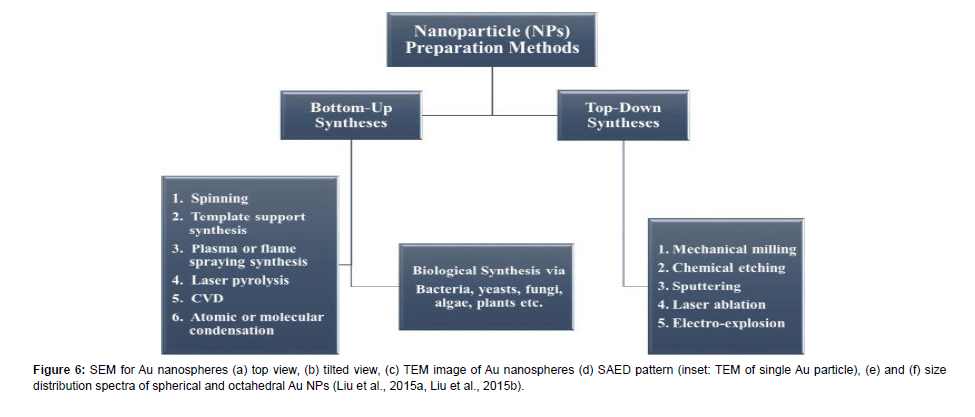
| Top–down method | Merits | Demerits | General Remarks |
|---|---|---|---|
| Optical lithography | Long-standing, micro/nanofabrication tool especially for chip production, sufficient level of resolution at high throughputs | Tradeoff between resist process sensitivity and resolution, involves state-of-the-art expensive clean room based complex operations | The 193nm lithography infrastructure already reached a certain level of maturity and sophistication, and the approach could be extended to extreme ultraviolet (EUV) sources to shrink the dimension. Also, future developments need to address the growing cost of a mask set |
| E-beam lithography | Popular in research environments, an extremely accurate method and effective nanofabrication tool for <20nm nanostructure fabrication with desired shape | Expensive, low throughput and a slow process (serial writing process), difficult for <5nm nanofabrication | E-beam lithography beats the diffraction limit of light, capable of making periodic nanostructure features. In the future, multiple electron beam approaches to lithography would be required to increase the throughput and degree of parallelism |
| Soft and nanoimprint lithography | Pattern transfer based simple, effective nanofabrication tool for fabricating ultra-small features (<10nm) | Difficult for large-scale production of densely packed nanostructures, also dependent on other lithography techniques to generate the template, and usually not cost-effective | Self-assembled nanostructures could be a viable solution to the problem of complex and costly template generation, and for templates of periodic patterns of <10nm |
| Block co-polymer lithography | A high-throughput, low-cost method, suitable for large-scale densely packed nanostructures, diverse shapes of nanostructures, including spheres, cylinders, lamellae possible to fabricate including parallel assembly | Difficult to make self-assembled nanopatterns with variable periodicity required for many functional applications, usually high defect densities in block copolymer self-assembled patterns | Use of triblock copolymers is promising to generate more exotic nanopatterns geometries. Also, functionalization of parts of the block copolymer could be done to achieve hierarchy of nanopatterning in a single step nanofabrication process |
| Scanning probe lithography | High resolution chemical, molecular and mechanical nanopatterning capabilities, accurately controlled nanopatterns in resists for transfer to silicon, ability to manipulate big molecules and individual atoms | limited for high throughput applications and manufacturing, an expensive process, particularly in the case of ultra-high-vacuum based scanning probe lithography | Scanning probe lithography can be leveraged for advanced bio nanofabrication that involves fabrication of highly periodic biomolecular nanostructures |
| Bottom–up method | Merits | Demerits | General remarks |
| Atomic layer deposition | Allows digital thickness control to the atomic level precision by depositing one atomic layer at a time, pin-hole free nanostructured films over large areas, good reproducibility and adhesion due to the formation of chemical bonds at the first atomic layer | Usually a slow process, also an expensive method due to the involvement of vacuum components, difficult to deposit certain metals, multicomponent oxides, certain technologically important semiconductors (Si, Ge, etc.) in a cost-effective way | Although a slow process, it is not detrimental for the fabrication of future generation ultra-thin ICs. The stringent requirements for the metal barriers (pure; dense; conductive; conformal; thin) that are employed in modern Cu-based chips can be fulfilled by atomic layer deposition |
| Sol gel nanofabrication | low-cost chemical synthesis process-based method, fabrication of a wide variety of nanomaterials including multicomponent materials (glass, ceramic, film, fiber, composite materials) | Not easily scalable, usually difficult to control synthesis and the subsequent drying steps | A versatile nanofabrication method that can be made scalable with further advances in the synthesis steps |
| Molecular self-assembly | Allows self-assembly of deep molecular nanopatterns of width less than 20nm and with the large pattern stretches, generates atomically precise nano systems | Difficult to design and fabricate nano systems unlike mechanically directed assembly | Molecular self-assembly of multiple materials may be an useful approach in developing multifunctional nano systems and devices |
| Physical and chemical vapor-phase deposition | Versatile nanofabrication tools for fabrication of nanomaterials including complex multicomponent nano systems (e.g. nanocomposites), controlled simultaneous deposition of several materials including metal, ceramics, semiconductors, insulators and polymers, high purity nanofilms, a scalable process, possibility to deposit porous nanofilms | Not cost-effective because of the expensive vacuum components, high-temperature process and toxic and corrosive gases particularly in the case of chemical vapor deposition | It provides unique opportunity of nanofabrication of highly complex nanostructures made of distinctly different materials with different properties that are not possible to accomplish using most of the other nanofabrication techniques. New advances in chemical vapor deposition such as ‘initiated chemical vapor deposition’ (i-CVD) provide unprecedented opportunities of depositing polymers without reduction in the molecular weights |
| DNA-scaffolding | Allows high-precision assembling of nanoscale components into programmable arrangements with much smaller dimensions (less than 10nm in half-pitch) | Many issues need to explore, such as novel unit and integration processes, compatibility with CMOS fabrication, line edge roughness, throughput and cost | Very early stage. Ultimate success depends on the willingness of the semiconductor industry in terms of need, infrastructural capital investment, yield and manufacturing cost |
Theranostics is defined as the combination of therapy and diagnosis. Nanoparticles are multifunctional theranostic agents. Theranostic nanoparticles assist in diagnosing the disease, reporting the location, identifying the stage of the disease, and providing information about the treatment response. Moreover, nanoparticles can carry and deliver a therapeutic agent to a tumor. Equivocally, also addressed nanoparticles by the portmanteau nano theranostics, indicating nanoparticles integrate the modalities of diagnostic imaging and therapy into a single package for the treatment of cancer. They reported nanoparticles not only can deliver treatment but simultaneously monitor therapy response in real-time, potentially reducing over or under-dosing patients. Tumor biomarkers are proteins found in blood, urine, stool, or tissues of cancer patients [7]. For example, human epidermal growth factor receptor 2 (HER2) is associated with breast cancer and folate receptors (FR) are found in several tumor types, more commonly, in ovarian and endometrial cancers. Measuring the levels of tumor biomarkers allows microbiologists to detect, diagnose, and manage cancer. Biomarkers are also used for monitoring the progression of the disease and its response to therapy, but most importantly detects disease early enough to decrease the death rate. In the United States, cancer accounts for almost one-fourth of all deaths. There are many types of cancer, contributing to the ever-growing list of biomarkers. The authors also reported folic acid binds to tumor cells 20 times more than epithelial cells, making folate receptors an attractive tumor biomarker. Nanoparticles can be coupled with folic acid to help in tumor imaging. Performed a study on the effects of both gold nanoparticles and folic acid-modified gold nanoparticles (FA-modified AuNP) as a contrast agent in CT imaging systems. The researchers experimented with different concentrations of nanoparticles at various tube currenttime products. From the CT images, it was found the cells which contained nanoparticles [8], modified or not, produced higher contrast enhancements in comparison to the cells not exposed to nanoparticles. Additionally, it was found the cells treated with modified FA-AuNPs exhibited significantly higher contrast enhancement than those of the cells exposed to non-modified gold nanoparticles. Moreover, they deduced from their experiments, by keeping the voltage at a constant 130 kVp and elevating the tube current-time product from 60 to 250 mAs, radiation dose increased by approximately 4.17 folds whereas the enhancement in image contrast was barely noticeable. In sharp contrast, without increasing the mAs or radiation dose, the simple incorporation of nanoparticles whether modified or not, drastically increased the image contrast, keeping the mAs as low as reasonably achievable.
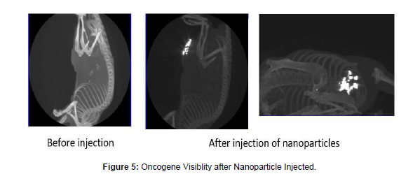
In similitude, conducted in vivo research in which they utilized folic acid (FA) gold nanoparticles through cysteamine (Cys) linking for the detection of human nasopharyngeal head and neck cancer by CT imaging. They found a small-sized tumor was undetectable by CT imaging, however, by incorporating gold nanoparticles the same tumor was visible at least 4.30 times more. Moreover, active tumor cell targeting nanoparticles (FA-Cys-AuNPs) demonstrated more specific and efficient images of the tumor at least 2.03 times than passive targeting gold nanoparticles. Therefore, nanoparticles present the potential for dose reduction and enhanced image contrast of CT images by using minimal mAs product. Furthermore, the utilization of FA-modified AuNPs provides a targeted CT imaging strategy for specific recognition of cancerous cells.
The fundamental basis for the administration of imaging contrast agents or therapeutic drugs is to attain a favorable diagnostic/ therapeutic outcome with minimal side effects. When therapeutic or diagnostic agents are injected, various bio barriers are preventing the agents from reaching the affected and targeted sites such as enzymatic degradation, inadequate marinating in the bloodstream, or inability to overcome vascular endothelium. stated only one out of 100,000 drug molecules successfully reach the intended targeted pathological site, resulting in 99.99% of the drugs being delivered to unintended sites, thus causing unwanted adverse reactions. Therefore, to achieve therapeutic efficacy, one must inject unreasonably high doses of the therapeutic agent, warranting detrimental cytotoxic side effects to healthy organ systems. However, nanoparticles are now being used as a diagnostic tool and therapeutic drug carrier which delivers drugs to intended targeted sites in a controlled manner.
Nanoparticles are microscopic and able to easily penetrate through blood vessels of tumor tissues, enabling them to gather within the cancerous cells. They possess high surface areas allowing them to carry therapeutic and imaging agents. Moreover, nanoparticles can selectively accumulate in diseased tissues, providing the functionality of site-specificity, reducing the issue of increased drug volume use and accumulation of drugs in healthy tissues described, one of the most important characteristics nanoparticles possess is the ability to possibly reduce side effects and damage to healthy tissues as well as organs compared with conventional cancer therapeutic drugs. Nanoparticles not only target cancerous cells with more accuracy and specificity but can deliver therapeutic drugs for treatment to the targeted organs difficult to reach such as the brain and pancreas.
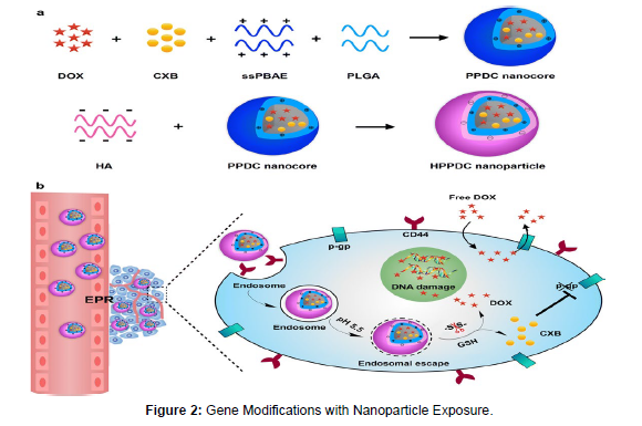
Methods for how nanoparticle works Nanoparticles satisfy the requirements to become effective drug carriers by penetrating through barriers, reaching the desired tumor site with little to no loss of drug volume and activity, and selectively killing cancerous cells without affecting normal cells. These features not only improve patient survival, but also the quality of life by increasing the concentration of drugs within the diseased cells and reducing dose-limiting toxicities concurrently. However, for the nanoparticles to remain in the bloodstream and not get caught during circulation by the reticuloendothelial system such as the spleen and the liver, its size and surface characteristics can be adjusted. Surface characteristics determine the lifespan and destiny of nanoparticles. To prevent being captured by macrophages, nanoparticles must have a hydrophilic surface. This can be accomplished by either coating the surface with a hydrophilic polymer or by forming nanoparticles from block copolymers with hydrophilic and hydrophobic domains. Moreover, the size of the nanoparticles can be adjusted. The size of the nanoparticles should be small enough to escape macrophages embedded into the reticuloendothelial system but should be large enough to prevent them from rapidly leaking out of blood vessels [9].
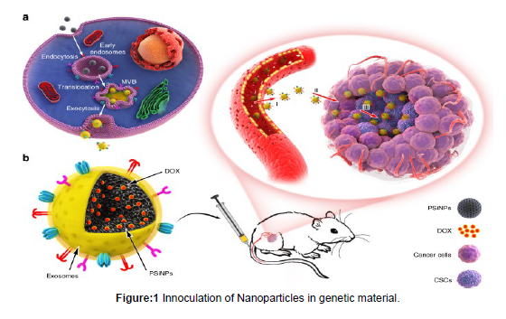
Nanoparticles deliver drugs in two ways: passive and self-delivery. Imaging of nanoparticle uptake in passive delivery, the inner cavity of the nanostructure is filled with the drug needed to be transported. Contrarily, in self-delivery, the drug is conjugated to the carrier nanoparticle. However, with self-delivery, timing is extremely crucial because if the drugs are not released at the right time, the drug will not reach the intended site, once nanoparticles reach the site of action, based on the type of delivery, the drug cargo is either released outside or the cells engulf the nanoparticles with the unloaded cargo by a process known as endocytosis. Ultimately, once the nanoparticles interact with the cytomembrane and enter the cells, they travel through the cell via various different pathways.
Although, nanoparticles offer multiple benefits in diagnosing and treating cancer using low radiation dose by CT and target-oriented delivery, insufficient knowledge about the toxicity caused by the nanoparticles is doubtfully worrisome. This warrants further work into designing nanoparticles with materials non-toxic to the human body. Especially since imaging agents are often used for diagnostic purposes, it is crucial to prevent unnecessary toxicity to patients who might turn out to be healthy. Therefore, cautiously designing nanoparticles could be beneficial in tackling the issues associated with their use. For nanoparticles to accumulate within the tumor, it requires long circulation times however, this is not ideal for imaging. Imaging can only be performed once contrast agents have cleared through the system.
Future Applications
Nanotechnology has tremendous potential and is constantly evolving. Trends are pointing towards field workers shifting their thinking to the smallest of technologies to solve the biggest problems in the healthcare field. These same field workers are prototyping new alternatives to perform tasks currently being executed by hand or equipment. They are currently developing programmable and controllable nano assemblers and nanorobots having the capabilities to reverse the effects of atherosclerosis and cardiovascular disease. These innovative new technologies are also demonstrating the capabilities of being able to fix genetic errors in cells. Furthermore, Routley (2019) emphasized how nanotechnology is the future of medicine and by 2024 the global market for nanotechnology will exceed 125 billion dollars. Specialized nanobots controlled by magnetic fields are being developed to perform a wide range of surgeries such as eye surgeries, clearing blocked arteries, and collecting biopsies. Micromotors, microscopic beads of magnesium and titanium, are currently being developed for treating stomach ulcers. However, very little is known about the long-term impacts of nanotechnology and much more resources are required. A developing concern is whether nanoparticles will accumulate in living tissues causing toxicity issues and whether it can be affordably manufactured at commercial scale. Nonetheless, nanotechnology constantly proves to be promising in the field of medicine and it is not long before nanotechnology moves from science fiction into the real world. Ilustrations for uptake of nanoparticles in oncogenic cell.
Conclusion
Cancer has been an existential threat for centuries. Nanoparticles may be the long-sought solution to finally combating this deadly disease and reversing its trend once and for all. In a world where bigger is often considered to be better, nanoparticles are an exception to conventional wisdom in their potentiality in becoming a revolutionary step in personalized medicine. Thus, when it comes to nanoparticles and their mission to stop cancer, it is time to stop thinking so big and start thinking small, but of course more mind work and resources needs to be invested to reach a conclusive diagnostic tool for future application.
References
- Retrieved from https://www.quantumrun.com/article/nanotechnology-future-medicine
- Naseri N, Ajorlou E, Asghari F, Pilehvar-Soltanahmadi Y (2017) An update on nanoparticle-based contrast agents in medical imaging. Artif Cells Nanomed Biotechnol 46: 1111-1121.
- Parvanian S, Mostafvi SM, Aghashiri M (2017) Multifunctional nanoparticle developments in cancer diagnosis and treatment. Int J Nanomedicine 13: 81-87.
- Patra JK, Das G, Fraceto LF, Campos EVR, Rodriguez-Torres MP, et al. (2018) Nano based drug delivery systems: recent developments and prospects. J Nanobiotechnology 16.
- Salatin S, Khosroushahi AY (2017) Overviews on the cellular uptake mechanism of polysaccharide colloidal nanoparticles. J Cell Mol Med 21: 1668-1686.
- Taghavi H, Bakhshandeh M, Montazerabadi A, Moghadam HN, Behnam S, et al. (2020) Comparison of gold nanoparticles and iodinated contrast media in radiation dose reduction and contrast enhancement in computed tomography. Iranian J Radiology 17.
- Routley N. The future of nanotechnology in medicine [Internet]. 2019. Available from: https://www.visualcapitalist.com/the-future-of-nanotechnology-in-medicine/
- Vizirianakis IS (ed). Handbook of personalized medicine advances in nanotechnology, drug delivery, and therapy [Internet]. Singapore: Jenny Stanford Publishing; 2014. Available from: https://www.jennystanford.com/9789814411196/handbook-of-personalized-medicine/
- Zarif ME, Florea DA, Grumezescu AM (2019) Chapter 1 introduction to cancer nanotherapeutics. Biomedical App Nanoparticles 1-29.
Indexed at, Google Scholar, Crossref
Indexed at, Google Scholar, Crossref
Indexed at, Google Scholar, Crossref
Indexed at, Google Scholar, Crossref
Citation: Hamza N (2022) Theranostics Nanoparticles: Revolutionizing Cancer and Imaging. Occup Med Health 10: 435. DOI: 10.4172/2329-6879.1000435
Copyright: © 2022 Hamza N. This is an open-access article distributed under the terms of the Creative Commons Attribution License, which permits unrestricted use, distribution, and reproduction in any medium, provided the original author and source are credited.
Share This Article
Recommended Journals
Open Access Journals
Article Tools
Article Usage
- Total views: 1022
- [From(publication date): 0-2022 - Mar 12, 2025]
- Breakdown by view type
- HTML page views: 822
- PDF downloads: 200

