The Utilization of Cone Beam CT Imaging in Dentistry
Received: 04-Aug-2022 / Manuscript No. roa-22-73182 / Editor assigned: 06-Aug-2022 / PreQC No. roa-22-73182 (PQ) / Reviewed: 20-Aug-2022 / QC No. roa-22-73182 / Revised: 24-Aug-2022 / Manuscript No. roa-22-73182 (R) / Published Date: 31-Aug-2022 DOI: 10.4172/2167-7964.1000395
Image Article
New imaging technologies advance in dentistry are turning out to be progressively well known for determination and treatment appraisal [1]. 3D computed tomography (CT) which is cross-sectional methodology, intrinsically dodges the superimposition and issues because of amplification and offers to visualize of the craniofacial structures with more accuracy than the 2D technique [2,3].
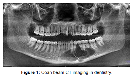
In dentistry, to get pictures from the beginning, medical CT (MDCT) was utilized for this reason. Concentrates on MDCT demonstrated the way that 3D CT examination can be performed exact and dependable evaluations for dental techniques [4]. Regardless of these benefits, the effective dose of MDCT is a lot higher than the customary radiographs which are utilizing dental and maxillofacial imaging. This delivers its utilization for routine assessments (particularly those for cephalometric investigation and growth assessments) unmerited. Additionally, MDCT is also expensive technique and scanners are not effectively open by dental specialists and small clinics for maxillofacial imaging (Figure 1).
Somewhat recently, another procedure called as cone beam computed tomography (CBCT) was proposed for the maxillofacial imaging which was first revealed in the writing by Mozzo P, et al [5]. A CBCT scan utilizes an alternate kind of obtaining that the tradition MDCTs. The x- ray source delivers a cone formed x- ray beam and this makes it conceivable catch the image in a solitary shot, as opposed to catching cut independently as in MDCT. The advantages of this imaging methodology are; substantially more lower radiation portion than MDCT, the chance of individualized overlap free reconstructions and DICOM information can be in-and exported for different applications. In addition, this imaging innovation permits 3D imaging and data on 3rd dimensional which offers to picture of the craniofacial and dental designs for maxillofacial careful applications and orthodontics use.
References
- Kumar V, Ludlow J, SoaresCevidanes LH, Mol A (2008) In vivo comparison of conventional and cone beam CT synthesized cephalograms. Angle Orthod 78: 873-879.
- Kumar V, Ludlow JB, Mol A, Cevidanes L (2007) Comparison of conventional and cone beam CT synthesized Cephalograms. Dentomaxillofac Radiol 36: 263-269.
- Sandler PJ (1988) Reproducibility of cephalometric measurements. Br J Orthod 15: 105-110.
- Swennen GR, Schutyser F, Barth EL, De Groeve P, De Mey A (2006) A new method of 3-D cephalometry Part I: the anatomic Cartesian 3-D reference system. J Craniofac Surg 17: 314-325.
- Mozzo P, Procacci C, Tacconi A, Martini PT, Andreis IA (1998) A new volumetric CT machine for dental imaging based on the cone-beam technique: preliminary results. Eur Radiol 8: 1558-1564.
Indexed at, Google Scholar, Crossref
Indexed at, Google Scholar, Crossref
Indexed at, Google Scholar, Crossref
Indexed at, Google Scholar, Crossref
Citation: Akirha T (2022) The Utilization of Cone Beam CT Imaging in Dentistry. OMICS J Radiol 11: 395. DOI: 10.4172/2167-7964.1000395
Copyright: © 2022 Akirha T. This is an open-access article distributed under the terms of the Creative Commons Attribution License, which permits unrestricted use, distribution, and reproduction in any medium, provided the original author and source are credited.
Select your language of interest to view the total content in your interested language
Share This Article
Open Access Journals
Article Tools
Article Usage
- Total views: 2060
- [From(publication date): 0-2022 - Dec 02, 2025]
- Breakdown by view type
- HTML page views: 1632
- PDF downloads: 428

