The Transmantle Sign: A Specific Sign of Taylor's Cortical Dysplasia (Type II)
Received: 03-May-2024 / Manuscript No. roa-24-138358 / Editor assigned: 06-May-2024 / PreQC No. roa-24-138358 / Reviewed: 20-May-2024 / QC No. roa-24-138358 / Revised: 27-May-2024 / Manuscript No. roa-24-138358 / Published Date: 31-May-2024
Abstract
Taylor's focal cortical dysplasia, first described by Taylor, is a cortical development anomaly and a prevalent cause of drug-resistant epilepsy in adults. Surgical intervention often yields a favorable prognosis if the lesion is entirely resected. Clinically, it presents with drug-resistant partial epilepsy from childhood, sometimes with an acquired, localizing neurological deficit. EEG typically shows continuous rhythmic spikes at the anomaly site. MRI scans fail to detect the lesion in about one-third of cases, making the identification of small dysplasias challenging. An appropriate MRI protocol and prior clinical and electrical localization are essential. The transmantle sign, marked by FLAIR and T2 hypersignal and T1 hyposignal in subcortical white matter extending to the ventricle, is highly characteristic and seen in 80% of cases. Isolated cortical thickening, showing dedifferentiation between white and gray matter, varies in size and is often located in the frontal lobe. Differentiating this from conditions like Bourneville's tuberous sclerosis and closed-lip schizencephaly is crucial.
Keywords
Taylor’s cortical dysplasia; epilepsy; MRI
Clinical Image
Taylor's focal cortical dysplasia, first described by Taylor, is a cortical development anomaly and a common cause of drug-resistant epilepsy in adults. Surgical treatment often results in a good prognosis if the lesion is completely resected [1].
This condition presents as a distinct clinical, electrical, and radiological entity. It typically involves partial epilepsy that has been drug-resistant since childhood, sometimes accompanied by an acquired, localizing neurological deficit. The EEG trace usually shows continuous or subcontinuous rhythmic spikes at the site of the anomaly [2].
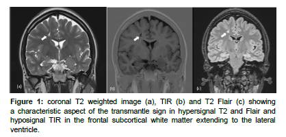
MRI scans fail to detect the dysplastic lesion in about one-third of cases [1], and identifying small dysplasias can be particularly challenging. Therefore, an appropriate protocol and prior clinical and electrical localization are crucial. The transmantle sign, characterized by signal anomalies including FLAIR and T2 hypersignal and T1 hyposignal in the subcortical white matter extending to the ventricle, is highly characteristic, observed in 80% of cases (Figure 1). The thickness of this sign correlates with the extent of subcortical signal abnormalities [3]. Isolated cortical thickening is sometimes seen, with dedifferentiation between white and gray matter, varying in size up to hemispheric forms, often located in the frontal lobe, opposite the central sulcus. It is important to differentiate this radiological sign from conditions like Bourneville's tuberous sclerosis and closed-lip schizencephaly [3].
Conflict of Interest
The authors declare that they have no ties of interest.
Funding
The author(s) received no financial support for the research, authorship, and/or publication of this article.
Author Contribution
All authors have accepted responsibility for the entire content of this manuscript and approved its submission
References
- Francine Chassoux, Elisabeth Landré, Charles Mellerio, Baris Turak, Michael W, et al. (2012)Type II focal cortical dysplasia: Electroclinical phenotype and surgical outcome related to imaging. Epilepsia 53: 349-358.
- Souirti Zouhayr, Oussous Siham, Belfkih Rachid (2013) Dysplasies corticales focales. NAMEEJ 2.
- Mellerio C, Labeyrie MA, Chassoux F, Daumas-Duport C, Landre E, et al. (2012)Optimizing MR Imaging Detection of Type 2 Focal Cortical Dysplasia: Best Criteria for Clinical Practice. Am J Neuroradiol 33: 1932-1938.
Indexed at, Google Scholar, Crossref
Citation: Messaoud O (2024) The Transmantle Sign: A Specific Sign of Taylor’sCortical Dysplasia (Type II). OMICS J Radiol 13: 568.
Copyright: © 2024 Messaoud O. This is an open-access article distributed underthe terms of the Creative Commons Attribution License, which permits unrestricteduse, distribution, and reproduction in any medium, provided the original author andsource are credited.
Select your language of interest to view the total content in your interested language
Share This Article
Open Access Journals
Article Usage
- Total views: 1421
- [From(publication date): 0-2024 - Nov 20, 2025]
- Breakdown by view type
- HTML page views: 1111
- PDF downloads: 310

