Make the best use of Scientific Research and information from our 700+ peer reviewed, Open Access Journals that operates with the help of 50,000+ Editorial Board Members and esteemed reviewers and 1000+ Scientific associations in Medical, Clinical, Pharmaceutical, Engineering, Technology and Management Fields.
Meet Inspiring Speakers and Experts at our 3000+ Global Conferenceseries Events with over 600+ Conferences, 1200+ Symposiums and 1200+ Workshops on Medical, Pharma, Engineering, Science, Technology and Business
Case Report Open Access
The Role of PET Camera Emission as an Alternative Method for Radioactivity Spillage Measurement during PET/CT Study
| Hishar Hassan*, Fathinul Fikri Ahmad Saad and Rosli Md. Yusof | |
| Faculty of Medicine and Health Sciences, Universiti Putra Malaysia, Serdang, Selangor 43400, Malaysia | |
| Corresponding Author : | Hishar Hassan Faculty of Medicine and Health Sciences Universiti Putra Malaysia, Serdang Selangor 43400, Malaysia Tel: +60389471641 FAX: +60389472775 E-mail: hishar.hassan@gmail.com |
| Received October 14, 2014; Accepted December 03, 2014; Published December 06, 2014 | |
| Citation: Hassan H, Saad FFA, Yusof RM (2014) The Role of PET Camera Emission as an Alternative Method for Radioactivity Spillage Measurement during PET/CT Study. OMICS J Radiol 3:174. doi: 10.4172/2167-7964.1000174 | |
| Copyright: © 2014 Hassan H, et al. This is an open-access article distributed under the terms of the Creative Commons Attribution License, which permits unrestricted use, distribution, and reproduction in any medium, provided the original author and source are credited. | |
Visit for more related articles at Journal of Radiology
Abstract
Urinary incontinence in patients undergoing PET/CT study using 18F-Fluorodeoxyglucose (18F-FDG) is a usual encounter during the normal examination. Nevertheless, the potential role of PET camera as an alternative method for radioactivity spillage measurement has never been reported.
| Keywords |
| 18F-FDG; PET/CT; PET noise-equivalent-count |
| Introduction |
| PET images are subjected to artefactual degradation by many factors; decontamination at the injection site, biological or technical factors [1-3]. Quantitative analysis by PET on the radiation noise level can be done via the noise-equivalent count [4]. Traditionally, the use of hand-held Geiger Muller survey meter is common in detecting the residual radioactivity in determining the radiation level to the decontaminated area. Nevertheless, this technique can pose direct hazard to the personnel given the proximity of the radiation source and the personnel. This report highlights the potential role of PET camera emission, the importance of adopting an appropriate decontamination technique and a time window to allow complete decontamination of the radiation hazard in ensuring subsequent PET image quality is not being affected. |
| Case Report |
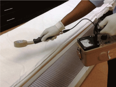
| A 57-year-old female presented with carcinoma of breast with bladder incontinence underwent PET/CT examination. Following the last bed position of the PET camera emission, there was an artefact seen inferior to the patient pelvis consistent with the 18F-FDG urine contamination as confirmed by the survey meter (Figure 1). The spillage image was acquired at the end of the PET/CT examination for quantification on the PET noise-equivalent-count (PET NEC) analysis at two time points (pre decontamination and post decontamination) made at 30 minutes interval. Following radiation decontamination technique, the image yield as quantified by mean of the PET NEC changes were found to be significantly better as the timely dedicated decontamination technique had profoundly minimised the image degradation. |
| Discussion |
| Patient was administered with 18F-FDG (dose of 300 MBq), 32 minutes prior to the scanning procedure. Prior to last bed position during PET acquisition, patient had experienced urinary incontinence. Immediate action was taken by allowing the patient to set clear from the scanning table facilitated by nuclear medicine technologists 1 (NMT) (Table 1). During the assistance, direct contact between NMT1 and patient was kept at very minimal and optimal distance was maintained. The patient was asked to change clothes and decontaminate herself under running water in the hot toilet. With a gloves on, NMT1 had the decontaminated scanning table sheet changed and had subjected it to the decontamination room. |
| NMT2 took the background reading and moved closer to the scanning table and floor before recorded them on form. Moving away from the decontamination point to the unaffected area, the area which the radioactivity level was deemed similar as background was marked and barricaded with the yellow tapes and radiation hazard signage. The spillage was not deemed as a major spills as the maximum activity level on scanning table and floor were less than 100 Bq/centimeter squared (cm2) [5]. |
| The radioactivity level at point 0 was 90 mR/hr. We investigated the potential role of PET camera to estimate the noise radioactivity count via PET NEC analaysis [3]. The NMT1 in control room obtained the PET image acquisition on the spillage area. Table 2 shows the spillage image acquired at time point 0, with the PET NEC of 5991 PROPCPPS unit. |
| The NMT2 then proceeded with the decontamination procedure. Wearing a full set of personal protective attire, he put down the absorbent papers, in a circular arrangement outside the spillage area. He moved all absorbent papers in-side the spillage area to avoid the radiation spillage spread to unaffected area. The utilised absorbent papers were then carefully removed and put onto the lead lined container. Steps were repeated using a Radiacwash. He sprayed it all over the spillage area before blotted dry. Radioactivity reading was taken and compared to the background reading. After the third time, the radioactivity reading had decreased. The NMT2 terminated the decontamination procedure to allow for the decay activity to take place. The radioactivity level was measured on the NMT2 using survey meter. Used personal protective attire were removed and put onto the lead line container prior from leaving the PET/CT room. The interval time of 30 minutes was allowed before next radioactivity readings were taken. |
| Following the post decontamination, the radioactivity reading was measured again and spillage images were also acquired PET camera. Table 2 shows the spillage image as acquired at time point two–30 minutes, with the PET NEC of 0 PROPCPPS unit (Table 2). |
| From our report, with the evidence from spillage image acquired at post decontamination, the interval time taken was rational. We anticipated that with the appropriate decontamination procedure, a time interval of 30 minutes is adequate for a PET/CT centre with high flow routine PET/CT studies. Significantly, this incident shows that PET camera has the potential role to record down the radioactivity level via PET NEC value. The use of PET camera emission could be suggested as an alternative method for the radioactivity counting should radioactive spillage occur on scanning table of PET/CT camera to ensure the personnel radiation exposure is kept at the lowest reasonable level. |
| Conclusion |
| Appropriate decontamination procedure, adequate time–point delay and the use of PET camera emission in quantifying the spillage area are essential in ensuring good PET image quality acquisition whilst minimizing inter personnel radiation burden during PET/CT examination. |
References |
|
Tables and Figures at a glance
| Table 1 | Table 2 |
Figures at a glance
 |
| Figure 1 |
Post your comment
Relevant Topics
- Abdominal Radiology
- AI in Radiology
- Breast Imaging
- Cardiovascular Radiology
- Chest Radiology
- Clinical Radiology
- CT Imaging
- Diagnostic Radiology
- Emergency Radiology
- Fluoroscopy Radiology
- General Radiology
- Genitourinary Radiology
- Interventional Radiology Techniques
- Mammography
- Minimal Invasive surgery
- Musculoskeletal Radiology
- Neuroradiology
- Neuroradiology Advances
- Oral and Maxillofacial Radiology
- Radiography
- Radiology Imaging
- Surgical Radiology
- Tele Radiology
- Therapeutic Radiology
Recommended Journals
Article Tools
Article Usage
- Total views: 13683
- [From(publication date):
December-2014 - Mar 29, 2025] - Breakdown by view type
- HTML page views : 9215
- PDF downloads : 4468
