Make the best use of Scientific Research and information from our 700+ peer reviewed, Open Access Journals that operates with the help of 50,000+ Editorial Board Members and esteemed reviewers and 1000+ Scientific associations in Medical, Clinical, Pharmaceutical, Engineering, Technology and Management Fields.
Meet Inspiring Speakers and Experts at our 3000+ Global Conferenceseries Events with over 600+ Conferences, 1200+ Symposiums and 1200+ Workshops on Medical, Pharma, Engineering, Science, Technology and Business
Short Communication Open Access
The Role of MRI in the Management of Cases of Mayer-Rokitansky- Kuster-Hauser (MRKH) Syndrome
| Rizwan Ahmad Khan*, Shagufta Wahab and Abhishek Kumar Varshney | |
| Jawaharlal Nehru Medical College and Hospital Aligarh, India | |
| Corresponding Author : | Rizwan Ahmad Khan Jawaharlal Nehru Medical College and Hospital Aligarh, India Tel: 0571 270 0920 E-mail: drrizwanahmadkhan@yahoo.co.in |
| Received: June 04, 2014; Accepted: August 22, 2015; Published: August 29, 2015 | |
| Citation: Khan RA, Wahab S, Varshney AK (2015) The Role of MRI in the Management of Cases of Mayer-Rokitansky-Kuster-Hauser (MRKH) Syndrome. J Preg Child Health 2:187. doi:10.4172/2376-127X.1000187 | |
| Copyright: © 2015 Khan RA, et al. This is an open-access article distributed under the terms of the Creative Commons Attribution License, which permits unrestricted use, distribution, and reproduction in any medium, provided the original author and source are credited. | |
| Related article at Pubmed, Scholar Google | |
Visit for more related articles at Journal of Pregnancy and Child Health
Abstract
Mayer- Rokitansky- Kuster- Hauser (MRKH) syndrome is a rare entity characterized by congenital aplasia of the uterus and the upper part of 2/3rd of vagina in normal karyotype females with normal development of secondary sexual characteristics. The incidence rate is about 1 in every 4500 females. We report a case series of three different patients who presented with different clinical picture to various outpatient departments of our hospital. Magnetic Resonance Imaging of the pelvis was done in all these patients.
| Keywords |
| Mayer–Rokitansky-Kuster-Hauser syndrome; Karyotype; Magnetic resonance imaging |
| Introduction |
| Mayer–Rokitansky-Kuster-Hauser syndrome is a very rare condition and the varying presentation of this entity often presents a diagnostic challenge [1]. But meticulous radiological assessment of the patient not only provides early and precise diagnosis but also helps in classifying the types of this syndrome, thus providing a skillful surgical approach. MRKH may be isolated (type I) but more frequently associated with renal, vertebral, auditory and cardiac defects (MRKH type II or MURCS association). The majority of are sporadic; however review of literature also demonstrates few cases depicting the familial nature of the disease. The mode of inheritance is autosomal dominant with an incomplete degree of penetrance and variable expressivity. Type II MRKH is more frequent than Type 1 MRKH [1]. We, hereby, present a case series of Mayer–Rokitansky-Kuster-Hauser syndrome in phenotypically normal female patients presenting with complaints of primary amenorrhea diagnosed on 1.5 Tesla MRI studies. |
| Cases |
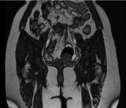
| 4.1 A 14 years female patient presented with complaints of abdominal pain. There was no history of fever, urinary complaints, swelling or lump in abdomen or history of any prolonged drug intake. On further enquiry it was revealed that patient has not achieved menarche yet. On clinical examination, the height was normal, secondary sexual characteristics were normal for age. The ultrasound examination of pelvis which revealed non visualization of uterus within the uterine fossa. For further evaluation and characterization, MRI pelvis was performed which confirmed the empty uterine fossa with no evidence of any abnormal increased signal intensity or mass lesion in the area with evidence of visualization of normal bilateral ovaries. So, the diagnosis of MEYER ROKITANSKY KUSTER HAUSER SYNDROME (TYPE I) was made (Figure 1). |
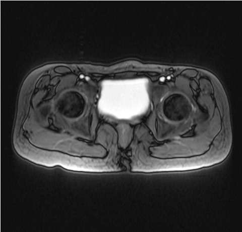
| 1.2 A 10 years female patient presented with complaints of abdominal pain off and on with failure to thrive. Patient had not achieved menarche. No other relevant findings were noted. On sonography of pelvis region, no definite evidence of visualization of uterus within the uterine fossa was noted. Thus, MRI pelvis was performed which confirmed the presence of empty uterine fossa with no evidence of visualization of ovaries. So, the diagnosis of MEYER ROKITANSKY KUSTER HAUSER SYNDROME (TYPE II) was made (Figure 2). |
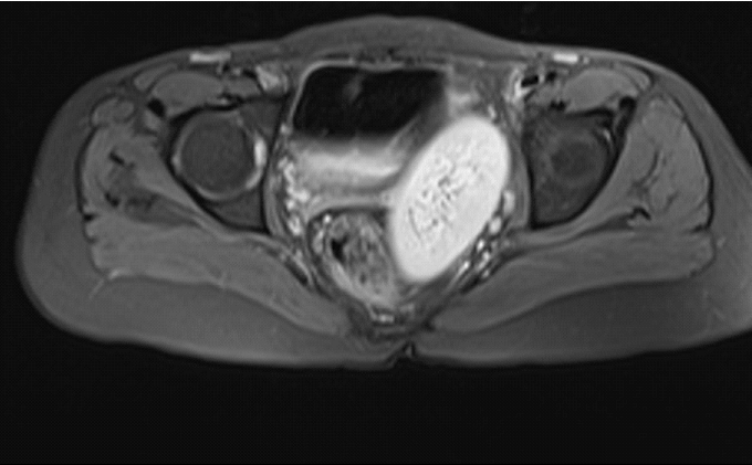
| 1.3 A 22 years female patient presented with complaints of primary amenorrhea. There was no significant past history. Clinical examination was normal. The patient was then subjected to ultrasound pelvis examination which revealed non visualization of uterus within the uterine fossa. For further evaluation and characterization, MRI pelvis was performed which confirmed the presence of empty uterine fossa with no evidence of any abnormal increased signal intensity or mass lesion within it and no evidence of visualization of ovaries. Also noted was the presence of fusion of bilateral kidneys seen located in pelvis region suggestive of pancake kidney confirming the diagnosis of MEYER ROKITANSKY KUSTER HAUSER SYNDROME (TYPE II) with associated renal anomaly (Figure 3). |
| Genetic evaluation of these patients revealed normal karyotype i.e. 46,XX. |
| Discussion |
| The Mayer- Rokitansky-Küster-Hauser (MRKH) syndrome or Rokitansky syndrome is a congenital malformation of the female genital tract and is the second most frequent cause of primary amenorrhea after gonadal dysgenesis. It is characterized by vaginal atresia, and uterine/tubal abnormalities including absence or hypoplasia of the uterus and fallopian tubes [1,2]. The patients have normal karyotype 46,XX and normal secondary sexual characteristics, as the ovaries are present and functional, but menstruation is absent. It occurs due to interrupted embryonic development of the Müllerian ducts at about 7 weeks after fertilization. The fallopian tubes, uterus, cervix and upper ¾ of vagina develop from Müllerian ducts usually develop between 8th to 12th gestational weeks [3]. Any developmental defect occurring at this stage leads to agenesis of Mullerian structures. Concomitantly, the development of kidneys, ureter, and bladder occurs between 6th to 12th weeks of gestation. There are two different forms of this syndrome: the typical form of this syndrome (type I) is characterized by congenital absence of the uterus and upper vagina with normal ovaries and fallopian tubes [4]. Secondly, the atypical form (type II) of the syndrome includes associated abnormalities of the ovaries and fallopian tubes and renal or vertebral anomalies. Renal anamolies are seen in 30-40% of the cases however, only 10% of the cases are associated with vertebral anomalies. Renal malformations include: unilateral agenesis, horseshoe kidney, renal hypoplasia, ectopic kidneys and hydronephrosis [4]. |
| High Resolution Sonography of pelvis helps in initial evaluation and assessment of the disease. Magnetic resonance imaging is the most sensitive and specific imaging modality in the evaluation of this syndrome. It provides acquisition of multiplanar images with or without fat saturation. It acts as a good predictor in evaluation of various anatomical alterations such as uterine agenesis, as well as evaluating ovaries, vagina and associated anomalies [2]. Laparoscopic examination of the pelvis can be helpful in cases where the evaluation of patients by different imaging modalities is uncertain. The final diagnosis and classification is made by correlating the findings of imaging and karyotyping. The treatment is usually multimodal because explanation of the disease process to the parents and the patient generates great amount of anxiety and psychological stress. In almost all the patients’ creation of neovagina is required. This can be achieved surgically and sometimes nonsurgically. Vaginoplasty allows these patients to have a normal sex life and avoid the risk of physchological trauma. Whenever surgical approach is chosen, consideration should be given to excision of uterine remnants to avoid risk of endometriosis in future life [5,6]. |
| Conclusion |
| MRI is a useful tool which can help in diagnosis and proper management of this rare syndrome. |
References
- Sultan C, Biason-Lauber A, Philibert P (2009) Mayer-Rokitansky-Kuster-Hauser syndrome: recent clinical and genetic findings. GynecolEndocrinol25:8-11.
- Sem KK, Kapoor A (2006) Mayer-Rokitansky-Kuster-Hauser syndrome. Ind J RadiolImag. 16: 805-807.
- Junqueira BLP, Allen LM, Spitzer RF (2009) Müllerian duct anomalies and mimics in children and adolescents: correlative intraoperative assessment with clinical imaging. Radiographics. 29:1085-1103.
- Muelle GC, Hussain HK, Smith YR (2007) Müllerian duct anomalies: comparison of MRI diagnosis and clinical diagnosis. AJR Am J Roentgenol. 189:1294-302.
- Bean EJ, Mazur T, Robinson AD (2009) Mayer-Rokitansky-Kuster-Hauser: sexuality, psychological effects, and quality of life. J PediatrAdolesc Gynecol. 22:339-346.
- Tarry WE, Duckett JW, Stephens FD (1986) The Mayer-Rokintansky Syndrome: pathogenesis, classification and management. J Urol 136: 648-652.
Figures at a glance
 |
 |
 |
| Figure 1 | Figure 2 | Figure 3 |
Post your comment
Relevant Topics
Recommended Journals
Article Tools
Article Usage
- Total views: 14226
- [From(publication date):
October-2015 - Apr 03, 2025] - Breakdown by view type
- HTML page views : 9649
- PDF downloads : 4577
