Research Article Open Access
The Radix Entomolaris and Paramolaris: A Review and Case Reports with Clinical Implications
Amit Parashar1*, Shikha Gupta2, Abhishek Zingade1 and Shashi Parashar3
1FAGE, FPFA Department of Periodontics, KLE VK Institute of Dental Sciences, Belgaum, Karnataka, India
2Department of Pedodontics, People's Dental Academy, People's University, Bhopal, India
3Division of Virology, Defence R&D Establishment (DRDE), Jhansi Road, Gwalior, MP, India
- Corresponding Author:
- Dr Amit Parashar
Department of Periodontics, KLE VK Institute of Dental Sciences
Belgaum, Karnataka, PIN-590010, India
Tel: +91-9685480824
E-mail: captamitparashar@gmail.com
Received Date: November 14, 2014; Accepted Date: December 16, 2014; Published Date: December 20, 2014
Citation: Amit Parashar, Shikha Gupta, Abhishek Zingade and Shashi Parashar (2015) The Radix Entomolaris and Paramolaris: A Review and Case Reports with Clinical Implications. J Interdiscipl Med Dent Sci 3:161. doi: 10.4172/2376-032X.1000161
Copyright: © 2015 Parashar A, et al. This is an open-access article distributed under the terms of the Creative Commons Attribution License, which permits unrestricted use, distribution, and reproduction in any medium, provided the original author and source are credited.
Visit for more related articles at JBR Journal of Interdisciplinary Medicine and Dental Science
Abstract
Normally the permanent mandibular first molar has two roots, mesial and distal. But mandibular molars may have an additional root located either buccally (radix paramolaris) or lingually (radix entomolaris). Understanding of the presence of an additional root and its root canal anatomy is essential for successful treatment outcome.The aim of this paper is to review the prevalence and morphology of radix entomolaris and to present two cases of permanent mandibular first molars with an additional third root (radix entomolaris) in Indian population. In this study we did clinical investigation of two case; One case of successful endodontic management of permanent mandibular first molar characterized as radix entomolaris. Whereas second one is a presentation of a case of severe bone loss around permanent first molar with an additional third root. Presence of an additional third root in permanent mandibular first molars may affect the prognosis of tooth if it is misdiagnosed. Thus, an accurate diagnosis and thorough understanding of variation in root canal anatomy is essential for treatment success.
Keywords
Permanent mandibular first molar; Radix entomolaris; Additional third root; Root canal anatomy
Introduction
The main aim of endodontic therapy is the elimination of bacteria from the infected root canals and prevention of further reinfection which is mainly achieved by thorough cleaning and shaping of root canals followed by tridimensional obturation with a coronal fluid tight seal. For this, clinicians must have an in-depth knowledge of the morphology of root canal systems and its variations that may complicate the procedure. The majority of mandibular first molars have two roots, mesial and distal with two mesial and one distal canal [1,2]. Many variations in root canal systems have been described. Fabra-Campos reported the presence of three mesial canals while Stroner reported three distal canals [3,4].
The number of roots in permanent mandibular first molar may also vary. A major variant is the presence of three roots in mandibular first molar, first mentioned in the literature by Carabelli known as radix entomolaris located in distolingual position [5]. When located on mesiobuccal surface, the anomaly is known as radix paramolaris. The external morphology of this anomaly having additional lingual or buccal root, are described by Carlsen and Alexandersen [6]. Radix entomolaris has a frequency of less than 5% in white Caucasian, African, Eurasian and Indian populations while it appears to be commonly present in races of Mongoloid traits such as the Chinese, Eskimos, and Native American populations with a frequency of 5-30% [7-10].
Radiographic diagnosis plays a pivotal role in successful endodontic treatment of tooth. One of the main reasons for failure of endodontic treatment is incomplete removal of pulp tissue and microorganisms from all the root canals. So, radiographs taken at different angulations give information about extra canals or roots [11]. Radix entomolaris has an occurrence of less than 5% in the Indian population an such cases are not routinely observed during dental procedures. Knowledge of variations in root canal anatomy can be beneficial for delivering treatment to the patients. In this paper, radix entomolaris was observed in permanent mandibular first molar of two patients, of which one was root canal treated while other was extracted because of poor periodontal prognosis.
Case Reports
Case 1
A 28 year-old Indian male patient reported with a chief complaint of pain in lower- right posterior tooth region of jaw since few days. Clinically, the lower right first molar tooth had distoproximal caries and was tender on vertical percussion. Mobility of the tooth was in physiologic limits.
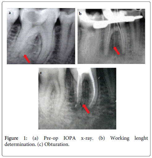
Radiographically, periapical radiolucency was seen in relation to both mesial and distal roots (Figure 1a). The presence of third additional root was also revealed on the distal side. The extra root was relatively straight and originated from distolingual aspect of the tooth. The tooth was unresponsive on electric pulp testing. A diagnosis of chronic apical periodontitis in relation to lower right first molar was made because of pulpal necrosis. The tooth was anesthetized. Access opening was made, and two mesial canal orifices (mesiobuccal, mesiolingual) and one distal canal orifice (distobuccal) were initially located. Another orifice was located on distolingual part of the pulpal floor on further exploration. The root canals orifices were enlarged using gates glidden drills Mani Inc., Japan) for a straight line access and the shape of access cavity was modified from a triangular form to a more trapezoidal form to better locate distolingual root. The root canals were explored with a K-file ISO number 15 and radiographic length of the root canals were determined (Figure 1b). Biomechanical preparation was carried out using the Pro Taper rotary files (Dentsply, Switzerland) in all the canals with intermittent irrigation using 1% sodium hypochlorite. Obturation of the root canals was performed using the Pro Taper gutta percha points and AH plus sealer (Dentsply, Switzerland). The access open cavity was then sealed with amalgam restoration (Figure 1c).
Case 2
A 48 year-old Indian male patient reported with a chief complaint of inability to chew with left lower back tooth region of jaw. Patient’s past medical history was non-contributory. On clinical examination, gingiva in relation to lower left permanent first molar was red, inflamed. Bleeding on probing was present. Periodontal pocket of around 8 mm was present on distal side. Grade III mobility was present. The involved tooth also exhibit grade III furcation involvement.
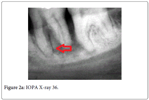
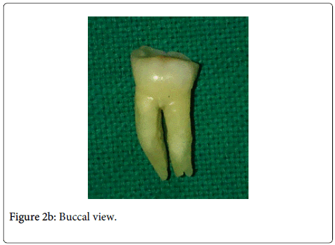
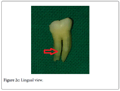
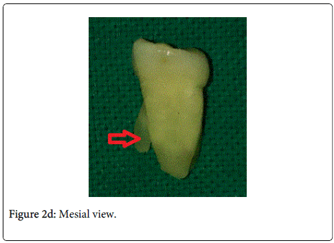
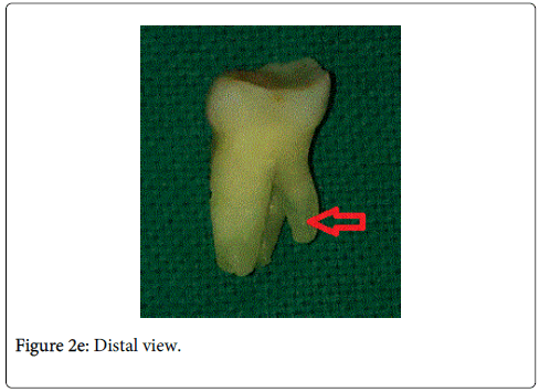
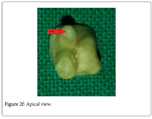
Radiographically, additional root was evident emerging from distolingual side of the tooth. Based on the literature evidence, this additional distolingual root was diagnosed as Radix Entomolaris. Periapical radiolucency was present in relation to all the roots, periodontal ligament space was widened and lamina dura was absent on distal side. Radiographic sign of moderate-severe bone loss was also evident around the involved tooth (Figure 2a). Periodontal prognosis was poor with the involved tooth. Hence, the tooth was extracted under local anesthesia. The extracted teeth exhibiting Radix Entomolaris morphology was also assessed clinically (Figure 2b, 2c, 2d, 2e, 2f). The extra root emerged from the lingual aspect of the distal root. The additional root was narrow, short and tapers towards the apex. Thus, care should be taken to avoid overpreparation and any inadvertent perforation of the root during root canal treatment. Access cavity was prepared on the extracted molar. Additional orifice was located on distolingual aspect of the pulpal floor making access cavity shape from triangular form to a trapezoidal form.
Discussion
Radix Entomolaris (additional root located lingually)
Prevalence: Anatomic variations in permanent mandibular first molars are documented in literature. Majority of mandibular first molars are two rooted; mesial and distal. Sometimes, an extra distobuccal or distolingual root may be encountered [12]. The aetiology for radix entomolaris is still unknown; it can be because of external factors during tooth formation or can be attributed to atavic gene or polygenic system [8,13].
It has also been suggested that “three-rooted molar” traits have a high degree of genetic predisposition as in Eskimos and in mixture of Eskimos with Caucasians [14]. The presence of radix entomolaris has been associated with ethical groups of mongoloid origin (>30%), rather low prevalence (<5%) in white Caucasian, African, Eurasian and Indian populations. The radix entomolaris may also be present in first, second and third molar; being less prevalent in second molar [15]. Bilateral occurrence of radix entomolaris has also been reported [10,16]. The relationship between radix entomolaris (RE), gender predilection and side distribution is not clear. Few studies have reported more of male predilection for RE while others reported no significant difference between gender and RE [17]. Similarly, no significant difference was reported for side distribution, despite few studies reporting it to be more on left side while others on right side [17]. Bilateral occurrence for RE have been reported to range from 37.14 - 67% [6,17-20].
Classification: Carlsen & Alexandersen (1990) classified radix entomolaris (RE) into four different types based on the location of its cervical part [6]:
1. Type A: the RE is located lingually to the distal root complex which has two cone-shaped macrostructures.
2. Type B: the RE is located lingually to the distal root complex which has one cone-shaped macrostructures.
3. Type C: the RE is located lingually to the mesial root complex.
4. Type AC: the RE is located lingually between the mesial and distal root complexes.
De Moor et al. (2004) classified radix entomolaris based on the curvature of the root or root canal [11]:
1. Type 1: a straight root or root canal.
2. Type 2: a curved coronal third which becomes straighter in the middle and apical third.
3. Type 3: an initial curve in the coronal third with a second buccally oriented curve which begins in the middle or apical third.
Song JS et al. (2010) further added two more newly defined variants of RE [21]:
1. Small type: length shorter than half of the length of the distobuccal root.
2. Conical type: smaller than the small type and having no root canal within it.
Morphology: The radix entomolaris is located distolingually ranging from short, conical extension to normal mature root length with its coronal third partially or completely fixed to distal root. Generally, the radix entomolaris is smaller than mesio- and distobuccal roots and may contain pulpal tissue [22]. Externally, the distal furcation is slightly lower (1 mm.) than the furcation between mesial and distal roots [23].
Clinically, tooth with additional distolingual root may present a more bulbous crown outline, an additional cusp, a prominent distolingual lobe or cervical prominence. These features can indicate the presence of additional root. Radiographically, third root is visible in 90% of cases [24]. Occasionally it may be missed because of its slender dimension or overlapping with distal root. Radiographs should be carefully inspected to reveal the presence of hidden radix entomolaris which might present as unclear outline of distal root or root canal. Additional radiographs taken from different horizontal projections, 20 degree from mesial and 20 degree from distal reveals the basic information about the anatomy of additional third root [25,26]. In addition to this, magnifying loupes, dental microscope or intraoral camera may also be useful. Recently, cone-beam computed tomography (CBCT) has emerged as a useful tool to aid in the diagnosis of teeth with complex root anatomies. However, cost and accessibility are the main limiting factors till now [27,28].
Radix paramolaris (additional root located buccally)
Prevalence: Bolk reported the occurrence of radix paramolaris [12]. Radix paramolaris is very rare and occurs less frequently than radix entomolaris [12]. Visser reported the prevalence of radix paramolaris to be 0% for mandibular first molars, 0.5% for second molars and 2% for third molars [15].
Classification: Carlsen & Alexandersen (1991) classified radix paramolaris (RP) into two different types [29]:
1. Type A: cervical part is located on the mesial root complex.
2. Type B: cervical part is located centrally, between the mesial and distal root complexes.
Morphology: The radix paramolaris (RP) is located mesiobuccally. The dimensions of RP may vary from short conical extension to a mature root which can be separate or fuse. Few observations can be made from various studies, i.e. an increased number of cusps is not necessarily related to an increased number of roots; however, an additional root is always associated with an increased number of cusps, and with an increased number of root canals [30-32].
Clinical Implications
Endodontic procedures: The presence of radix entomolaris has clinical implications in root canal treatment. Accurate clinical and radiographic diagnosis can avoid failure of root canal treatment because of missed canal in distolingual root. Most important basic principle for successful root canal treatment is the principle of ‘straight-line access’ [33]. Ultimate objective is to provide access to the apical foramen. As the orifice of radix entomolaris is distolingually located, the shape of access cavity should be modified from classical triangular form to trapezoidal or rectangular form in order to better locate the orifice of distolingual root. The root canal orifices follow the laws of symmetry which help in locating the radix entomolaris. Canal orifices are equidistant from a line drawn in a mesiodistal direction through the pulpal floor and lie perpendicular to this mesiodistal line across the centre [17,33-35]. Straight line access is essential as majority of radices entomolaris are curved. Care must be taken to avoid excessive removal of dentin or gauging during access cavity preparation as this may weaken the tooth structure.
Oral surgical procedures: Radix entomolaris may pose difficulty during extraction and orthodontic procedure. Tooth should be thoroughly luxated during extraction, as distolingual root might fracture as it is smaller than mesial and distobuccal roots.
Orthodontic procedures: During orthodontic procedure, presence of distolingual root and its curvature makes tooth movement difficult.
Conclusion
The oral health care professionals should be aware of this variation in anatomy of permanent mandibular first molars. The initial diagnosis is of utmost importance, to facilitate the endodontic procedure and to avoid treatment failures. Proper interpretation of radiographs taken at different horizontal angulations may help to identify number of roots and their morphology. Once diagnosed, the conventional triangular cavity should be modified to a trapezoidal form distolingually to locate the orifice of the additional root.
References
- Vertucci FJ (1984) Root canal anatomy of the human permanent teeth.Oral Surg Oral Med Oral Pathol 58: 589-599.
- Barker BC, Parsons KC, Mills PR, Williams GL (1974) Anatomy of root canals. III. Permanent mandibular molars.Aust Dent J 19: 408-413.
- Fabra-Campos H (1989) Three canals in the mesial root of mandibular first permanent molars: a clinical study.IntEndod J 22: 39-43.
- Stroner WF, Remeikis NA, Carr GB (1984) Mandibular first molar with three distal canals.Oral Surg Oral Med Oral Pathol 57: 554-557.
- Carabelli G (1844) SystematischesHandbuch der Zahnheilkunde (2 edn). Braumuller und Seidel, Vienna, Italy. p114.
- Carlsen O, Alexandersen V (1990) Radix entomolaris: identification and morphology.Scand J Dent Res 98: 363-373.
- Tratman EK (1938) Three-rooted lower molars in man and their racial distribution. British Dental Journal. 64: 264-274.
- Curzon ME, Curzon JA (1971) Three-rooted mandibular molars in the Keewatin Eskimo.J Can Dent Assoc (Tor) 37: 71-72.
- Turner CG 2nd (1971) Three-rooted mandibular first permanent molars and the question of American Indian origins.Am J PhysAnthropol 34: 229-241.
- Yew SC, Chan K (1993) A retrospective study of endodontically treated mandibular first molars in a Chinese population.J Endod 19: 471-473.
- De Moor RJ, Deroose CA, Calberson FL (2004) The radix entomolaris in mandibular first molars: an endodontic challenge.IntEndod J 37: 789-799.
- Bolk L (1914)WelcherGebibreihegehoren die Molaren an? Z MorpholAnthropol 17: 83-116.
- Walker RT (1988) Root form and canal anatomy of mandibular second molars in a Southern Chinese population. Endodontics& Dental Traumatology 14: 325-329.
- Curzon ME (1974) Miscegenation and the prevalence of three-rooted mandibular first molars in the Baffin Eskimo.Community Dent Oral Epidemiol 2: 130-131.
- Visser JB (1948)BeitragzurKenntnis der menschlichenzahnwurzelformen. Hilversum:Rotting. 49-72.
- Steelman R (1986) Incidence of an accessory distal root on mandibular first permanent molars in Hispanic children.ASDC J Dent Child 53: 122-123.
- Abella F, Patel S, Durán-Sindreu F, Mercadé M, Roig M (2012) Mandibular first molars with disto-lingual roots: review and clinical management.IntEndod J 45: 963-978.
- Tu MG, Huang HL, Hsue SS, Hsu JT, Chen SY, et al. (2009) Detection of permanent three-rooted mandibular first molars by cone-beam computed tomography imaging in Taiwanese individuals.J Endod 35: 503-507.
- Tu MG, Tsai CC, Jou MJ, Chen WL, Chang YF, et al. (2007) Prevalence of three-rooted mandibular first molars among Taiwanese individuals.J Endod 33: 1163-1166.
- Vivekananda Pai AR, Jain R,ColacoAS (2014) Detection and endodontic managament of radix entomolaris: Report of case series. Saudi Endod J 4: 77-82.
- Song JS, Choi HJ, Jung IY, Jung HS, Kim SO (2010) The prevalence and morphologic classification of distolingual roots in the mandibular molars in a Korean population.J Endod 36: 653-657.
- Reichart PA, Metah D (1981) Three-rooted permanent mandibular first molars in the Thai.Community Dent Oral Epidemiol 9: 191-192.
- Gu Y1, Zhou P, Ding Y, Wang P, Ni L (2011) Root canal morphology of permanent three-rooted mandibular first molars: Part III--An odontometric analysis.J Endod 37: 485-490.
- Walker RT, Quackenbush LE (1985) Three-rooted lower first permanent molars in Hong Kong Chinese.Br Dent J 159: 298-299.
- Klein RM, Blake SA, Nattress BR, Hirschmann PN (1997) Evaluation of X-ray beam angulation for successful twin canal identification in mandibular incisors.IntEndod J 30: 58-63.
- Ingle JI, Heithersay, GS, Hatwell GR (2002) Endodontic Diagnostic Procedures. BC Decker, London, UK.
- Gopikrishna V, Reuben, J., Kandaswamy, D (2008) Endodontic management of a maxillary first molar with two palatal roots and a single fused buccal root diagnosed with spiral computed tomography-a case report. Oral Surgery Oral Medicine, Oral Pathology, Oral Radiology and Endodontology105: e74-e8.
- Aggarwal V, Singla M, Logani A, Shah N (2009) Endodontic management of a maxillary first molar with two palatal canals with the aid of spiral computed tomography: a case report.J Endod 35: 137-139.
- Carlsen O, Alexandersen V (1991) Radix paramolaris in permanent mandibular molars: identification and morphology.Scand J Dent Res 99: 189-195.
- Sperber GH, Moreau JL (1998) Study of the number of roots and canals in Senegalese first permanent mandibular molars.IntEndod J 31: 117-122.
- Carlsen O, Alexandersen V (1999) Radix paramolaris and radix distomolaris in Danish permanent maxillary molars.ActaOdontolScand 57: 283-289.
- Brabant H, Klees, L, Werelds, RJ (1958) Anomalies, mutilations ettumeurs des dents humanies. Editions JulienPrelat ed. Paris, France.
- Calberson FL, De Moor RJ, Deroose CA (2007) The radix entomolaris and paramolaris: clinical approach in endodontics.J Endod 33: 58-63.
- Krasner P, Rankow HJ (2004) Anatomy of the pulp-chamber floor.J Endod 30: 5-16.
- Gu Y, Lu Q, Wang H, Ding Y, Wang P, et al. (2010) Root canal morphology of permanent three-rooted mandibular first molars--part I: pulp floor and root canal system.J Endod 36: 990-994.
Relevant Topics
- Cementogenesis
- Coronal Fractures
- Dental Debonding
- Dental Fear
- Dental Implant
- Dental Malocclusion
- Dental Pulp Capping
- Dental Radiography
- Dental Science
- Dental Surgery
- Dental Trauma
- Dentistry
- Emergency Dental Care
- Forensic Dentistry
- Laser Dentistry
- Leukoplakia
- Occlusion
- Oral Cancer
- Oral Precancer
- Osseointegration
- Pulpotomy
- Tooth Replantation
Recommended Journals
Article Tools
Article Usage
- Total views: 23848
- [From(publication date):
February-2015 - Apr 03, 2025] - Breakdown by view type
- HTML page views : 18553
- PDF downloads : 5295

