Case Report Open Access
The Neglected Phase in Orthodontic Diagnosis and Treatment
Maen Mahfouz*
Lecturer, Department of Orthodontics and Pediatric Dentistry, Arab American University, Jenin, Palestine
- *Corresponding Author:
- Maen Mahfouz
Lecturer, Department of
Orthodontics and Pediatric Dentistry
Arab American University, Jenin, Palestine
Tel: 00972599752555
E-mail: maenmahfouz@gmail.com
Received Date: May 23, 2014; Accepted Date: July 26, 2014; Published Date: August 02, 2014
Citation: Mahfouz M (2014) The Neglected Phase in Orthodontic Diagnosis and Treatment. J Oral Hyg Health 2:147. doi:10.4172/2332-0702.1000147
Copyright: © 2014 Mahfouz M. This is an open-access article distributed under the terms of the Creative Commons Attribution License, which permits unrestricted use, distribution, and reproduction in any medium, provided the original author and source are credited.
Visit for more related articles at Journal of Oral Hygiene & Health
Abstract
The leading factors for orthodontic patients to attend orthodontic office is either due to esthetic concern , functional concern or/and oral health concern. In this paper the light has been thrown on one of fundamental aspects in orthodontic diagnosis and treatment that is function by presenting a clinical case which is demonstrating the importance of this phase in orthodontic diagnosis and treatment.
Keywords
Neglected phase; Orthodontic; Diagnosis; Treatment
Introduction
The successfully corrected dental malocclusion is usually described as one that is excellent in appearance, excellent in function and stable or permanent in result. These are indeed the objectives that are desirable, but they are in a sense, somewhat intangible at least insofar as the exactness of the evaluation is considered [1]. The phrase form and function reflects a concept of interrelating the shape or attributes of something with its function. In dentistry the phrase may indicate the entire masticatory system acting as a biomechanical engine for the reduction of food [2]. For some clinicians the general principle that becomes operant is form follows function, meaning for examples include the dependant relationship between esthetics and optimum occlusion [3] physical forces and the periodontal ligament [4] arch form and beta function [5] and the controversial relationship between the morphology and function of the disco-muscular apparatus of the human temporomandibular joint. The masticatory system is a functional unit composed of the teeth; their supporting structures, the jaws; the temporomandibular joints; the muscles involved directly or indirectly in mastication (including the muscles of the lips and tongue); and the vascular and nervous systems supplying these tissues. Functional and structural disturbances in any one of the components of the masticatory system may be reflected by functional or structural disorders in one or more of its other components [6]. However, there is a lot of evidence that the masticatory system has ability to the wide range of adaptive modalities. These adaptations can be functional and/or structural and may respond to transient and/or permanent demands. Therefore, this system, like any biological system, cannot be viewed as a rigid and immutable [7]. Knowledge of how the mandible moves during mastication has greatly influenced procedures in clinical dentistry. Historically, an understanding of mandibular movement was considered important in removable prosthodontics. Later, this information was used in the design and setting of articulators, and in the design of the dentures and denture teeth themselves. Today the importance of jaw movements has become apparent in fixed prosthodontics, periodontics, orthodontics, and in the diagnosis and treatment of pain disorders of the masticatory system[8,9]. In regard to the occlusion of the teeth there are two concepts to be recognized the first is the morphological or anatomical concept, as the name implies this is an occlusion based on the forms of the teeth while the second is the physiologic concept. The physiologic occlusion is a living-dynamic as it participates as one of the components of the masticatory system. Abnormal functional relationship of the teeth may vary in degree of abnormality from mild to extreme. In the first instance certain teeth may occlude slightly in advance of the other teeth. This is premature contact of the teeth. This condition may also exist in the functional movements of the mandible during mastication [1]. In this paper the light is thrown on one of fundamental aspects in orthodontic diagnosis and treatment that is function by presenting a clinical case which is demonstrating the importance of this phase in orthodontic diagnosis and treatment.
Case History
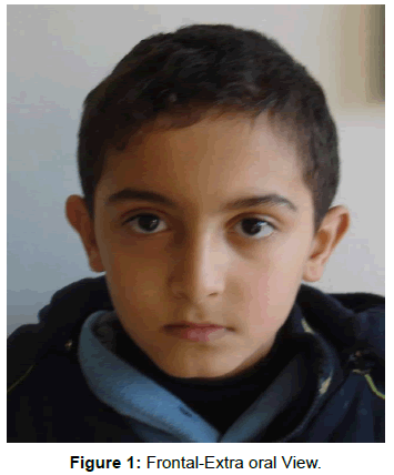
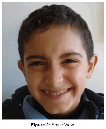
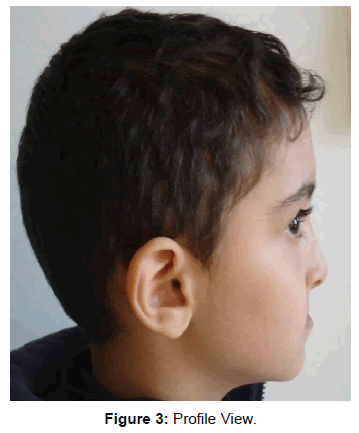
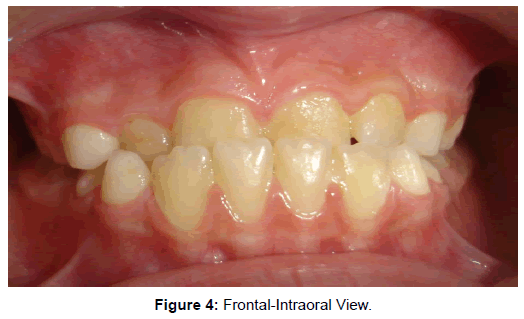
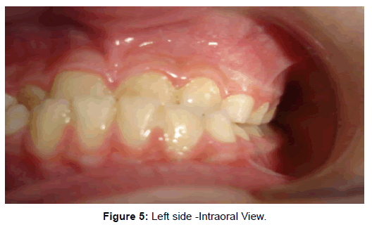
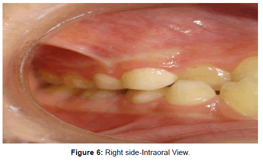
A 7 year old boy attended to my private orthodontic clinic with chief compliant of lower anterior teeth overlapping of upper anterior teeth. He was in early mixed dentition stage. In the extra oral the patient profile was straight while lip profile was reversed giving the appearance of class III (Figures 1-3). In the intraoral examination revealed a forward shift of the mandible, with a marked class III molar relationship and an anterior cross bite (Figures 4-6).
Diagnosis
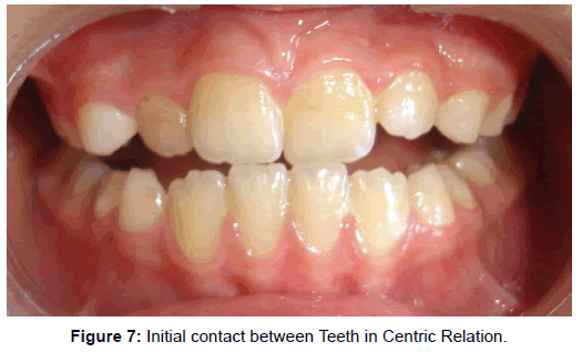
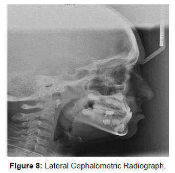
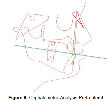
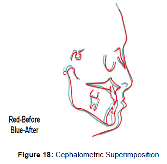
The clinical examination revealed retruded upper lip with protruded lower lip giving the view of deficient mid face as seen in class III. The upper incisors retroclined and spaced while lower incisors slightly proclined. An anterior cross bite in the presence of a forward mandibular displacement and functional shift to the left side due to premature contact between upper and lower central incisors, Class III malocclusion with reverse overjet and negative deep overbite. The starting point in diagnosis and treatment of this case was by establishing centric relation through guiding the mandible into centric relation rather than centric occlusion and then initial contact with the teeth occurs so an edge to edge anterior incisor contact with posterior open bite indicating Pseudo Class III (Figures 3-8). Cephalometric analysis indicated a mild Class III malocclusion characterized by a little mandibular protrusion (ANB =-1 degree, Wits=-6 mm) (Figures 8 and 9).
Treatment Objectives
Forward movement of 11 maxillary incisors, eliminating functional shift, mandibular displacement, premature contact, enhancing normal lip profile, achieving class I molar relationship and canine relationship with ideal over jet and overbite.
Treatment plan
Fixed orthodontic treatment by using protrusive arch wire for the forward movement of 11 without raising the bite.
Treatment procedure
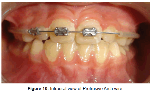
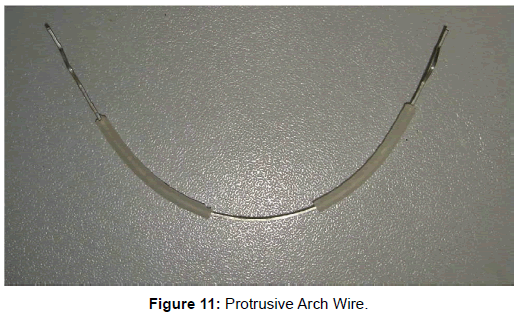
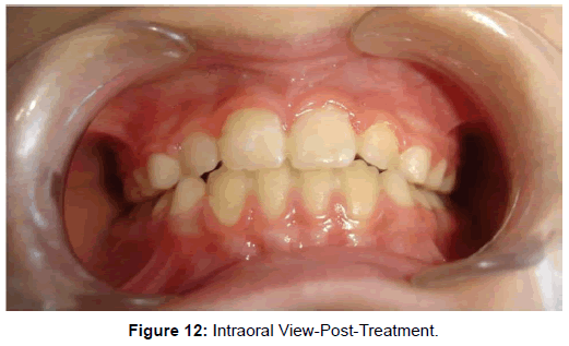
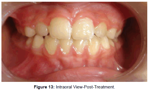
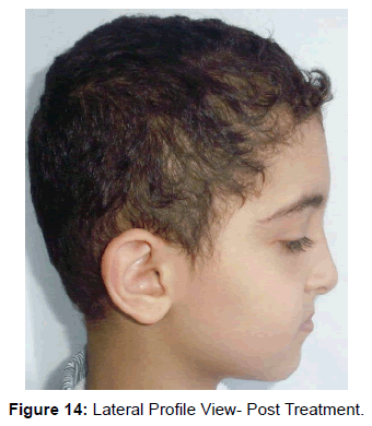
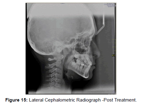
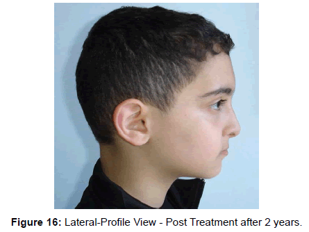
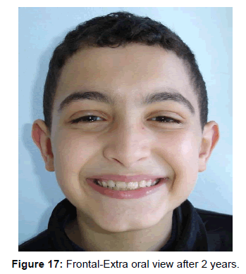
Treatment started so a bondable tubes placed on the buccal surfaces of upper first permanent molars on both sides with brackets (slot 22) placed on the labial surface of the upper central incisors. Then placement of upper Niti arch wire rectangular (0.016x0.022) (Protrusive Arch Wire) was customized to the arch form of the patient A gable bend was made mesial to the bondable tubes on first permanent molars. The exposed part of arch wire coated with sleeve to prevent irritation to cheek (Figures 10 and 11). After two weeks orthodontic evaluation has been done of the retroclined upper central incisors which were orthodontically adjusted so the patient could bite in the new situation. The total treatment time was two weeks and the appliance was removed on the end of the second week (Figures 12 and 13). Two years follow up for this case with good stability of occlusion, optimal functional and aesthetical states. Final superimpositions showed improvements in ANB and Wits values (+1 degree, -1 mm) respectively. The slight maxillary incisor protrusion coupled with the clockwise mandibular rotation produced an overall improvement of the patient’s aesthetic appearance (Figures 14-18).
Discussion
Typically, an orthodontic case begins with a basic evaluation of the patient’s occlusion in static state. The view of occlusion has been enlarged so that it must be thought of as it functions as an integral part of the dynamic masticatory system. An orthodontist should compare a patient’s centric relation (CR) and centric occlusion (CO). Ideally, CR and CO are in harmony. It is important to note that centric relation is no longer considered to be the most posterior and superior position of the condyle in the glenoid fossa. Currently, centric relation is defined as the condyle in the most superior position. Centric occlusion is the initial point of contact in centric relation. An obvious example of where a discrepancy between CR and CO exists is in pseudo-Class III patients, who are typically able to bite with their incisors in an edge-to-edge position. However, these patients must protrude the mandible in order for the posterior teeth to occlude. It is important to begin orthodontic treatment on pseudo- Class III cases at an early age in order to prevent the mandible from growing asymmetrically. A more subtle example of a CR-CO discrepancy is found when anterior teeth create orthopedic instability. Protrusive and excursive movements should also evaluate in the practice of orthodontics. The orthodontist’s goal is to establish anterior guidance during protrusion and canine guidance during left and right excursive movements. The tooth interference may be more pronounced than premature contact and the mandible may be deflected from its normal functional path, now more than the teeth and supporting tissues are involved and the abnormal function may extend into the temporomandibular joints and musculature [1]. If the interferences cannot be avoided comfortably by mandibular displacement in chewing and swallowing by neuromuscular mechanisms (functional adaptation), if the tooth becomes mobile and is moved out of position (structural adaptation) and or if the patient cannot ignore the discomfort or the presence of change even for a short period (behavioral adaptation) overt symptoms of dysfunction of the muscles, joints, periodontium or teeth (pulp) may occur [2]. In the current case ,as the upper central incisors are retroclined , so there is premature contact which avoided by forward mandibular displacement associated with functional shift to the left to achieve more comfortable bite in centric occlusion (functional adaptation). In contrast there is an edge to edge relationship between upper and lower teeth with posterior open bite which is not comfortable in centric relation. In the presented case the method used to eliminate the premature contact was not occlusal adjustment by selective grinding which involves the selective reshaping of tooth surfaces that interfere with normal jaw function, it was an orthodontic occlusal adjustment by correction of the position of the retroclined upper central incisors to procline them forward by using the protrusive arch wire [10,11].
References
- John R. Thompson (1956) Function-The Neglected Phase of Orthodontics. The Angle Orthodontist26: 129-143
- Major M.Ash,Jr.StanleyJ.Nelson (2003) Wheeler’s Dental Anatomy ,Physiology ,and Occlusion.(8thEdn) Elsevier Science.
- Sorenson DA (1998) Form follows function: occlusion based rationale for esthetic dentistry. J Indiana Dent Assoc 77: 25-30.
- McCulloch CA, Lekic P, McKee MD (2000) Role of physical forces in regulating the form and function of the periodontal ligament. Periodontol 2000 24: 56-72.
- Braun S, Hnat WP, Fender DE, Legan HL (1998) The form of the human dental arch. Angle Orthod 68: 29-36.
- Ash MM, Ramfjord S (1995) Occlusion. (4thEdn) W.B. Saunders Company.
- Mohl ND (1988) A Textbook of Occlusion. In: Mohl ND, Zarb GA, Carlsson GE, Rugh JD(Eds) Introduction to Occlusion. Quintessence books.
- Rugh JD, Smith BR (1988) A Textbook of Occlusion. In: Mohl ND, Zarb GA, Carlsson GE, Rugh JD (Eds) Mastication. Quintessence books.
- Soboleva U, Laurina L, Slaidina A (2005) The masticatory system- an overview. Stomatologija. Baltic Dental and Maxillofacial Journal 7:77-80.
- Mahfouz M (2014) Pseudo class III treatment in 2-year-old children. Open Journal of Stomatology 4: 10-13.
- Mahfouz M (2014) Pseudo Class III correction in early mixed dentition by using protrusive arch wire. Journal of Orthodontic Research,in press.
Relevant Topics
- Advanced Bleeding Gums
- Advanced Receeding Gums
- Bleeding Gums
- Children’s Oral Health
- Coronal Fracture
- Dental Anestheia and Sedation
- Dental Plaque
- Dental Radiology
- Dentistry and Diabetes
- Fluoride Treatments
- Gum Cancer
- Gum Infection
- Occlusal Splint
- Oral and Maxillofacial Pathology
- Oral Hygiene
- Oral Hygiene Blogs
- Oral Hygiene Case Reports
- Oral Hygiene Practice
- Oral Leukoplakia
- Oral Microbiome
- Oral Rehydration
- Oral Surgery Special Issue
- Orthodontistry
- Periodontal Disease Management
- Periodontistry
- Root Canal Treatment
- Tele-Dentistry
Recommended Journals
Article Tools
Article Usage
- Total views: 16119
- [From(publication date):
September-2014 - Jul 05, 2025] - Breakdown by view type
- HTML page views : 11433
- PDF downloads : 4686

