The Modified Anterior Drawer Test (MADT): A New Clinical Test for Assessing Subtalar Instability a Cadaveric and Clinical Assessment
Received: 04-Jun-2018 / Accepted Date: 14-Jun-2018 / Published Date: 21-Jun-2018 DOI: 10.4172/2329-910X.1000271
Abstract
Background: Chronic lateral ankle instability describes multiple pathologies affecting the tibiotalar or talocalcaneal articulations of the foot. One quarter of cases are caused by isolated or concomitant subtalar instability. The standard anterior drawer test (ADT) is used in routine clinical practice to assess abnormal movement at the tibiotalar joint. However, a positive test does not discriminate whether excessive translation is at the tibiotalar or subtalar joint. We suggest a modification to the standard ADT which separates the ankle joint translation by manually immobilising the tibiotalar joint in order isolate anterior translation at the subtalar joint. In the presence of a positive ADT, the modified anterior drawer test (MADT) would help discriminate if the pathological translation is at the tibiotalar or the subtalar joint or both.
Methods: Our clinical study was an observational analysis of a cohort of 12 patients who had presented to an outpatient foot clinic in east of England. In addition, a cadaveric study was used to assess subtalar anterior translation.
Results: In 50% of patients who had a clinically positive ADT, the MADT was negative which correlated 100% with Stress Broden radiographic views. Cadaveric analysis was done to show an increase in anterior translation with sequential sectioning of supporting ligaments. We demonstrated an absolute reduction in anterior translation in subtalar instability with the additional posterior force (MADT).
Conclusion: The clinical and cadaveric data supports the use of the MADT as a valuable adjunct in the diagnosis and exclusion of subtalar instability as a cause of symptoms in addition to stress radiography.
Keywords: Subtalar instability; Anterior drawer test; Ankle instability; Subtalar joint
Introduction
Chronic lateral ankle instability is a relatively uncommon condition presenting to foot and ankle clinics. It is an umbrella term which describes multiple pathologies affecting the tibiotalar or talocalcaneal (subtalar) articulations [1]. Subtalar instability can occur in one quarter of cases of chronic lateral instability and is often over looked [2]. The presentation can be varied and symptoms include ankle swelling, pain, clicking or giving way. The pathology is equally varied and injury to a number of ligaments can give rise to these symptoms [3]. The anatomy is described below. Therefore ankle instability is a vast entity, and the main cause of the symptoms must be investigated to ensure effective treatment is provided. The standard anterior drawer test (ADT) is a clinical test which is routinely used to assess abnormal movement at the ankle joint in the presence of instability symptoms [4]. The hind foot is held in neutral/up to 20 degrees plantar flexion and an anterior translation and internal rotation force is applied. It is considered positive if asymmetrical and excessive translation of the calcaneum is elicited. However, it is a composite movement of both the tibiotalar articulation and subtalar articulation as this manoeuvre provides anterior translation force at the tibiotalar and subtalar joint.
Whilst it is assumed the majority of the anterior translation derives from the tibiotalar joint it does not differentiate the excess movement from the subtalar joint and therefore fails to ascertain which the pathological joint is [4].
There is currently no satisfactory clinical test for subtalar instability [5]. We suggest a modification to the standard anterior drawer test which separates ankle joint translation by manually immobilizing the tibiotalar joint using manual thumb pressure in order isolate anterior translation at the subtalar joint. In the presence of a positive ADT, the modified anterior drawer test (MADT) would help discriminate if the pathological translation is at the tibiotalar or the subtalar joint or combined. By negating the effect of anterior translation of the tibiotalar joint by applying thumb pressure over the anterolateral talar neck, any residual anterior translation must be due to isolated subtalar instability/pathology. Radiographic anterior translation at the subtalar joint with anterior stress has already been described in conventional foot and ankle textbooks. Therefore, in the presence of a positive ADT, a negative MADT would help rule out associated subtalar instability. A high negative predictive value of this clinical test would help prove this. This would then influence subsequent clinical management.
The study was conducted in two parts. First a clinical study based on patients presenting to an orthopaedic department in east of England. Secondly, a cadaveric study analyzing subtalar anterior translation and hence instability on fresh frozen cadavers. Both aspects of the study are discussed below.
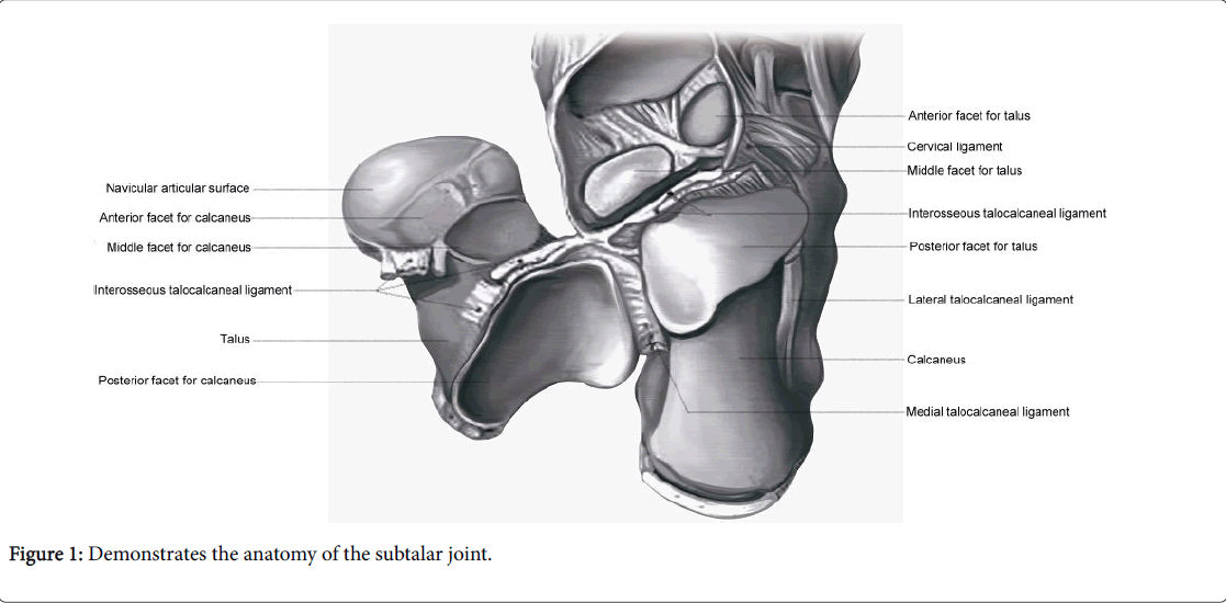
The subtalar joint is composed of an anterior, middle and posterior facet. The posterior facet is the largest and is a convex surface on the calcaneus. It is separated from the smaller middle facet by the sinus tarsi. The middle and anterior facets of the calcaneus are concave and articulate with the head of talus. The middle facet is on the sustentaculum tali whereas the anterior facet is situated more laterally. They are frequently confluent, therefore the subtalar joint can be regarded as a two-faceted joint [6] as shown in Figure 1.
The subtalar articulation is a stable joint owing to the presence of five ligaments (medial and lateral talocalcaneal, interosseous, cervical and calcaneofibular ligaments). The interosseous ligament is the strongest structure between the talus and the calcaneus and was first described by Jones W [7]. It remains unclear which ligament would produce the greatest anterior translation when divided.
Subtalar instability was first described by Rubin et al. The initial diagnosis was based upon stress tomograms [8]. The presentation can be similar to chronic lateral instability or recurrent ankle sprains and hence overlooked.
Combined injury to the tibiotalar articulation and subtalar joint can make the diagnosis challenging [9] and clinically it can be difficult to differentiate between ankle and subtalar instability [5,10]. The exact etiology and incidence of subtalar ligament injury is unknown. Patients report a variety of symptoms, including giving way, recurrent sprains, pain, swelling and stiffness [1]. Pain at the site of the anterior talofibular ligament (ATFL) or anterolateral gutter can help determine the presence of tibiotalar instability. The presence of a sulcus sign can be suggestive of tibiotalar instability [11]. Patients with the suggestion of subtalar instability in isolation or in conjunction with tibiotalar instability can present with pain over the sinus tarsi or deep pain in the subtalar area. This however is not diagnostic. Internal rotation and forward displacement of the calcaneus as described by the anterior drawer test will reveal overall instability, but this can be caused by instability leading to anterior translation at both tibiotalar and subtalar articulations [4]. There are no other described tests which assess stability of each joint individually.
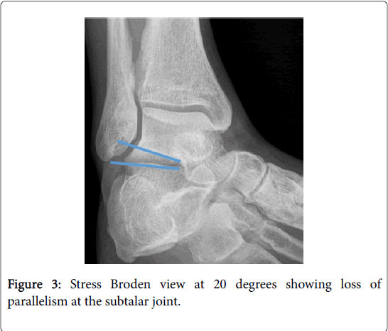
Radiological assessment follows a logical sequence. Standard anteroposterior (AP) radiographs of the ankle are initially performed to exclude any underlying bony abnormality. Patients with subtalar instability rarely have any abnormal findings on this view. There may be divergence of the subtalar joint on this view and separation of greater than 7 mm is considered indicative of subtalar instability [12]. However, subtalar instability is more likely to be noted on dynamic investigations [13]. The lateral radiograph with an anterior stress test applied at the calcaneum can show anterior translation respectively, but this is less specific to the subtalar joint and is often overlooked. The stress broden radiographic view is used to identify instability of the subtalar joint if there is loss of parallelism with an angle of >5 degrees. We have used this as the gold standard for diagnosing subtalar instability. Brantigan et al. found a significantly increased subtalar inversion angle in patients with clinically diagnosed subtalar instability [14]. Radiologic diagnoses and classification systems have been evaluated thoroughly by Laurin et al. through cadaveric based studies [15].
Magnetic resonance imaging (MRI) has become a more commonplace investigation to perform in the assessment of ankle instability. It is extremely useful in assessing the anatomical structures and the collateral ligaments [16]. However, this is a static investigation and provides clue to ligamentous disruption but does not provide an indication of the degree of instability the joint as would be obtained when put under stress. Subtalar instability ranks high on possible differential diagnoses in patients sustaining high-energy injury. We accept all radiological signs have to be correlated with clinical findings in order to establish subsequent treatment.
The Modified Anterior Drawer Test (MADT)
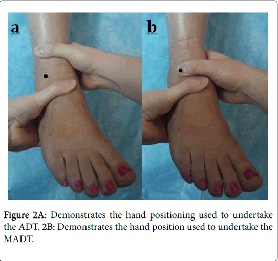
The ADT assesses the anatomical restraint to anterior translation and internal rotation of the hind foot on the tibia (the anterior talofibular ligament is the primary restraint [4]). The pathological translation may be at the tibiotalar or subtalar articulations, or both. It is considered positive if there is greater than 8mm of displacement [11]. More usefully it is a subjective phenomenon and of increased laxity when compared with the contralateral side is more useful than the absolute displacement [4]. The ADT traditionally hinges on an intact medial deltoid ligament although this is not a pre-requisite for a positive test [17]. Injury to the calcaneofibular ligament may affect the distance of the anterior drawer test and help signify the presence of subtalar instability. In the presence of subtalar instability there is often an increased translation compared to the unaffected side [18]. The ADT is performed by firmly securing the distal lower limb with one hand and applying an anterior translational force on the heel with the other whilst maintaining a little plantar-flexion, as shown in Figure 2A. The MADT relies on the same position of the lower extremity, but the proximal thumb of the hand holding the tibia is used to immobilize the tibiotalar joint. This is performed by placing the thumb on the lateral anterior aspect of the talus, distal to the joint at the position just medial to the anterolateral gutter, as shown in Figure 2B. This results in the talus and tibia becoming a single unit and negates the anterior translation of the talus from the tibia. All movement is therefore from the subtalar joint alone, thus isolating the anterior translation of this joint. We believe this modification has a high negative predictive value for ruling out subtalar instability.
Methods
Clinical study
Our study was a retrospective analysis of a cohort of 12 patients who had presented to an outpatient foot clinic in East of England. The recruitment period was 2 years overall. All patients had symptoms and clinical signs of unilateral ankle instability (tibiotalar, subtalar, or both). They were identified on the basis of history and clinical examination. All patients had clinical signs and symptoms of instability in the hind foot with a proven anterior draw test and no signs or symptoms of syndesmotic instability. The demographics of these patients are shown in Table 1. All patients were clinically assessed using the ADT and MADT, as described above. This was performed by a single senior consultant foot and ankle surgeon (CP). Standard broden stress view radiographs (described below) formed the gold standard for radiographic diagnosis of subtalar instability and were performed as a routine part of clinical practice in all cases. In addition, all patients underwent ankle MRI scan to identify the presence of ATFL rupture and the structural cause of the subtalar instability. We analysed the correlation, specificity and sensitivity of the MADT compared to the Broden stress views.
| Demographics of patients | n = 12 | % |
|---|---|---|
| Female | 3 | 25 |
| Male | ||
| Age (range) | 40 (19 - 63) | |
| Left foot | 3 | 25 |
| History of ankle injury1 | 2 | 17 |
| 1The pathology of the 2 patients considered to have relevant ankle injury or conditions are as follows: 1 sprain, 1 previous twisting injury | ||
Table 1: Clinical characteristics of patient cohort.
The Broden views are performed with an inversion stress being applied to the subtalar joint and the X-ray beam tilted 20 degrees and 40 degrees caudal to cephalad. This provides an enhanced view of the subtalar joint and reveals loss of parallelism if subtalar instability is present, as shown in Figure 3. At our institution, the view is obtained by positioning the patient supine with the knee held in mild flexion, rested on a sandbag. Inversion stress opens the subtalar joint if there is instability. We considered the Broden view positive if there were >5 degrees of subtalar opening [19]. However, in almost all our cases this was much greater. Normal values of talocalcaneal displacement or angulation are not well established within the literature to define subtalar instability; although an attempt has been made using radiographic anterior drawer testing [20].
Cadaveric Study
Nine cadavers were used to analyze anterior translation in subtalar instability. The demographics of the cadavers are as shown in Table 2.
| n = 9 | % | |
| Female | 8 | 89 |
| Age (range) | 74 (59 – 89) | |
| Left laterality | 3 | 33 |
| BMI (range) | 20 (15 – 25) | |
Table 2: Demographics of cadaveric cohort.
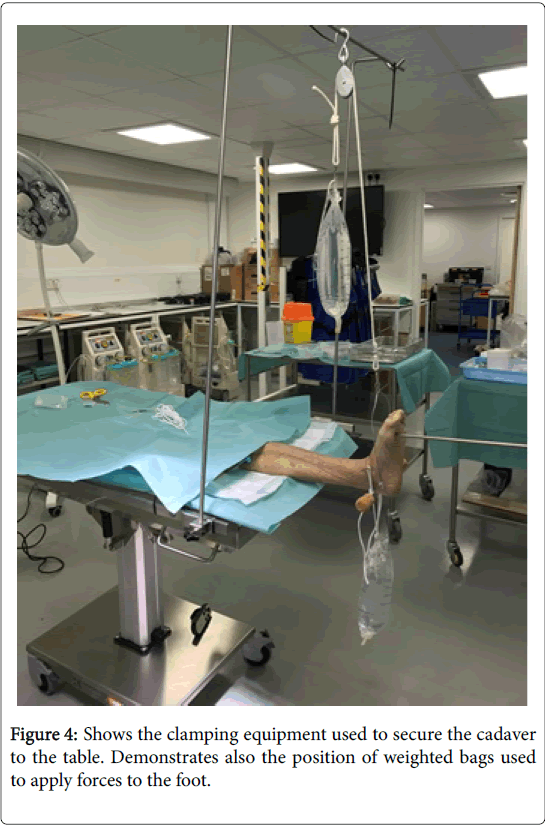
All cadavers were tested to ensure that there was no pre-existing laxity at the subtalar or tibiotalar articulations clinically. All cadavers used were fresh frozen and defrosted 36 hours prior to use in the research. Cadavers were secured to a dissecting table using clamps, as shown in Figure 4 proximally. This would help ensure that the cadavers would not move vertically when applying loads.
2 mm K-wires were then inserted into a fixed point of the lateral malleolus and proximally on into the fibula. A ruler was then placed on this and a further K-wire was fixed onto this to ensure that it did not move. This would be a fixed reference point laterally on the leg. If the cadaver would move anteriorly then so would the reference point thus negating the effect of any macro movement of the cadaver on the table that may move when loads are applied. A wire was then put into the anterior process of the calcaneum. This would prove a reference point in the calcaneum. This wire would move forward when an anterior load was applied to the calcaneum. The shortest distance between the ruler and the k-wire in the calcaneum could then be measured. The wire in the anterior process of the calcaneum would span both subtalar and tibio-talar joints. This wire with fixed bony landmarks that the cadavers were used to measure any anterior translation.
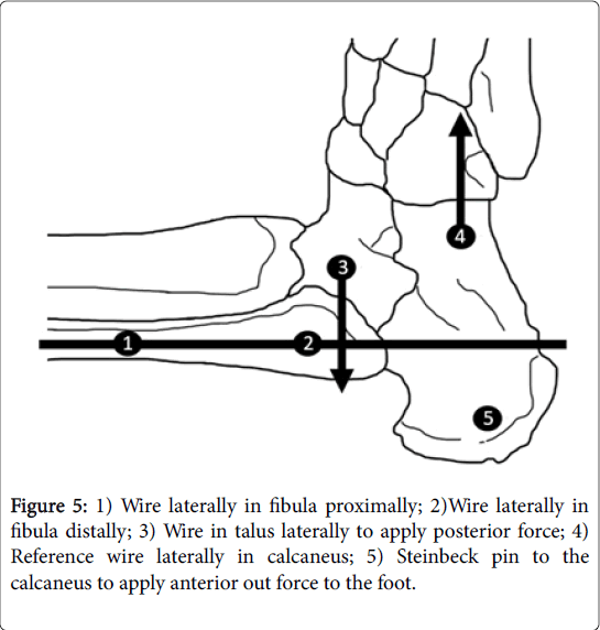
A trans-fixation Steinman pin was applied through the calcaneus. A pulley was applied to lateral part of this to simulate the force that the hand on the calcaneum would make when applying an anterior force in the anterior draw test. A further pin was then placed into the talar neck under image intensifier (II) guidance laterally, as shown in Figure 5. This would simulate the thumb pressure applied to the anterior talus in the modified anterior draw test. A force applied through this would neutralize/counteract the anterior force through the tibiotalar joint. Any residual anterior translation would therefore be due to subtalar joint anterior translation only.
The setup was as described below
An anterior force was applied through the ankle joint in each sample (bag 1) and then an additional opposing posterior force through the talar neck was applied (bag 2). This posterior force on the talar neck would reduce the talus back into joint and therefore any residual anterior translation can only arise from the subtalar joint. Subsequently the anterior talofibular ligament (ATFL) and calcaneofibular ligament (CFCL) were then surgically divided from each cadaver to demonstrate subtalar instability.
We applied a force of 30 newton’s/3 kg mass through each bag except bag 1 which was a 10 newton/1 kg mass. We accepted that during the experiment we felt a larger force applied would help achieve peak strain in the ligaments loaded and not obtain a mid-strain reading and therefore a decision to apply a larger load was used after the testing of the first cadaver to ensure that this would have occurred. Any further load above 3 kg would not realistically mimic true to life forces applied.
Forces were applied in the following scenarios.
Ligaments in the ankle were dissected in the following manner.
• Ligaments intact.
• ATFL sectioning.
• ATFL and CFCL sectioning.
• ATFL and CFCL and lateral talocalcaneal ligament.
• ATFL and CFCL and lateral talocalcaneal ligament and cervical ligament.
• ATFL and CFCL and lateral talocalcaneal ligament and cervical ligament and interosseous ligaments divided.
The ligament spanning the subtalar joints was surgically divided from a superficial to deep fashion as stated above.
Readings were taken with the intact cadaver with a load applied in bag 1 and bag 2. All readings were then taken in the above scenarios with a load applied through bag 1 and the bag 2. The aim was to find out if there was any additional translation in the cadavers due to each sequential sectioning. Forces were then applied to see the translation with successive dissection. Anterior translation was measured using calipers. The initial distance between the k-wires was then subtracted to ensure that the residual anterior translation distances were obtained.
One cadaver was not included in the final analysis due to preexisting subtalar instability on examination. Subsequent analysis of the clinical data revealed this individual had left hemiparesis following a cerebrovascular accident.
Results
Of the 12 patients, the ADT was positive in all cases. The MADT was positive in six cases; of these five had positive stress Broden views, with a mean opening angle of 11.4 degrees. Of the other six patients where MADT was negative, all cases had negative Broden stress views. This highlights the negative predictive value (NPV) of the test (NPV=1). This is the ability of a test to rule out a condition. Full sensitivity and specificity calculations are outlined in Table 3.
| Stress Broden radiographs (Gold standard for diagnosis) | |||
|---|---|---|---|
| Positive | Negative | ||
| Modified anterior drawer test | Positive | 5 (True positive) | 1 (False positive) |
| Negative | 0 (False negative) | 6 (True negative) | |
| Sensitivity = 1; Specificity = 0.86 | |||
Table 3: Modified anterior drawer test.
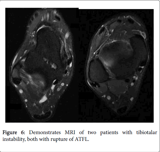
In the case of the patient where the broden view was negative and MADT test positive. This may indicate a false positive MADT or a potential variation of pathology causing subtalar instability, which was not detected radiographically. The exact entity has not been fully understood. Subsequent MRI scans of all 12 patients revealed all patients with tibiotalar instability had positive MRI findings of ATFL rupture, as shown in Figure 6. MRI failed to diagnose any disruption of the subtalar ligamentous complex in all cases of subtalar instability.
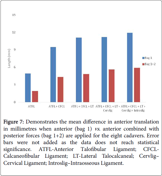
The cadaveric data shows greater anterior translation distance with the ATFL and CFCL divided in all cadavers. This was then reconstituted to the original anterior translation when the talus was put under pressure. With successive translation of the deeper subtalar ligaments there was increased amount of anterior translation overall, as shown in Figure 7 and Table 4 and Table 5.
Figure 7: Demonstrates the mean difference in anterior translation in millimetres when anterior (bag 1) vs. anterior combined with posterior forces (bag 1+2) are applied for the eight cadavers. Error bars were not added as the data does not reach statistical significance. ATFL-Anterior Talofibular Ligament; CFCLCalcaneofibular Ligament; LT-Lateral Talocalcaneal; Cervlig– Cervical Ligament; Introslig–Intraosseous Ligament.
| S1 | S2 | S3 | S4 | S5 | S6 | S7 | S8 | Mean | STDEV | |
|---|---|---|---|---|---|---|---|---|---|---|
| ATFL Bag 1 | 4.6 | 8.1 | 6.6 | 5.4 | 6.9 | -2.6 | 5.2 | 4.7 | 4.9 | 3.3 |
| ATFL Bag 1+2 | -3 | 5.5 | 3.5 | 3.1 | 0.4 | -0.3 | 3.8 | 2.6 | 1.9 | 2.7 |
| ATFL+CFCL Bag 1 | 8.3 | 10.9 | 7.1 | 7.3 | 11.9 | -1.6 | 22.3 | 9.5 | 9.5 | 6.6 |
| ATFL+CFCL Bag 2 | -1.4 | 9.3 | 5.2 | 3.3 | 1 | -1 | 19 | -1.2 | 4.3 | 7 |
| ATFL+CFCL+ LT Bag 1 | 10 | 13.7 | 7.1 | 8.2 | 12 | -1.1 | 21.8 | 17.4 | 11.1 | 6.9 |
| ATFL+CFCL+LT Bag 2 | -2.1 | 11 | 4.7 | 3.6 | 2.8 | 0.6 | 19 | -1.1 | 4.8 | 7 |
| ATFL+CFCL+LT+Cervlig Bag 1 | 8.6 | 12.9 | 7.4 | 8.6 | 13 | 0.1 | 21.7 | 17.6 | 11.2 | 6.6 |
| ATFL+CFCL+LT+Cervlig Bag 1+2 | -1.7 | 12.2 | 4.1 | 3.8 | 3.8 | 2.4 | 20 | -0.1 | 5.6 | 7.1 |
| ATFL+CFCL+LT+Cervlig+Introslig Bag 1 | 8.7 | 13.8 | 7.3 | 9.3 | 14.1 | 1.3 | 22.9 | 18.2 | 11.9 | 6.8 |
| ATFL+CFCL+LT+Cervlig+Introslig Bag 1+2 | -1.1 | 12.4 | 4.8 | 4.1 | 3 | 4.3 | 18.8 | 1 | 5.9 | 6.5 |
| ATFL-Anterior Talofibular Ligament; CFCL-Calcaneofibular Ligament; LT-Lateral Talocalcaneal; Cervlig – cervical ligament; Introslig-Intraosseous ligament. | ||||||||||
Table 4: Demonstrates the differences between the anterior vs. anterior+posterior forces applied together. Values shown represent the mean from 3 samples tested. These values are measured in millimeters and represent difference in length when taken away from baseline reading.
| Joint causing instability | |||
|---|---|---|---|
| Clinical test | Tibiotalar | Subtalar | Combined |
| ADT | + | + | ++ |
| MADT | - | + | + |
| ADT-Anterior Drawer Test; MADT-Modified Anterior Drawer Test; - joint dose not contribute to instability; + moderate joint contribution to instability; ++ significant joint contribution to instability | |||
Table 5: Comparison of ADT and MADT tests.
Discussion
Ankle instability is a poorly defined entity and patients can present with a number symptoms. The symptoms can arise from a range of structures around the ankle; therefore it is important to identify the joint(s) affected in order to be able to treat the problem effectively. We noticed that in six patients who had a positive ADT, the MADT was negative. Clinically therefore we had a high correlation between the radiographic presence of subtalar instability and the MADT. More importantly the absence of an MADT strongly correlated with a negative stress Broden view. Therefore it can be used to rule out the subtalar joint instability as the cause of symptoms. This highlights the value of the MADT in excluding subtalar instability in the presence of tibiotalar instability. There is currently no accepted clinical test to assess the stability of the subtalar joint in the outpatient department. We believe the MADT is a useful clinical tool when assessing ankle instability and if positive may change subsequent clinical management.
The anterior translation of the talus can be a combination of movements of the tibiotalar or subtalar joint movement depending on if one or both are present. However, our MADT helps localize the instability of the subtalar joint, if present, by excluding translation at the tibiotalar joint. When negative, it rules out subtalar instability. Moreover, when positive, it strongly indicates the presence of subtalar instability. This may also require treatment when addressing tibiotala instability and therefore influences subsequent management. The overall subjective feel of the anterior draw with associated subtalar instability is different to the anterior draw of tibiotalar instability. There is a clinical sensation of greater anterior translation. This is however difficult to prove and has been the experience of the senior author. In the presence of isolated subtalar instability there would be no difference in the MADT and ADT. The clinical feel of the test whilst a subjective phenomenon would be identical. We accept that this would take time to develop and appreciate. In the presence of combined instability there would be an anterior draw on both but less so with the MADT. In the absence of subtalar instability then the MADT would be negative, as shown in Table 4. We accept that there is a subjective feel to these components and just as there is a learning curve in performing the ADT there would be a learning curve in performing the MADT. The test would however be a useful adjunct to the clinical history and radiographic imaging to both initially suspect a wider injury and further influence management. It is both a relatively simple modification of the existing tests and easy to perform in the clinic setting and provides a high sensitivity and negative predictive value. Clinical applications may be more useful in chronic instability which is what we based our study upon and its value is unproven in the acute setting. We accept its use in the acute setting has to be further determined as pain inhibition from the acute injury may mask signs. It would also be of value in the post-operative setting to assess the success of surgical intervention.
The choice of radiographic investigation is also very important in assessing ankle instability. MRI is useful in assessing the stabilizing structures around the ankle, but it is a static investigation. We highlighted that MRI failed to show subtalar joint involvement in the five cases where MADT and Broden stress views revealed subtalar instability despite review by a musculoskeletal radiologist. However, the MRI scans did reveal injury to the ATFL in all 12 cases. This highlights that dynamic investigations can be more sensitive in assessing subtalar instability. If MRI alone is relied upon, subtalar instability would have been missed in these five patients. This may lead to suboptimal treatment of instability due to poor recognition of a subtalar component in static imaging alone.
Nevertheless, the MADT resulted in a false positive where the translation noted did not result in a positive broden stress view. This may simply be a false positive or it could represent a variation of pathology leading to clinical symptoms, but not loss of parallelism radiographically. We hypothesis there may be more than one type of subtalar instability that has not been recognized. In order to evaluate this, a subsequent cadaveric study may be useful in determining which ligament(s) in the subtalar complex contribute the most to the stability of the joint and which ligaments would lead sectioning would lead to visualization on the stress broden view which is derived from applying a medial force to the foot and which ligament sectioning would result in an increase MADT which is derived from an anterior translation force to the foot. We accept that both the radiological broden view and the MADT are not derived from the same forces applied to the subtalar joint.
Due to the limited number of cadavers (n=8) it has hard to achieve statistical significance in this cadaveric cohort and we accept that in the cadaveric models the analysis failed to show a significant difference. A larger number of cadavers would have reduced inconsistencies that may have arisen as a result of micro motion, which may skew the translation that we were trying to measure.
This was however due to the small measurements and the wide standard deviations. A greater number of cadavers would be needed to reduce the standard deviations and demonstrate significance. However, on average each cadaver there was an increase in anterior translation with successively more ligaments divided and an absolute reduction in anterior translation in subtalar instability with the additional posterior force applied at the talus. This cadaveric data supports the clinical data that the modified anterior draw test may be of clinical use when assessing for subtalar instability as an adjunct to stress radiography. The data collected so far supports the use of MADT as an improved method of assessing the degree of subtalar instability in patients. We accept that the cadaveric model focused on anterior translation of the foot as a measure of instability and that subtalar instability may exist in different forms. This may be more readily appreciated when an inverting force is applied as demonstrated on radiographic studies. The cadaveric analysis would have been more powerful if the sectioning of the ligaments and measurements were carried out in reverse order. However, during the cadaveric model prove of concept stage, we found that this was neither practical nor reliable to perform. Moreover, attempting to divide the deeper structures would have invariably damaged some of the more superficial structures.
Of the 9 cadavers used one had to be sacrificed as there was preexisting weakness and significant deformity in the foot. The history later revealed that the patent did have stroke affecting that foot leading to significant disuse atrophy, weakness and deformity. This was therefore not used in the study. We accept that the demographics of the cadavers, which had a strong female preponderance, were different to the clinical study and that younger cadavers are not commonly available. However, we do not believe that this would have changed the outcome.
Since subtalar instability is an uncommon presentation to our foot and ankle clinics, we acknowledge the number of participants in the study is small. In addition, like all tests, the reliability would be influenced by examiner experience. There is also a learning curve associated with mastering the technique. However, the MADT is a useful test, with a high negative predictive value. We feel it should be used regularly in addition to the anterior draw test to identify a subtalar component in ankle instability. We have demonstrated the MADT has high sensitivity & specificity regarding subtalar instability. We also recommend broden stress views should be obtained in ankle instability as they are easily performed and provide a useful dynamic assessment of the subtalar joint.
Conclusion
In conclusion we believe that the MADT can be a useful adjunct to help detect the presence of subtalar instability. We accept that further validation studies would need to be done to assess the intraobserver and interobserver reliability of the study.
Funding
The work was supported by LEDA orthopaedics.
References
- Barg A, Tochigi Y, Amendola A, Phisitkul P, Hintermann B, et al. (2012) Subtalar instability: Diagnosis and treatment. Foot Ankle Int 33: 151-60.
- Aynardi M, Pedowitz DI, Raikin SM (2015) Subtalar Instability. Foot Ankle Clin 20: 243-252.
- Bonnel F, Toullec E, Mabit C, Tourne Y, Sofcot (2010) Chronic ankle instability: Biomechanics and pathomechanics of ligaments injury and associated lesions. Orthop Traumatol Surg Res 96: 424–432.
- Croy T, Koppenhaver S, Saliba S, Hertel J (2013) Anterior talocrural joint laxity: Diagnostic accuracy of the anterior drawer test of the ankle. J Orthop Sports Phys Ther 43: 911-919.
- Zwipp H, Rammelt S, Grass R (2002) Ligamentous injuries about the ankle and subtalar joints. Clin Podiatr Med Surg 19: 195–229.
- Standring S (2008) Gray's Anatomy 40th Edn, Churchill Livingstone, Elsevier, pp: 1434-1437.
- Wood Jones F (1944) The talocalcaneal articulation. Lancet 24: 241–242.
- Rubin G (1962) The subtalar joint and the symptom of turning over on the ankle. Am J Orthop 4: 16-19.
- Keefe DT, Haddad SL (2002) Subtalar instability. Etiology, diagnosis, and management. Foot Ankle Clin 7: 577–609.
- Karlsson J, Bergsten T, Lansinger O, Peterson L (1989) Surgical treatment of chronic lateral instability of the ankle joint. A new procedure. Am J Sports Med 17: 268-273.
- Orr JD, Robbins J, Waterman BR (2014) Management of chronic lateral ankle instability in military service members. Clin Sports Med 33: 675-692.
- Heilman AE, Braly WG, Bishop JO, Noble PC, Tullos HS (1990) An anatomic study of subtalar instability. Foot Ankle 10: 224–228.
- Sijbrandij ES, van Gils AP, van Hellemondt FJ, Louwerens JW, de Lange EE (2001) Assessing the subtalar joint: The Broden view revisited. Foot Ankle Int 22: 329-334.
- Brantigan JW, Pedegana LR, Lippert FG (1977) Instability of the subtalar joint: Diagnosis by stress tomography in three cases. J Bone Joint Surg Am 59: 321-324.
- Laurin CA, Ouellet R, St-Jacques R (1968) Talar and subtalar tilt: An experimental investigation. Can J Surg 11: 270-279.
- Tochigi Y, Yoshinaga K, Wada Y, Moriya H (1988) Acute inversion injury of the ankle: Magnetic resonance imaging and clinical outcomes. Foot Ankle Int 19: 730-734.
- Watanabe K, Kitaoka HB, Berglund LJ, Zhao KD, Kaufman KR, et al. (2012) The role of ankle ligaments and articular geometry in stabilizing the ankle. Clin Biomech 27: 189-195.
- Pijnenburg (2012) Chronic ankle instability. Springer Berlin Heidelberg pp: 627-633.
- Langer P, Nickisch F, Spenciner D, Fleming B, DiGiovanni CW (2007) In vitro evaluation of the effect lateral process talar excision on ankle and subtalar joint stability. Foot Ankle Int 28: 78–83.
- Dowling LB, Giakoumis M, Ryan JD (2014) Narrowing the normal range for lateral ankle ligament stability with stress radiography. J Foot Ankle Surg 53: 269-273.
Citation: Pasapula C, Memarzadeh A, Devany A, Parmar D, West J, et al. (2018) The Modified Anterior Drawer Test (MADT): A New Clinical Test for Assessing Subtalar Instability a Cadaveric and Clinical Assessment. Clin Res Foot Ankle 6: 271. DOI: 10.4172/2329-910X.1000271
Copyright: © 2018 Pasapula C, et al. This is an open-access article distributed under the terms of the Creative Commons Attribution License, which permits unrestricted use, distribution, and reproduction in any medium, provided the original author and source are credited.
Share This Article
Recommended Journals
Open Access Journals
Article Tools
Article Usage
- Total views: 6907
- [From(publication date): 0-2018 - Apr 25, 2025]
- Breakdown by view type
- HTML page views: 6026
- PDF downloads: 881

