Review Article Open Access
The Mechanisms Maintenance Stability Internal Energy and Internal Medium an Organism in Norm and in Quasi-Stationary Pathologic States
| Ponizovskiy MR* | |
| Head of Laboratory of Biochemistry Toxicology, Kiev Regional p/n Hospital, Kiev, Ukraine | |
| *Corresponding Author : | Ponizovskiy MR Head of Laboratory of Biochemistry Toxicology Kiev Regional p/n Hospital, Voksalna str. 8 Vasilkov district, Kiev, Ukraine Tel: (49911)-653-78-1; (0038-04471)-3-12-03 E-mail: ponis@online.de |
| Received May 20, 2013; Accepted August 06, 2013; Published August 09, 2013 | |
| Citation: Ponizovskiy MR (2013) The Mechanisms Maintenance Stability Internal Energy and Internal Medium an Organism in Norm and in Quasi-Stationary Pathologic States. Biochem Physiol 2:115. doi:10.4172/2168-9652.1000115 | |
| Copyright: © 2013 Ponizovskiy MR, et al. This is an open-access article distributed under the terms of the Creative Commons Attribution License, which permits unrestricted use, distribution, and reproduction in any medium, provided the original author and source are credited. | |
Visit for more related articles at Biochemistry & Physiology: Open Access
Abstract
There were elucidated mechanisms maintenance stability Internal Energy and Internal Medium as an organism as well as cells an organism in norm and in quasi-stationary pathologic states. These mechanisms are exerted by three levels of regulative mechanism: highest level regulation, high level regulation and low level regulation. Also, cells from all sections of an organism (blood, lymph, neurolymph, tissues) are connected with one another due to remote reactions across distance, as the results of their cellular capacitors operation via resonance waves, maintaining common stability of Internal Energy both in their section and in an organism. Role of autophagy for maintenance stability Internal Energy and Internal Medium is elucidated in various situations. The mechanisms of maintenance stability Internal Energy and Internal Medium are failed due to aging as cells of an organism, as well as an organism that leads to various quasi-stationary pathologic states. Differences between mechanisms various quasi-stationary pathologic states were explained considering excessive shifts balance catabolic and anabolic processes of low level regulation into either catabolic inflammatory pathway or anabolic proliferative pathway. Thus, mechanisms as normal Stationary State an organism, as well as Quasi-stationary pathologic States were explained from the point of view of thermodynamics, biophysics and biochemistry. There were made reviews from the point of view of offered concepts concerning results of some researches of pathological processes, and eliminated some doubts which were expressed by the authors of the some experiments, studying defensive immune mechanisms of an organism.
| Keywords |
| Anabolic endoergonic processes; Catabolic exoergonic processes; Cellular capacitors; Cellular variable capacitors; Cellular remote reactions; Cellular contact reaction; Cellular factors; Cellular signals; Apoptosis; Authopagy; Cellular cycle; Proliferative processes; Degenerative processes |
| Introduction |
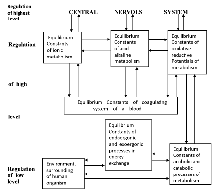
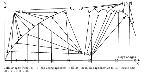
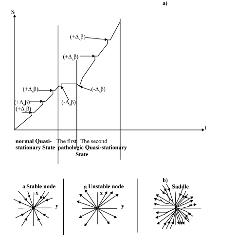
| According to first law of thermodynamics [Q=ΔU+Wint+Wext], Stationary State of open non-equilibrium non-linear thermodynamic system of an able-bodied organism and also Quasi-stationary State of open non-equilibrium non-linear thermodynamic system of a sick organism are characterized by stability of Internal Energy (ΔU) (stable temperature 36.0°C-36.9°C by which all enzymes operate etc.). The mechanisms stability of Internal Energy (ΔU) are maintained by Internal Works (Wint) and External Works (Wext), which generate the Total Heat Energy (Q) dissipating into Environment for maintenance of stability Internal Energy (ΔU). Just stability of Internal Energy promotes stability of Internal Medium (constant concentration substances in blood and in neurolymph) (Figure 1). Mechanisms maintenance stability of Internal Energy (ΔU) and Internal Medium an organism are based on mechanisms regulation of biophysical and biochemical processes, which depend on Internal Works (Wint) and External Works (Wext) of thermodynamic system an organism. Resistances of Stationary State or Quasi-stationary State of an organism to environmental influences are realized by External Works (Wext) of an organism. Hormonal and immune systems are the links of the compound mechanism of an organism’s regulative system. Development of an organism during its life occurs via moderate oscillating shifts of balance anabolic endoergonic and catabolic exoergonic processes into anabolic pathway, and into catabolic pathway which are caused by Internal Works (Wint) of an organism in normal Stationary State [1,2]. These moderate oscillating shifts of balance anabolic endoergonic and catabolic exoergonic processes into anabolic processes and into catabolic processes correspond to positive fluctuations entropy (+Δxβ), and lead to linear graph of development thermodynamic system of an organism, according to Glansdorff–Prigogine stability criterion (Figure 2). Shifts of balance anabolic endoergonic and catabolic exoergonic processes into excessive anabolic processes and into excessive catabolic processes correspond to negative fluctuations entropy (-Δxβ), and lead to non-linear graph of development into pathologic Quasi-stationary State of a sick organism, according to Glansdorff–Prigogine stability criterion [1,2] (Figure 3). Considering interactions between a human organism and environment in normal Stationary State and transition into pathologic Quasi-stationary States, operations of External Works (Wext) of an organism depend on operations of Internal Works (Wint), which is directed by mechanisms regulation of maintenance stability Internal Energy and Internal Medium of an organism [3]. Defensive reactions of an organism against the influences of Environment are realized by External Works (Wext) of an organism, and are maintained by Internal Works (Wint), i.e. the mechanism regulation of maintenance stability Internal Energy and Internal Medium [1-3]. The mechanisms violations of regulative system in some pathological processes were elucidated from the point of view of offered concepts [4-6]. These explanations of pathologic processes gave possibility to eliminate some doubts, and/or queries which were expressed by the authors of the some experiments. Study from the point of view of thermodynamics, biophysics and biochemistry both pathologic processes and normal processes will help to substantiate new approaches for treatment of some pathological processes [4-6]. |
| Structure Regulatory Processes for Maintenance Stability as Stationary State in Norm As Well As Quasistationary States in Pathology |
| Common mechanism of maintenance stability as Stationary State in norm as well as Quasi-stationary States in pathology is divided into three levels of regulative mechanism: highest level regulation, high level regulation and low level regulation (Figure 1). |
| There are the important footnotes: |
| Hormones cause influences on cellular processes and are the links in all levels Regulations: Highest level Regulation, High level Regulation and Low level Regulation of an organism. |
| Enzymes operate in biochemical reactions of all levels Regulations: Highest level Regulation, High level Regulation and Low level Regulation. |
| Low level of regulatory mechanism for maintenance stability as Stationary State in norm as well as Quasi-stationary States in pathology |
| Moderate fluctuating shifts of balance anabolic endoergonic and catabolic exoergonic processes in anabolic pathway and in catabolic pathway occur in low level of regulatory mechanism for maintenance stability Stationary State an organism (Figure 1). Low level regulation stability Stationary State consists of “Equilibrium Constant of energy exchanges” and “Equilibrium Constant of metabolism” [1-3]. “Equilibrium Constant of energy exchanges” reflects mechanism of balance exoergonic and endoergonic processes, and is substantiated indirectly owing to some indices: a) stable index of temperature 36.0°C-36.9°C by which all enzymes operate; b) stable index of pH 7.35 in blood and in neurolymph; c) stable index of osmotic pressure -285 ± 5 mil-osm/kg H2O, corresponding to 0.14-0.15 molar sodium chloride or the other univalent ions; d) stable index of colloidal-oncotic pressure 18-25 mm Hg, corresponding to human serum albumin solution up to 300 grams per liter etc. “Equilibrium Constant of metabolism” is substantiated indirectly owing to balance anabolic and catabolic processes resulting in stable concentrations indices of all substances in blood and in neurolymph. |
| High and highest levels of regulatory mechanisms for maintenance stability as Stationary State in norm as well as Quasi-stationary States in pathology |
| Low and high levels regulations are mutual influenced on each other that cause stability Internal Medium and Internal Energy as an organism, as well as cells of an organism (Figure 1) [3]. Regulative mechanisms of high level regulation consists of “Equilibrium Constant of ionic metabolism”, “Equilibrium Constant of acid-alkaline metabolism”, “Equilibrium Constant of oxidative-reduction Potentials of metabolism” and “Equilibrium Constant of coagulating system of blood” [3] (Figure 1). “Equilibrium Constant of ionic metabolism” is substantiated indirectly owing to balance cations and anions providing stable concentration indices as cations K+, Na+, Ca2+, Mg2+, H+, etc., as well as anions Cl- and General hydrocarbonates {HCO3- (95.8-132.0 mg %)} in blood and in neurolymph. “Equilibrium Constant of acid-alkaline metabolism” is substantiated indirectly owing to stable index pH 7.35 in blood and in neurolymph. “Equilibrium Constant of oxidationreduction Potentials of metabolism” is substantiated indirectly owing to such stable indices: a) stable index of respiratory coefficient 0.7-1.0; b) stable index of ratio partial pressure Oxygen (O2) to partial pressure carbon dioxide (CO2) that is in 4 times more in arterial blood than in venous blood; c) stable index of Mayerhof coefficient from 3 up to 6 of oxygen consumption as by normal tissue, as well as by cancer tissues etc. [7]. “Equilibrium Constant of coagulating system of blood” is substantiated indirectly owing to stable normal indices of blood coagulation. Mutual influences of high level regulation and low levels regulation occur via mutual influences between “Equilibrium Constant of oxidation-reduction Potentials of metabolism” and “Equilibrium Constant of metabolism” [3] (Figure 1). These influences maintain stability both temperature 36.6ºC-37.3ºC in an organism, and constant concentrations of substances in blood and in neurolymph of an organism (Figure 1). All above mentioned Equilibrium Constants are contributed to mechanisms stability as Stationary State in norm, and as well as Quasi-stationary States in pathology. Central nervous system is highest level regulation, which influences as on high level regulation, as well as on low level regulation through high level regulation [3] (Figure 1). |
| Biochemical and Biophysical Processes in Cells Promoting as Stability Stationary State in Norm As Well As Stability Quasi-stationary States in Pathologic States of an Organism |
| Considering mechanism maintenance stability Internal Energy and Internal Medium of an organism as in Stationary State as well as in some Quasi-stationary States, it is necessary to estimate mechanisms occurring in cells which maintain stability of basophilic chemical potential of cellular cytoplasm (μ) [1,2,4]. Interactions between cellular mechanisms and organism’s mechanism for maintenance stability Internal Energy and Internal Medium of an organism operate via moderate oscillating shifts into catabolic pathway, and into anabolic pathways in norm. Excessive shifts into catabolic pathway and into anabolic pathways cause different Quasi-stationary States of an organism. Besides they can promote either excessive inflow substances into an organism with excessive endocytosis or outflow substances from an organism with excessive exocytosis. |
| Participation pro-factors and anti-factors in mechanisms maintenance stability as Stationary State in norm as well as Quasi-stationary States in pathologic States of an organism |
| It is known that anabolic processes operate in G1/S phases of cellular cycle, and both anabolic processes and catabolic processes operate in G2/M phases of cellular cycle. Thus, studying the violations of processes tissues growth, it is necessary to consider, that both pro-proliferative factors and anti-proliferative factors can operate as in G1/S phase of cellular cycle, as well as in G2/M phase of cellular cycle. However, proapoptotic/ pro-autophagy factors and anti- apoptotic/anti-autophagy factors operate in quiescent G0 phase of cellular cycle, which exhibits Stationary State due to balance anabolic and catabolic processes in it. On the one hand, as pro-autophagy factors (Beclin 1 and regulators of autophagy phosphoinositide 3-kinases (PI3Ks) class I and class III etc.), as well as pro-apoptotic factors (pro-apoptotic proteins assemblage of BH3 proteins, Bak, Bax, caspase 3 and caspase 7), operate in catabolic exoergonic pathway. On the other hand, anti-apoptotic factors (Bcl- 2, Bcl-xL, Mcl-1 and also complex AKt/PI3K) reacts as in catabolic pathway, and as well as in anabolic pathway. Generating energy due to processes transphosphorylation via ATP/ADP/AMP, catabolic anaerobic processes of glycolysis are the primer for development of both catabolic exoergonic processes and anabolic endoergonic processes [2,4]. Stimulating glycolysis, AKt pathway is also the primer for both catabolic processes and anabolic processes [2,4]. Catabolic anaerobic processes of glycolysis produce energy, which are divided in “Nodal point of bifurcation of anabolic and catabolic processes in Acetyl-CoA” [NPBac] into anabolic endoergonic pathway and catabolic exoergonic pathway [2-4]. Therefore, glycolysis is the primer all above mentioned as pro-factors as well as anti-factors. |
| Mechanisms maintenance stability Stationary State during a life as each cell of an organism as well as an organism |
| The open thermodynamic systems of each cell and of an organism develop in advance of age terms, moving their metabolisms to the different stationary states: from young ages up to elderly age and up to old age [1] (Figure 2). At young ages, they show positive fluctuations entropy (+Δxβ), according to Glansdorff–Prigogine stability criterion. Progressive interactions of balanced catabolic and anabolic processes contribute to generating energy via catabolic processes and consumption energy into anabolic processes with moderate shifts into anabolic processes, that create positive fluctuation entropy (+Δxβ), and ascending flow of linear graph of Stationary State at young age as cell, as well as an organism [1,2] (Figure 2). Transition into Quasi-stationary pathologic States provokes mainly acute inflammatory processes due to excessive expression catabolic processes at young age. At middle ages, balanced interactions catabolic and anabolic processes are fixed by equilibriums between generating energy with dissipation energy into environment and preserving energy for processes biosynthesis that maintains stability of balance catabolic and anabolic processes. Stability of balance catabolic and anabolic processes contribute to horizontal flow of linear graph of Stationary State at middle ages because equilibrium positive fluctuations entropy (+Δxβ) is drawn side by side with negative fluctuation entropy (-Δxβ) suppressing negative fluctuation entropy (-Δxβ) [1] (Figure 2). Transition into Quasistationary pathologic States provokes mainly chronic inflammatory processes due to excessive expression both catabolic inflammatory processes and anabolic defensive immune processes at middle age. At elderly age and old age, it takes place regressive interactions of balance catabolic and anabolic processes, in which occurs moderate prevalence of catabolic processes with moderate dissipation energy. Such moderate dissipation energy contributes to moderate negative fluctuation entropy (-Δxβ), leading to descending shifts of linear graphs of Stationary State according to Glansdorff–Prigogine stability criterion (Figure 2). Such descending shifts of linear graphs of Stationary State can promote either transiting into excessive negative fluctuation entropy (-Δxβ), leading to Quasi-stationary pathologic States due to decrease of defensive system an organism (immune system, hormonal system etc.), or to natural death due to total exhaustion of energy, i.e. increase gain of entropy according to Prigogine theorem [1] (Figure 2 and 3). At elderly age such transition into Quasi-stationary pathologic States can lead to chronic infectious diseases. Also, transition into Quasi-stationary pathologic States can often provoke malignant Proliferative processes due to excessive expression anabolic processes at elderly age. The life of each cell advances from youth age till old age. At youth age, it occurs as increase production and consumption energy leading to moderate oscillating balance anabolic and catabolic processes, as into anabolic processes as well as into catabolic processes. At old age, it occurs that decrease production and consumption energy leading to decrease as anabolic processes, as well as catabolic processes and finishing in cell death, in which arise gain of entropy due to total exhaustion of energy according to Prigogine Theorem [1]. |
| Role of Autophagy in mechanism maintenance stability Stationary State as each cell of an organism as well as an organism |
| Autophagy takes part in defensive mechanism of an organism, which develops according to changes of fluctuation entropy advancing as an organism life as well as a cell life from infancy till old age. Normal distinctions between Stationary States in various ages of an organism, connecting with the moderate fluctuation entropy, contribute to normal development of an organism according to Glansdorff–Prigogine stability criterion [1,2]. Also, each cell has the specified term of its life which is ended with cell death [1,2] (Figure 2). In the beginning of termination cell life, there are occurred apoptotic processes, which exhibit degradations of nucleus and mitochondria leading to damage of balance anabolic processes and catabolic processes in cytoplasm with ruining stability of cellular basophilic chemical potential (μ) in cytoplasm, i.e. stability Internal Energy and Internal Medium of cell [2-4]. Just cellular chemical potential (μ) determines mechanisms of cellular capacitors operation due to balance chemical potentials (μ) between intracellular and extracellular mediums [1]. Degradation of nucleus is divided into phase nucleus depression, phase nucleus condensing (pyknosis), and phase nucleus fragmenting (karyorrhexis), which lead to destruction of proliferative processes due to damage of anabolic processes. Degradation of nucleus violates nucleus capacitor due to violation balance chemical potentials (μ) between intra-nucleus and extra-nucleus mediums, which induce formation of antiproliferative anti-growth factors. Degradations of mitochondria show degradation of catabolic oxidative processes in cell. Degradations of mitochondria violate the mitochondria capacitors due to violation balance chemical potentials (μ) between intra-mitochondrion and extra-mitochondrion mediums, which cause formation of supplemental pores in mitochondrial membranes, making them more permeable. Cytochrome C is released from mitochondria due to formation of MAC (mitochondrial apoptosis-induced channel) and VDACs (Voltage dependent anion channels) in the outer mitochondrial membrane, inducing formation of pro-apoptosis factors (Bax and/or Bak also caspases 3 and 7) [8-11]. Machinery of Apoptosis leads to cell death and to arising of Autophagy for maintenance stability of Internal Energy and Internal Medium of an organism. The mechanism of transition from Apoptosis into Autophagy occurrs due to such conditions: All living cells of the certain section of an organism (blood, lymph, neurolymph, tissues etc.) are connected between each other via common resonance waves of their capacitors [1], maintaining common stability of Internal Energy both in their section and in an organism. The dead cells do not generate resonance waves due to broken capacitors of cells’ walls. Therefore, the dead cells are out of this common relative resonance waves, and are perceived by living cells as how strange objects. It is necessary to consider that biochemical processes in cells occur after remote reaction across distance due to cellular capacitors operation on the changes of chemical potentials (μ) in surrounding medium caused by strange objects/processes [1]. Remote cellular reactions induce attraction cells to strange substances, as the result of cellular capacitors operation via resonance waves that provokes the cellular contact biochemical reactions [1]. Thus, living cells react via the resonance waves only to the complex substances into the dead cell, but not to the dead cell. These resonance waves provoke autophagy system via rearrangement and adjustment of macrophages, as well as attraction of macrophages to dead cell [1]. Then the remote cellular reactions across distance transit into the contact cellular reactions of autophagy [1]. The three levels autophagy regulations are determined in contact cellular reactions: The first level autophagy regulation promotes formation of autophagosome with double membrane around this organelle. The formation of autophagosome occurs due to attraction dead cell, as Ligand, to variable capacitor of macrophagocyte’s Receptor, with forming complex Receptor-Ligand and internalization of the complex Receptor-Ligand into macrophage through macrophage’s wall. So, it is formed the vacuole with the components of macrophage’s membranes. The transition of the vacuole into autophagosome, which recruits biochemical and biophysical mechanisms, occurs via certain factors with initial pathway at mTOR in first level of regulation and terminating in ATg machinery [8]. Major selective substrate or cargo receptor p62 is recruited to autophagosome’s membrane through interaction with LC3 protein, i.e. through LC3 protein which takes place as the dielectric in autophagosome’s capacitor [1]. Then the protein NBR1, related to p62, is localized into endoplasmic reticulum (ER) of macrophage which participates in autophagosome formation, and is placed independently of LC3. Also, phosphatidylinositol 3-phosphate (PI3-P) binding to ER takes part in autophagosome formation. At second level of regulation, following factors participate how class III phosphoinositide-3-kinase, Vps34, Beclin-1, and so on. Thus autophagosome obtains the own capacitor property in its wall to interfere in the common resonance waves of intracellular medium of macrophage. Then lysosome capacitors react to strange resonance waves of the autophagosome’s capacitor and attract autophagosome in the second level of regulation [8]. It should be drawn attention to membranous proteomics of autophagosome as latex bead-containing (LBC), LC3-II etc., which take place into autophagosome’s capacitor as integrative proteins with dielectric properties. Also, it should be drawn attention to lysosomal-associated membrane protein-1 (LAMP1), which takes place into lysosomal’s capacitors as integrative protein with dielectric property [1,8-11]. Thus, the remote reaction occurs between lysosome capacitors and autophagosome capacitors. Third level of regulation exhibits first maturation of autophagosome and then ruining of autophagosome owing to lysosomal enzymes operation [11,12]. At last stage of Autophagy, there are occurred exocytosis of autophagosome’s ruining Products, and then excretion these Products from an organism for maintenance stability Internal Energy and Internal Medium of an organism [1,4]. Autophagy operates in terminal period of cells life releasing an organism from dead cells [13-17]. Thus, Autophagy takes part in an able-bodied organism as normal physiological cleaning process, which is the link of mechanism maintenance stability Internal Energy and Internal Medium of an organism. Generally, Autophagy processes occur in catabolic exoergonic pathway, i.e. oxidizing processes of decomposing molecules of dead cells. Researching autophagosome formation Itakura and Mizushima [15] and Axe et al. [16] have found that nonselective engulfment cytoplasmic substances of dead cell is created by macrophages of selective recognizing of autophagosome membrane. Really, the recognition cytoplasmic substances of dead cell located in autophagosome occurs by capacitors of macrophages, which capacitance defines as the complex of specific proteins and organelles in autophagosome, as well as the change of the chemical potential (μ) of autophagosome’s medium defining the charge on internal membrane of the autophagosome [1]. Then, the contact reactions of the decomposing substances of autophagosome occur in macrophage. Oscillating shifts of balance catabolic and anabolic processes into anabolic pathway, and into catabolic pathway, define oscillations anti-apoptosis/antiautophagy processes and pro-apoptosis/pro-autophagy processes, which produce relevant factors: anti-apoptotic proteins - Bcl-2, Bcl-xL, Mcl-1 and pro-apoptotic proteins-assemblage of BH3 proteins, Bak, Bax, caspase 3 and caspase 7; and also pro-autophagic protein-Beclin 1 and regulators of autophagy phosphoinositide 3-kinases (PI3Ks) class I and class III, etc.. There are the normal cellular reactions which promote maintenance stability Internal Energy and Internal Medium of an ablebodied organism. Strict sequences of all these processes during life both an organism and cells of an organism are preserved by permanent influences of environment, which can cause permanent moderate changes of external chemical potential (μ external), with changing charges on outer cellular membranes (OCM) of cells walls and violating operation of cellular capacitors. However, shift balance catabolic and anabolic processes into catabolic exoergonic processes induce resistance to these environmental influences, preserving stability chemical potential of cytoplasm (μcytopl) and preventing changing charges on outer cellular membranes (OCM) of cells walls. Intracellular biochemical reaction with oxidizing processes of decomposing strange objects leads to exocytosis and excretion the strange objects from an organism into environment [1-4]. |
| Maintenance Stability Internal Energy And Internal Medium in Pathologic Quasi-stationary State of an Organism |
| Biochemical and biophysical driving mechanisms for maintenance stability of Stationary States of an able-bodied organism differ from driving mechanisms which maintain stability Internal Medium and Internal Energy of an organism in cases of pathologic processes (Figure 3) [18-20]. Excessive negative fluctuation entropy (-Δxβ), according to Glansdorff and Prigogine theory, plunge an organism into pathological Quasi-stationary State [1-6] (Figure 3). |
| Maintenance stability Internal Energy and Internal Medium by defensive mechanisms in inflammatory processes and infection diseases of an organism |
| Stability of Internal Medium and Internal Energy both an organism and cells of an organism is maintained by balance catabolic and anabolic processes, which determine cellular capacitors operations via interactions between intracellular and extracellular balances of catabolic and anabolic processes defining the charges on inner cellular membrane (ICM) and on outer cellular membrane (OCM) for cellular capacitors operation [1]. Catabolic exoergonic processes promote maintenance stability temperature of an organism 36.0°C-36.9°C, by which all enzymes operate, via dissipation of generating energy into environment and carry out cleaning role of Internal Medium an organism, as from strange cells as well as from strange substances and microorganisms, etc. However, unlike the normal situation of Autophagy, an organism in pathologic states is compelled to include especial mechanisms as against the strange objects, as well as against the harmful substances (toxins, harmful ions, harmful enzymes etc.), in order to defend balance catabolic and anabolic processes preserving biochemical structures of Internal Medium and Internal Energy an organism [21-24]. This defensive function fulfils immune system an organism, as via humoral mechanisms (immune antibodies, etc.), as well as via cellular mechanisms (phagocytosis). Immune system creates excessive shifts of balance catabolic and anabolic processes into catabolic processes for destruction of strange substances, and then excretion of oxidized decomposed product into environment. These excessive shifts of balance catabolic and anabolic processes into catabolic processes promote huge dissipation energy and substances into environment, causing huge increase of body temperature in processes of inflammation or infectious diseases [2]. Penetration of strange agent (virus, microbe, etc.) provokes the changes of chemical potential (μ), as in some cells of an organism, as well as in Internal Medium of an organism, and induces cellular capacitors to react to this strange agent that leads to expression of the defensive immune reactions: both phagocytosis and humoral immune reactions [2]. Strange objects (microbes or viruses) should be eliminated from an organism for maintenance stability Internal Energy and Internal Medium of an organism. Immune processes of cleaning Internal Medium an organism are carried out by immune responded cells, named phagocytes. Just cellular variable capacitors in the cellular Receptors of phagocytes find out these strange objects owing to remote reactions across distance and cause attraction phagocytes to the strange objects. The strange object, named as Ligand, is bound with cellular Receptor of phagocyte, making complex Receptor-Ligand. Then, it is occurred the contact reactions: internalization complex Receptor-Ligand into phagocyte; releasing lysosome enzymes due to remote reaction of lysosomes’ capacitors to the complex Receptor- Ligand; lysis of the strange object by lysosome enzymes via oxidizing processes; exocytosis Products of oxidizing processes. Processes decomposing molecules of the strange object occur in catabolic exoergonic pathways. Oka et al. [25] and Chiang et al. [26] researched influences on Mitochondria damage of Autophagy/lysosome system in pathologic processes of cardiomyocytes, and also pro-inflammatory factors (prostaglandins and leukotrienes) and specialized pro-resolving mediators (SPMs) in microorganisms. Just huge dissipation of energy into environment promotes expression of entropy, which can cause mitochondria damage of Autophagy/lysosome system in pathologic processes of cardiomyocytes, inducing pro-inflammatory factors. It is necessary to consider that biochemical processes in cells occur after remote reaction of cellular capacitors on strange objects. Such remote cellular reactions sensibilize macrophages-phagocytes to strange objects and induce attraction phagocytes to strange objects, creating contact biochemical cellular reactions of decomposing these strange objects [1]. Excessive shifts of balance catabolic and anabolic processes into catabolic processes cause excessive exocytosis, which transits into excessive endocytosis promoting increase anabolic processes. Increase anabolic processes causes increase defensive reaction of an organism via promoting biosynthesis of immune antibodies [1,2,4]. As opposed to autophagosome in healthy organism, phagosome consists of lipid body around this organelle, which can be formed either as defense of phagosome from ruining mechanisms of phagocyte, or as defense of phagocyte from the harmful substances of phagosome (toxins, harmful ions, harmful enzymes, etc.) [27]. Phagocytes produce either free antibodies into blood (IgM, IgG, antibodies’ Anti-peptidoglycan), or the special antibodies against phagosome’s pathogenic agent due to cellular variable capacitors operation. These processes are induced by phagocytes’ Fcy receptors and toll-like receptors 4, which are stimulated by microorganism’s cellular capacitors with LPS protein as dielectric into the microorganism’s cellular wall [28-31]. Moreover a phagocyte preserves capability to produce complement which possess of esterase properties and can ruin lipid body of phagosome [27,28]. Final mechanism eliminating strange objects from Internal Medium of an organism can occur either via immune processes of catabolic oxidizing processes with excretion of the products oxidizing processes. However, if formed, high-molecular substances cannot be decomposed via catabolic oxidizing processes by enzymes of an organism, alternative excretion of these high-molecular substances occurs within phagocytes, which are ruined owing to the damaged structure of their cells. Thus, it is formed abscess with pus, which must be broken releasing an organism from the pus and from the strange high-molecular substances or microorganisms. |
| Mechanisms maintenance stability Internal Energy and Internal Medium in cancer cells and cancer tissue |
| Excessive shifts of balance catabolic and anabolic processes into anabolic processes promote the huge consumption of Acetyl-CoA with energy for anabolic processes and partial suppression catabolic processes causing malignant processes (cancer, sarcoma, leukemia etc.), which exhibit Apoptosis Resistance due to irrepressible proliferative processes [2,4]. Unlike inflammation and infection processes, the shifts balance catabolic and anabolic processes into excessive anabolic endoergonic processes cause excessive advance of cellular cycle, exhibiting excessive proliferative oncologic processes. Macrophages’ cellular capacitors do not perceive cancer cells as the strange objects, despite of existence of oncogenic viruses in these cells, and these diseases exhibit very weak immune reactions against malignant cells. Mechanisms of proliferative oncologic processes show the huge expression of anabolic endoergonic processes, with partial suppression of catabolic exoergonic processes. As a matter of fact, catabolic processes of glycolysis carry out peculiar functions, unlike the subsequent catabolic processes after bifurcation of anabolic and catabolic processes in “nodal point of bifurcation anabolic and catabolic processes [NPBac]” [4]. Just catabolic processes of glycolysis generate energy. This energy is divided into anabolic and catabolic processes in NPBac, but the part of this energy is accumulated into Lactic acids for abundance anabolic processes of cancer metabolism [4]. Some quantity energy is generated in catabolic pathway of Krebs cycle maintaining stability of Internal Energy as basophilic chemical potential in cytoplasm (μ) for survival of cancer cell. However, even partial suppression of catabolic anaerobic processes in cancer tissue leads to accumulation of excessive quantity oxygen in cancer cells mitochondria due to permanent aerobic catabolic processes via cellular mitochondrial cytochrome and system of haemoglobin in an organism. Catabolic aerobic pathway occurs in mitochondria via system cytochrome, which accept oxygen and promote oxidation of the products metabolism down to CO2 and H2O with dissipation energy in environment for maintenance stable temperature 36.6ºC- 37.2ºC by which all enzymes operates. Thus, even partial suppression anaerobic catabolic processes leads to forming of excessive quantity Reactive Oxygen Species [ROS] in mitochondria of cancer cells, unlike moderate quantity Reactive Oxygen Species [ROS] in able-bodied cells. Generated by Reactive Oxygen Species [ROS], the excessive quantity of superoxide [O2*] produce free radicals (*OH), which can ruin mitochondria of cancer cells. However, cancer cells metabolism demonstrates property of Apoptosis Resistance. It is meant that free radicals generated by ROS are neutralized in some metabolic processes. It can be realized either by the redox transformation of glutathione disulfide for free radical’s elimination [32], or by the use of free radicals for advance cellular cycle, causing irrepressible proliferative processes of cancer cells [33-36]. Thus, the mechanism of neutralization free radicals occurs due to their participation in mechanism of nuclear DNA replication. Free radicals (*OH) promote separation nuclear DNA from new DNA in process of nuclear DNA replication, and were neutralized in process of nuclear DNA replication. Excessive quantity ROS in cancer cells’ mitochondria, as opposed to normal cells’ mitochondria, cause irrepressible proliferative processes in cancer cells metabolism [4,33-36]. ROS in small quantity fulfils the same role in able-bodied cells of G2 phase cellular cycle exerting normal process of nuclear DNA replication. Just accelerating cellular cycles in cancer tissue consume a lot of substances and energy for excessive anabolic processes in G1/S phases cellular cycles, and then it occurs, distribution this energy due to nuclear DNA replication in G2 phase cellular cycle transiting in M phase of cells division [4]. |
| Benefits of using prolonged medical Starvation for the new approach to cancer therapy |
| Taking into account mechanism of suppression catabolic anaerobic processes in cancer tissue, the new method cancer disease treatment, which use Prolonged medical Starvation 42 -45 days, gives chance to suppress anabolic processes due to increase of catabolic processes, both in an organism metabolism and in cancer metabolism. This method cancer treatment causes suppression cancer metabolism via targeting Warburg effect leading to depression of cancer development that gives possibility to use efficient cancer therapy, with considerably decreased dosage of cytotoxic drugs for preserving immune and hormonal systems of an organism and prevention of recurrences cancer disease after long treatment with cytotoxic therapy [5,6]. |
| Reviews From The Point of View of the Offered Concepts of Results of Some Researchers Studying Mechanisms Pathologic Processes |
| Reviews of results researches studying inflammatory processes and Infectious diseases |
| Studying mechanisms development inflammatory processes in immune response to Gram (+) infection, Sun et al. [29] described the role of anti-peptidoglycan antibodies and Fcy receptors as key mediators of inflammation in Gram-positive sepsis. Studying immune response to Gram (-) infection, Guha and Mackman [37] described Lipopolysaccharide (LPS (endotoxin)), the component of bacterial Gram-negative outer membrane, which is recognized by monocytes and macrophages of innate immune system. Indeed, both these researches showed role of remote reactions, causing by cellular capacitors, in immune processes of an organism on intrusion as Gram-positive microbes as well as Gram-negative microbes. Just anti-peptidoglycan antibodies and Fcy receptors are components of organism’s cellular capacitors, and bacterial Lipopolysaccharide (LPS (endotoxin)) is recognized by cellular capacitors via remote reaction and resonance waves of monocytes [1,29,37]. Biddie et al. [38] have written that Ligand-dependent transcription creating by nuclear glucocorticoid receptor (GR) is mediated by activator protein 1 (AP1), i.e. AP-1 is a major partner for productive GR-chromatin interactions. Then, they have noted that these interactions remain unclear. Researching inflammatory processes induced by Escherichia coli, Lu et al. [39] studied immunoglobulin-like receptor, subfamily A, member 2 (LILRA2), which induce pro-inflammatory cytokines, and queried “whether LILRA2 functions distinct from other receptors of the innate immunity, including Toll-like receptor (TLR) 4 and FcγRI remains unknown”, in spite of modulation functions of TLR7/9 by LILRA2 receptor in dendritic cells. They also noted that the effects of LILRA2 on TLR4 and FcγRI-mediated monocyte functions are not elucidated [39]. Besides, Lu et al. [39] showed that LILRA2 activates monocytes and increased GM-CSF production, i.e. stimulating hematopoietic systems of granulocyte, T-cells, B-cells, macrophages, fibroblasts by glycoprotein as growth factor. Simultaneously, they noted that LILRA2 induces similar amounts of IL-6, IL-8, G-CSF and MIP-1α, but lower levels of TNFα, IL-1β, IL-10 and IFNγ, and does not induce IL-12 and MCP-1 production up-regulating by LPS, and suggested that LPS functions distinct from TLR4, i.e. the other LPS is not recognized by the Toll-like receptor4 (TLR4) [39]. It is meant that antigen LPS in outer membrane of Gram-negative microbes is the other antigen than in outer membrane of Gram-positive microbes. Taking into account data of Biddie et al. [38] and Lu et al. [39] researches, it is possible to offer concept of different pathways inflammatory mechanisms induced by either Gram-positive microbes or Gram-negative microbes. Grampositive microbes, being carrier of strange substances as antigens, because violation of both “Equilibrium Constants ions exchanges” and “Equilibrium Constants oxidative-reduction exchanges” that leads to shift of balance catabolic and anabolic processes in “Equilibrium Constant of metabolism” into excessive catabolic processes or to acute inflammatory processes (Figure 1). The capacitors of cellular receptors as neutrophil leucocytes and eosinophil leucocytes, as well as T-lymphocytes and NK cells are rearranged to antigenic properties of Gram-positive bacteria. Then, it occurs interaction between cellular capacitors of immune cells and Ligand-dependent transcription creating by the nuclear glucocorticoid receptor (GR), mediated by the activator protein 1 (AP1) for GM-CSF production [38]. These interactions cause remote reactions with resonance waves between the immune cells and antigens PGN (Peptidoglycan) of Gram-positive bacteria wall and create attraction of these cells to Gram-positive bacteria, as result of remote reaction and resonance waves operation between the immune cells and the bacteria’s substances [1,2,29]. Then, contact reactions are carried out by phagocytosis of Gram-positive bacteria due to excessive catabolic processes of immune defensive reactions. The shift of balance catabolic and anabolic processes into excessive catabolic processes causes violation stability Internal Energy of an organism showing excessive increase of temperature of an organism. Such acute inflammatory processes can happen either via expression of general inflammatory processes or via formation of local isolated suppurative focus in an organism. General inflammatory processes can cause septic state of an organism. Local isolated suppurative focus isolates phagocytes with microbes into pus, and connective tissue barrier separates suppurative focus from an organism. These acute inflammatory processes have different mechanisms of contact biochemical reactions. The mechanism of excessive catabolic processes stimulates expression of anabolic processes that contribute to some decrease of temperature an organism, and to expression of immune defensive system, maintaining stability Internal Energy and Internal Medium of an organism. Expressions of anabolic processes stimulate action cellular Fcy receptor’s capacitors of B-leucocytes and monocytes, which induce syntheses IgG antibodies exerting immune process in humoral liquids of an organism (blood, lymph, neurolymph, tissue liquid) [2,29]. Gram-negative bacteria can cause as acute inflammatory processes, and as well as chronic inflammatory processes. The mechanisms of acute inflammatory processes induced by Gram-negative bacteria are the same as induced by Gram-positive microbes. Also, immune cells are rearranged corresponding to antigenic properties of Gram-negative bacteria due to interaction between cellular capacitors of immune cells and Ligand-dependent transcription factor, creating by the nuclear receptor glucocorticoid receptor (GR), mediated by the activator protein 1 (AP1) [38]. However, these processes progress less vigorously because Gram-negative bacteria have antigens which exhibit similar biochemical properties to the active pro-/anti-inflammatory factors of an organism [37]. Moreover, Gram-negative bacteria can also cause violation of both “Equilibrium Constants ions exchanges” and “Equilibrium Constants oxidativereduction exchanges”, shifting into excessive reduction processes that leads to shift of balance catabolic and anabolic processes into excessive anabolic processes, unlike Gram-positive microbes (Figure 1). Then, the excessive anabolic processes, induced by Gram-negative bacteria, exert the proliferative pathway, and Gram-negative bacteria are located within new maturated cells forming chronic inflammatory processes. Besides, Gram-negative bacteria inducing excessive anabolic processes stimulate biosynthesis of antibodies against antigens of Gram-negative bacteria. Thus, the mechanisms of inflammatory processes, induced by Gram-negative bacteria, have the several pathways: acute infectious disease, chronic infectious disease, bacteria carrying and immunity against these Gram-negative microbes. Thus, immunoglobulin-like receptor (LILRA2), inducing pro-inflammatory cytokines, Toll-like receptor (TLR) 4 and FcγRI receptor can operate via different pathways of chronic inflammatory processes [39,40]. |
| Reviews of results researches studying oncological diseases |
| Studying mechanisms of proliferation in malignant cells, Elstrom et al. [41] has surprised that proliferation of malignant cells did not increase in culture with the activated serine/threonine AKT kinase, though there was stimulation of glucose consumption in the transformed cells, without affecting the rate of oxidative phosphorylation. Indeed, the activated serine/threonine AKT kinase stimulates glycolysis, which is the primer for both anabolic processes and catabolic processes. The stimulation of glucose consumption in the transformed cells indicates expression anabolic processes with partial suppression of catabolic oxidative phosphorylation. However, anabolic processes occur also in the stationary state of normal tissue in cellular quiescence G0 phase cellular cycle, which does not require a lot of energy for the moderate biosynthesis of simple substances, which can be excreted via oxidizing and exocytosis processes [2,4]. Unlike cellular quiescence G0 phase cellular cycle, the malignant proliferative processes require considerably more energy accumulated in lactic acids for biosynthesis of compound high-molecular substances in G1/S phases cellular cycle, which cannot be excreted from the cell via exocytosis [2,4]. The exocytosis of the high-molecular substances via “Alternative excretion” occurs in malignant proliferative processes, owing to division cell through G2/M phases cellular cycle and distribution these nighmolecular substances among the new cells [2,4]. Driving mechanisms exerting anabolic processes for advance of cellular cycle in proliferative processes are located in nuclei of cells. There are two mechanisms of the driving mechanisms in nuclei: a) The result of normal changes nucleus metabolism due to natural development cellular cycles with moderate shift metabolism into moderate anabolic processes, which influence on development normal cellular cycle, i.e. each cell is limited with only 50 time divisions. b) The result of incorporation into nucleus the strange gene, i.e. oncogene, which changes nucleus proliferative program of cellular cycle into viral accelerative proliferative program, and viral accelerative proliferative program causes shift metabolism into excessive anabolic processes in cancer tissue. Resistance between these driving mechanisms defines pathways of proliferative processes: either v-oncogene is acclimatized into nucleus changing nucleus proliferative program into excessive anabolic processes with partial suppression catabolic processes, or v-oncogene is not acclimatized into nucleus because insufficiency of respiratory mechanism in cells culture leads to preservation certain level of catabolic processes inhibiting excessive increase of anabolic processes. Therefore, expression anabolic processes of malignant cells in cellular culture occur only in moderate level without overload “nodal point bifurcation anabolic and catabolic processes [NPBac]–in Acetyl-CoA” due to sufficiently of Acetyl-CoA. Hence, anabolic processes of malignant cells induce only biosynthesis of some simple substances in cells culture [2,4]. Bellacosa et al. [42] have exclaimed surprise that tumor cells rarely display increase size in comparison to their normal counterpart, in spite of the mTOR/cIF4E pathway that is often activated in human tumours. However, additional increases of quantity Acetyl-CoA are not formed in these mechanisms, and therefore, it fails strengthening of anabolic endoergonic processes and increase of proliferation [2,4]. Bonnet et al. [43] studied aerobic glycolysis of Warburg effect, and have found high mitochondrial membrane potential and low expression of the K+ channel, contributing to apoptosis resistance. Really, the excessive anabolic processes in cancer tissue promote some increase mitochondrial membrane potential (μ) for cancer cells proliferation, causing their survival in condition of suppression catabolic anaerobic processes. Suppression of K+ channel due to increase endocytosis substances for development cellular cycle in G1/S phases results in proliferative processes leading to growth tumor [2-6]. Proliferative processes distribute intracellular high-molecular substances between new cells that promote apoptosis resistance of cancer tissue and contribute to mechanism of metastasis, creating “Alternative excretion” these substances within metastatic cells [2,4]. So, the excessive anabolic processes cause apoptosis resistance accumulating energy in lactic acid for anabolic processes of cancer tissue, unlike catabolic processes which disseminate energy into environment and increase entropy contributing to apoptosis [1,2,4]. Plas et al. [44] and Plas and Thompson [45] researchers are connected with influence of AKt, and also Bcl-xL on growth processes and on cells survival. They have found that AKt and Bcl-xL cause anti-apoptotic effect by the different mechanisms, although both AKt and Bcl-xL stimulate growth processes. So AKt increases glucose transporter expression, glycolytic activity, mitochondrial potential and cell size, maintaining a physiologic mitochondrial activity and mitochondrial potential [44]. On the contrary, Bcl-xL creates the smaller cells, which have lower mitochondrial potential, reduced glycolytic activity, and are less dependent on extracellular nutrients [44]. Such different mechanisms of anti-apoptotic effects between AKt and Bcl-xL characterize difference development cancer processes in different tissues of an organism. Really, the necessity of extracellular nutrients and correspondingly glycolytic activity are differed by cells in different tissues of an organism even in norm. Hence, the cells of the tissues with increased as glycolytic activity, as well as necessity of extracellular nutrients and mitochondrial activity (expressed mitochondrial potential) have bigger cell size than the cells of the tissues with decreased as glycolytic activity, as well as necessity of extracellular nutrients and mitochondrial activity (reduced mitochondrial potential), even in norm. However, the mechanisms of proliferative processes are the same in different tissues, in spite of the different level of metabolism in them. These peculiarities of different tissues reflect also on different development cancer diseases in different tissues an organism. Shift of balance anabolic and catabolic processes into excessive anabolic processes consume huge quantity energy and Acetyl-CoA, partial suppressing catabolic processes, that accelerate cellular cycle and promote proliferative processes, i.e. growth processes, in cancer cells. However the activity catabolic oxidative processes in mitochondria are also remained for survival cancer cells, requiring maintenance stability temperature 36.6ºC-37.5ºC for enzymes operating, and as well as for advance of G2/M cellular cycle due to ROS/O2*/H2O2/free radicals (*OH) activity in nDNA replication [4]. However, the small cells with low expression of AKt and glycolytic activity cannot use AKt for stimulation anti-apoptotic mechanisms. Therefore, these cells use the other mechanism with Bcl-xL factor for anti-apoptotic effect. Investigating Akt activity in growth effect, Plas DR, Thompson CB note that the Akt/PKB protein kinase family activity increase cells size, metabolism and cells survival, as through phosphorylation-dependent inactivation of tumor suppressors, as well as by stimulating mTORdependent increases in protein translation via transport and metabolism of both glucose and amino acids [45]. Indeed, Akt/PKB protein kinase family stimulates glycolysis, which is the primer of both anabolic processes and catabolic processes. Cancer metabolism causes excessive anabolic endoergonic processes via supply glucose for glycolysis, filling G1/S phases cellular cycle with substances and energy and stimulating mTOR-dependent increases in translation processes of biosynthesis protein in G1/S phases cellular cycle [2,4]. Also, excessive anabolic processes in cancer metabolism cause partial suppression catabolic exoergonic processes, remaining some of these processes for both oxidative phosphorylation-energy generating and cells survival via dissipation energy into environment for maintenance stability temperature 36.6ºC-37.5ºC, by which all enzymes operate, and as well as for G2/M cellular cycle development [2]. This mechanism elucidates also the results of Pedersen PL experiments that mitochondria interact with the activated hexokinase 2 (HK-2) in cancer metabolism, resulting in suppression of cell death while supporting cell growth via enhanced glycolysis keeping cell life, even in the presence of oxygen (Warburg effect) [46]. Kim JW, Dang CV studied the mechanism pyruvate dehydrogenase (PDH) and pyruvate kinase (PDK) interaction, and came to conclusion: PDK1 is identified as a direct HIF-1 target gene in hypoxic cells [47]. PDK1 phosphorylates and inactivates the mitochondrial pyruvate dehydrogenase (PDH) complex. Suppression of PDH by PDK1 partial inhibits the conversion of pyruvate to Acetyl- CoA, thereby attenuating mitochondrial respiration function. So, Kim JW, Dang CV hypothesize that PDK1 levels may be up-regulated in nonhypoxic tumor cells by HIF, which would divert pyruvate from PDH and results in the increased lactate production [47]. Indeed, the shift into anabolic metabolism is characteristic for cancer metabolism. Anabolic processes prevail considerably over catabolic processes in cancer tissue. Therefore, the excessive anabolic processes contribute to suppression of PDH by PDK1 for partial inhibition the conversion of pyruvate to Acetyl-CoA, providing the endoergonic conversion pyruvate into lactic acids, in which energy were accumulated for huge anabolic processes, in condition of glycolysis metabolism in cancer tissue [4]. As concern with regard to mitochondria, full attenuating mitochondrial aerobic functions are not occurred. Mitochondrial aerobic function leads to forming excessive Reactive Oxygen Species [ROS] in cancer tissue due to some suppression of catabolic anaerobic processes. Oxygen superoxide (O*) with free radicals (*OH) from ROS are used in G2/M phases for advance of nDNA replications in proliferative processes and irrepressible tumor growth [4]. Investigating role glycolytic enzymes in various processes an organism, Kim JW, Dang CV noted that the multifaceted roles of glycolytic proteins link glycolysis and transcription processes, which are established directly through enzymes [48]. Just mechanism operating any enzyme is subjected to the certain Equilibrium Constant of chemical reaction according to Guldberg–Waage formula and “Law of action mass” and also Michaelis–Menten formula [2]. However, any enzyme is subjected to enzyme activators and enzyme inhibitors according Michaelis– Menten formula. The expression of glycolytic enzymes in glycolysis is induced by their activators, but the expression of anabolic processes is induced by the suppression of glycolytic enzyme PDH by PDK inhibitor for lactic acid cumulation. The stability of new balance between anabolic and catabolic processes in cancer tissues is formed and maintained by the shift to excessive anabolic processes for proliferative processes, which contribute to Apoptosis Resistance of cancer tissue. However, all processes in an organism were regulated via three levels regulation for maintenance stability of pathologic Quasi-stationary State of cancer disease in the organism (Figure 1 and 3). The final violation of stability of Quasi-stationary State is resulted in the changes in high level regulation of Equilibrium Constants especially “Equilibrium Constant of oxidative–reductive Potentials”, which were transmitted into low level regulation of balance catabolic exoergonic and anabolic endoergonic processes in critical stage of cancer disease leading to organism death (Figure 1). Hence, multifaceted roles of glycolytic enzymes maintain these regulative pathways: The stable balance anabolic and catabolic processes in new condition of diseases influence as on catabolic processes of glycolysis for energy generating, as well as on anabolic processes for energy consumption in proliferative processes. Studying Warburg effect, Dang CV described functions both Myc gene, which encodes Myc protein and is a transcriptional factor, and hypoxia-inducible factors (HIFs), which induce suppression mitochondrial respiration via the inducing glycolysis and expression pyruvate dehydrogenase kinase 1 (PDK1), which convert glucose into lactic acids with the decrease in mitochondrial respiration [49]. The cause of rivalry between lactate production and mitochondrial respiration is the cancer metabolism with mechanism of Warburg effect: The oncogene operations results in the shift balance catabolic exoerginic and anabolic endoergonic processes into the excessive anabolic endoergonic processes, which consume a lot of energy and Acetyl-CoA for anabolic processes, simultaneously accumulating energy in lactate for huge anabolic processes in condition of glycolysis, overloading “nodal point of bifurcation of anabolic and catabolic processes [NPBac]” due to lack Acetyl-CoA, and partial suppressing advance metabolism through Krebs cycle to mitochondrial respiration for expression proliferative processes of cancer irrepressible growth [4]. However, the some catabolic exoergonic processes remain for cancer cells survival, exhibiting aerobic glycolysis of Warburg effect [4]. |
| Conclusions |
| 1. Mechanism of maintenance stability of Internal Energy and Internal Medium an organism is exerted by the mechanism of regulation biophysical and biochemical processes which structure consists of the three levels: highest level, high level and low level. |
| 2. Living cells of the certain sections of an organism are connected between each other via relative resonance waves of their capacitors, maintaining common stability of Internal Energy and Internal Medium both in their sections, and in an organism. |
| 3. Dead cells do not generate cellular waves due to broken system of cellular capacitors, and remote cellular reactions macrophages of autophagy system induce attraction to dead cells of macrophages, and create contact biochemical cellular reactions of Autophagy for decomposition and excretion these substances, cleaning of Internal Medium an organism. |
| 4. Remote reactions of phagocytes’ capacitors to a strange pathologic object lead to phagocytes’ sensibilization for the expression of defensive immune reactions, which transit into contact immune reactions, either phagocytosis or humoral immune reaction for decomposing pathologic object. |
| 5. Excessive shifts balance catabolic and anabolic processes either into catabolic processes or into anabolic processes define transition of an organism from normal development into pathologic development, either into inflammation/infection, or into pathologic proliferative malignant processes. |
| 6. Distinction between the mechanisms of inflammation/infection induced by Gram(+) microbes and mechanisms forming chronic diseases, bacteria carrying and immunity inducing by Gram-negative bacteria is depended on distinctions of integrative proteins properties between cellular capacitors of Gram(+) and Gram(-) microorganisms. |
| 7. Excessive shift balance of anabolic and catabolic processes to catabolic exoergonic processes leads to inflammatory processes, with dissipation energy into Environment and increase of a body temperature, expression phagocytosis causing also damage both infectious agent and phagocytes, which are characteristic diseases as infectious processes, inflammations, pyesis, etc. |
| 8. Excessive shift balance of anabolic and catabolic processes to catabolic exoergonic processes leads to inflammatory processes with expression defensive immune reactions owing to the transit of catabolic exoergonic processes into anabolic endoergonic processes. |
| 9. Excessive shift of balance anabolic and catabolic processes to anabolic processes in cancer tissue results in formation excessive proliferative processes, irrepressible cancer growth, unhealed cancer wounds, metastasis and cancer cells Apoptosis Resistance. |
| Acknowledgement |
| This article is dedicated to the memory of TM Ponizovskaya. |
References
|
Figures at a glance
 |
 |
 |
| Figure 1 | Figure 2 | Figure 3 |
Relevant Topics
- Analytical Biochemistry
- Applied Biochemistry
- Carbohydrate Biochemistry
- Cellular Biochemistry
- Clinical_Biochemistry
- Comparative Biochemistry
- Environmental Biochemistry
- Forensic Biochemistry
- Lipid Biochemistry
- Medical_Biochemistry
- Metabolomics
- Nutritional Biochemistry
- Pesticide Biochemistry
- Process Biochemistry
- Protein_Biochemistry
- Single-Cell Biochemistry
- Soil_Biochemistry
Recommended Journals
- Biosensor Journals
- Cellular Biology Journal
- Journal of Biochemistry and Microbial Toxicology
- Journal of Biochemistry and Cell Biology
- Journal of Biological and Medical Sciences
- Journal of Cell Biology & Immunology
- Journal of Cellular and Molecular Pharmacology
- Journal of Chemical Biology & Therapeutics
- Journal of Phytochemicistry And Biochemistry
Article Tools
Article Usage
- Total views: 15648
- [From(publication date):
November-2013 - Apr 03, 2025] - Breakdown by view type
- HTML page views : 11069
- PDF downloads : 4579
