The Mastitis Carcinomatosa, a Case Report
Received: 18-Dec-2021 / Manuscript No. roa-21-50169 / Editor assigned: 20-Dec-2021 / PreQC No. roa-21-50169(PQ) / Reviewed: 12-Jan-2022 / QC No. roa21-50169 / Revised: 17-Jan-2022 / Manuscript No. roa-21-50169(R) / Published Date: 24-Jan-2022 DOI: 10.4172/2167-7964.1000360
Abstract
The mastitis carcinomatosa or inflammatory breast is an aggressive form of mammary Tumors, it can be found in the usual different histological types with a low survival rate compared to that of other types of cancer, The diagnosis is mainly based on clinical and radiological criteria, MRI is more reliable in assessing tumor size than clinical examination, The most common presentation is a poorly limited heterogeneous infiltrating mass associated with thickening. The 5-year survival rate range 19-59% depends on the tumor stage. The treatment of inflammatory breast cancer is multidisciplinary, combining neoadjuvant chemotherapy followed by loco regional treatment.
Keywords
Mastitis; Carcinomatosa; Breast; Cancer; Inflammatory
Introduction
The mastitis carcinomatosa or inflammatory breast cancer (IBC) is an aggressive form of mammary tumors, with a capacity for embolization of vascular and lymphatic structures, which would explain the low survival rate compared to that of other types of cancer.
Case Report
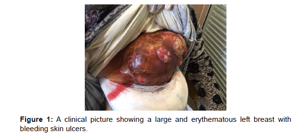
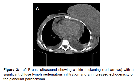
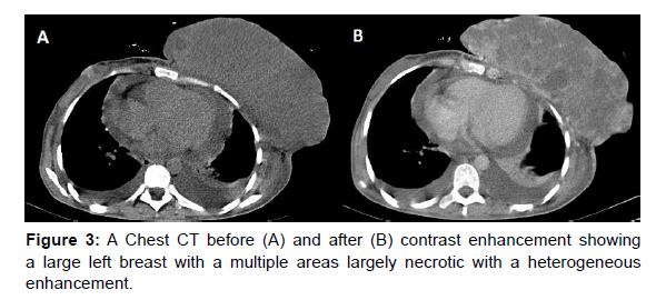
We report a case of 30-year-old female patient with no particular history ,she is followed in our department for an Invasive Ductal Carcinoma of the left breast, having benefit from neoadjuvant chemotherapy, the evolution was marked by the installation of a large, hot, erythematous, and painful breast with skin ulcers (Figure 1), Breast ultrasound finds a skin thickening with significant diffuse lymph oedematous infiltration of the breast (Figure 2), chest- CT shows a large left breast with a multiple necrotic areas and a heterogeneous enhancement without individualization of a solid mass. (Figure 3)
Discussion
The Carcinomatous mastitis or inflammatory breast cancer (IBC) is a serious and aggressive form of mammary Tumors, It is a rare unit, represents between 1 and 5% of breast cancers, with incidence constantly increasing for several decades and Africa North is individualized by a higher rate [1].
Sir Charles Bell was the first to describe it in 1814. Lee and Tenenbaum were the first to use the term “inflammatory breast cancer” in 1924 [2].
Several theories have been described in its pathogenesis mainly the viral theory including the mammalian mouse virus, which is a ubiquitous virus in North Africa transmitted by the orofecal route. Several patients with inflammatory breast cancer have been found to carry this virus. The hormonal theory and genetic theory seem unconfirmed and no specific abnormalities have been identified.
The IBC is not a specific of histological subtype and can be found in the usual different histological types. It may be present such as invasive ductal, lobular or medullary carcinoma and generally is a poorly differentiated tumor of high prognostic grade, with a low survival rate compared to the other types of cancer. IBC has a high capacity for early invasion of vascular and lymphatic structures, particularly in the dermis, which would explain the high metastatic potential, and a great number of patients die within 1 to 2 years, with disseminated metastases. The average age of diagnosis varies between 45 and 57 years,
The rate of initially metastatic inflammatory breast tumor is very high. It varies from 17 to 36% depending on the study, which remains significantly higher than in other breast Tumors
Although patient survival has largely improved with a combined therapeutic approach compared to local therapy alone, it remains very low compared to that of other types of breast cancer. With an overall survival ranging from 30 to 50% at 5 years depending on the series [3, 4]
For too many decades, the diagnosis has been resting on the presence of an erythema, edema, pain/tenderness, enlargement, induration, and warmth this clinical approach, merely based on observation. The diagnosis of inflammatory breast cancer is mainly based on clinical criteria’s; the average age of diagnosis varies between 45 and 57 years. It presents in the form of a large, heavy, hot, erythematous temporal breast. Painful palpation hampered by the edema may not individualize a tumor mass, sometimes associated with skin ulcerations and skin permeation nodules [1, 2].
Mammography it difficult to perform in front of a large painful breast. It finds skin thickening with an increase in breast density which reflects glandular edema hindering the analysis of micro calcifications. Nevertheless mammography can be normal. Sonographic demonstrates a localized or diffuse skin thickening over the entire gland with significant lymphatic dilation and interstitial edema. Lymphedema infiltration it responsible for an increased echogenicity of the subcutaneous fat and diffuse modification of the echo texture of the gland. Sometimes the tumoral mass as a solid hypo echoic formation with axillary lymphadenopathy may be visualized [5, 6].
CT and MRI are used to assess the extension to the skin as well as the adhesion to the deep plane towards the pectoral muscle.
The most common aspect is a poorly limited heterogeneous infiltrating mass associated with thickening of the skin and Cooper’s ligaments, with pathological enhancement of punctiform and early skin contrast uptake which reflects tumor infiltration of dermal lymphatic vessels. MRI is more reliable in assessing tumor size than clinical examination [1, 6, and 7]. The PET scan is being evaluated in inflammatory breast cancer. However, the inflammatory component increases the uptake. Some schools use a skin biopsy to look for dermal invasion; however the biopsy can cause the migration of the tumoral embolus with the risk of spreading into the general circulation [1, 6].
In terms of prognosis, inflammatory breast cancer represents the most aggressive form of loco regionally advanced breast cancer. The clinical factors of poor prognosis are mainly the size of the skin erythema, the number of palpable lymph nodes and their adherence with the deep plane and the age greater than 50 years. Several biological factors are likely to affect the prognosis of inflammatory breast cancer, notably the overexpression of the protein linked to the p53 gene or the mutation in the p53 level resulted in these patients at a relative risk of death. The presence of oestrogen receptors is a good prognostic factor; conversely, the absence of oestrogen or progesterone receptors is a factor of poor prognosis. The favourable factors related to the initial chemotherapy treatment on survival are the complete clinical response at the end of treatment and the complete regression of the erythema after three cycles of chemotherapy. [1, 6].
The differential diagnoses are essentially benign mastopathies, in particular infectious mastitis [1].
The treatment of inflammatory breast cancer is multidisciplinary. Combining neoadjuvant chemotherapy aimed at reducing inflammatory signs and reducing tumor mass, followed by loco regional treatment. The combination of surgery and radiotherapy has improved relapse-free survival [1, 8]. The 5-year survival rate for people with inflammatory breast cancer is estimated to 41%. It can depend on the stage, tumor grade, for distant spreading to the body 19% and for regional lymph’s nodes 56% [9].
Conclusion
The mastitis carcinomatosa is an aggressive form of mammary Tumors, with a low survival rate compared to that of other types of cancer. The diagnosis of inflammatory breast cancer is mainly based on clinical criteria, the differential diagnosis arises mainly with benign mastopathies, in particular infectious mastitis, and the treatment of IBC is multidisciplinary, combining neoadjuvant chemotherapy followed by loco regional treatment. The 5-year survival rate range 19- 59% depends on the tumor stage.
References
- El Hatimi H (2009) Adjuvant chemotherapy in inflammatory breast cancer reported 111 cases at the National Institute of Oncology. Faculty of Medicine and Pharmacy of Rabat. Doctoral thesis in medicine.
- Lee B, (1924) Tenenbaum Inflammatory carcinoma of the breast: A report of twenty-eight (28) cases from the breast clinic of Memorial Hospital Surg. Gynecol. Obstet 76:359-385.
- Jaiyesimi IA, Buzdar AU, HortobagyiG.J (1992) Inflammatory breast cancer: a review J Clin Oncol 10: 1014-1024.
- Chevalier B, Asselain B, Kunlin A (1987) Inflammatory breast cancer. Determination of prognostic factors by univariate and multivariate analysis. Cancer 60: 897-902.
- Tardivon AA, Viala J, Corvellec Rudelli A, (1997) Mammographic patterns of inflammatory breast carcinoma: a retrospective study of 92 cases. Eur J Radiol 24:124-130.
- Féger C, Leconte I, Fellah L (2006) Imageries des cancers du sein inflammatoires. Imagerie de la femm 16: 181-190.
- Renz DM, Baltzer PAT, Böttcher J. (2008) Inflammatory breast carcinoma in magnetic resonance imaging: a comparison with locally advanced breast cancer. Acad Radiol 15:209-221.
- Swain SM, Lippmann ME Lippincott (1989) Treatment of patients with inflammatory breast cancer important advances in oncology 7:129-150.
- Mamouch F, Berrada N, Aoullay Z, (2018) Inflammatory Breast Cancer: A Literature Review. World J Oncol 9:129-135.
Indexedat Google Scholar Crossref
Indexedat Google Scholar Crossref
Indexedat Google Scholar Crossref
Citation: Abdelilah ADM, Habibchorfa S, Rachida SEL, Youssef O, Matara K, et al. (2022) The Mastitis Carcinomatosa, a Case Report. OMICS J Radiol 11: 360. DOI: 10.4172/2167-7964.1000360
Copyright: © 2022 Abdelilah ADM. This is an open-access article distributed under the terms of the Creative Commons Attribution License, which permits unrestricted use, distribution, and reproduction in any medium, provided the original author and source are credited.
Share This Article
Open Access Journals
Article Tools
Article Usage
- Total views: 6745
- [From(publication date): 0-2022 - Mar 29, 2025]
- Breakdown by view type
- HTML page views: 6071
- PDF downloads: 674


