Research Article Open Access
The Imagined Muscle Contraction Strengths did not affect the Changes of Spinal Motor Neurons Excitability
Yoshibumi Bunno1,2*, Chieko Onigata2 and Toshiaki Suzuki1,21Graduate School of Health Sciences, Graduate School of Kansai University of Health Sciences, Japan
2Clinical Physical therapy Laboratory, Kansai University of Health Sciences, Japan
- *Corresponding Author:
- Yoshibumi Bunno
Graduate School of Health Sciences
Graduate School of Kansai University of Health Sciences
2-11-1, Wakaba, Kumatori, Sennan, Osaka 590-0482, Japan
Tel: +81 72-453-8374
Fax: +81 72-453-8798
E-mail: bunno@kansai.ac.jp
Received date: June 10, 2016; Accepted date: July 04, 2016; Published date: July 14, 2016
Citation: Bunno Y, Onigata C, Suzuki T (2016) The Imagined Muscle Contraction Strengths did not affect the Changes of Spinal Motor Neurons Excitability. J Nov Physiother S3:008. doi:10.4172/2165-7025.S3-008
Copyright: © 2016 Bunno Y, et al. This is an open-access article distributed under the terms of the Creative Commons Attribution License, which permits unrestricted use, distribution, and reproduction in any medium, provided the original author and source are credited.
Visit for more related articles at Journal of Novel Physiotherapies
Abstract
Background: Motor imagery can facilitate the spinal motor neurons excitability. However, it is unclear whether the imagined muscle contraction strengths affects the changes of spinal motor neurons excitability. Methods: The F-waves of the left thenar muscles were recorded in 10 healthy volunteers while the muscle was relaxed. In motor imagery trial, subjects imagined the thenar muscles activity at maximal voluntary contractions of 10%, 30%, 50%, and 70% while holding the sensor of the pinch meter; In post-imagery trial, immediately after motor imagery, the F-waves was recorded under resting. Results: Persistence and F/M amplitude ratio during motor imagery under all imagined muscle contraction strengths were significantly increased than at rest. However, there were no significant differences in the relative values of persistence, F/M amplitude ratio, and latency under all motor imagery conditions. Conclusions: Motor imagery under maximal voluntary contractions of 10%, 30%, 50%, and 70% can increase the spinal motor neurons excitability, but excitability does not vary with the imagined muscle contraction strengths.
Keywords
Motor imagery; F-wave; Imagined muscle contraction strength
Introduction
Motor imagery (MI) is the mental rehearsal of a movement without any overt movement or muscle contraction [1]. Recently, MI has gained importance as an effective component of rehabilitation. MI may improve various aspects of motor function, such as muscle strength [2] and range of motion [3], in patients with damage to central nervous system [4]. Neurophysiological studies demonstrated the similar brain activity pattern during motor execution and MI, such as primary motor cortex, supplementary motor area, premotor cortex, parietal lobule, primary somatosensory cortex, basal ganglia, and cerebellum [5-8]. Enhanced corticospinal excitability may explain the increase in motor evoked potential (MEP) amplitude induced by transcranial magnetic stimulation (TMS) during MI [9]. However, the previous Hreflex and F-wave measurements could not established the changes of spinal motor neurons excitability during MI [9-11].
To improve motor function, it has the potential to facilitate both central and spinal motor neurons excitability. Our ultimate goal is to find the most effective MI protocols to enhance the excitability of motor pathways, including spinal motor neurons. We previously reported that the spinal motor neurons excitability was significantly increased during MI of the isometric thenar muscles activity under 50% maximal voluntary contraction (MVC) [10]. About the spinal motor neurons excitability during MI under different imagined muscle contraction strengths, Hale et al. [12] suggested that the difference in imagined muscle contraction strength is not involved in the change in spinal motor neurons excitability. Also, our previous study demonstrated that the spinal motor neurons excitability during MI under 50% MVC was similar to that under 10% and 30% MVC [13,14].
In our previous study, the selected imagined muscle contraction strengths were smaller than 50% MVC [10,14]. While Cowley et al. [13] suggested that higher imagined muscle contraction strength results in greater facilitation of the spinal motor neurons excitability. Therefore, we hypothesized the imagined muscle contraction strength greater than 50% MVC may more increase the spinal motor neurons excitability. The novel point of this study is that we included the imagined muscle contraction strength greater than 50% MVC. Specifically, to investigate the influence of difference in the imagined muscle contraction strengths on the spinal motor neurons excitability in more detail, 10%, 30%, 50%, and 70% MVC were adopted. We assessed the spinal motor neurons excitability during MI by analyzing the F-wave.
Materials and Methods
Participants
The 10 healthy volunteers (five males and five females; mean age, 28.7 ± 4.5 years) participated in this study, and written informed consent was obtained prior to study commencement. This study was approved by the Research Ethics Committee of Kansai University of Health Sciences. The experiments were conducted in accordance with the Declaration of Helsinki.
Procedure
Subjects were lied supine on a bed and instructed to fix one eye on a pinch meter display [Digital indicator F304A (Unipulse)] throughout the test. The skin impedance was kept below 5 kΩ. The temperature was maintained at 25°C. A Viking Quest electromyography machine (Natus Medical Inc.) was used for F-waves recording.
We recorded F-waves of the left thenar muscles using a pair of disks attached with collodion to the skin over the belly and the bones of the metacarpophalangeal joint of the thumb, after stimulating left median nerve at the wrist.
The stimulating electrodes comprised the cathode placed over the left median nerve 3 cm proximal to the palmar crease and the anode was placed 2 cm more proximally. The maximal stimulation intensity was determined by delivering 0.2-ms square-wave pulses of increasing intensity to elicit the largest compound muscle action potential.
The stimuli of supramaximal intensity (20% above that inducing the maximum response) were delivered at 0.5 Hz for acquisition of Fwaves. The bandwidth filter was ranged 2 Hz to 3 kHz.In the resting trial (Rest), we recorded the F-wave during relaxation. Next, we measured MVC by asking the subjects to apply maximum pressure to the pinch meter sensor for 10 s. Subsequently, the subjects were required to learn isometric thenar muscles activity under 10% MVC for 1 min. The subjects were instructed to keep the 10% MVC value which was numerically recorded on the digital pinch meter display.
For MI trial, the subjects were instructed to imagine 10% MVC thenar muslces activity while holding the sensor between their thumb and index finger with no applied pressure. F-waves were measured both during (10% MI) and immediately after 10% MI (Post). The above process was defined as the MI using 10% MVC condition (10% MI condition). This training process was repeated for MI of 30%, 50%, and 70% of MVC, and F-waves were recorded as described. Trials under these conditions were performed in random order on different days.
Data analysis
The F-wave results from backfiring of spinal anterior horn neurons following distal antidromic electrical stimulation of α-motor neurons [15-17], in this case the median nerve. The F-waves from 30 stimuli were analyzed with respect to the persistence, F/M amplitude ratio, and latency. Persistence was defined as the number of measurable F-wave responses divided by 30 supramaximal stimuli. The F/M amplitude ratio was defined as the mean amplitude of all responses divided by Mwave amplitude.
Latency was defined as the mean latency from the time of stimulation to onset of a measurable F-wave. Persistence reflects the number of backfiring anterior horn cells. The F/M amplitude ratio reflects the number of backfiring anterior horn cells and the excitability of individual cells [15,16]. Therefore, these parameters are considered the indexes of the spinal motor neurons excitability.
Statistical analysis
The normality of F-wave data was confirmed using the Kolmogorov–Smirnov and Shapiro–Wilk tests. The persistence, F/M amplitude ratio, and latency among three trials (Rest, MI, Post) under each MI conditions (10% MI, 30% MI, 50% MI, and 70% MI conditions) were compared using the Friedman test and Scheffe’s post hoc test. The relative values among the four MI conditions were compared using the Friedman test. We used IBM SPSS statistics ver.19 for all statistical analysis.
Results
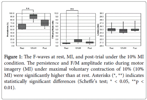
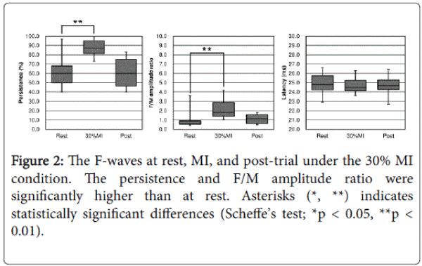
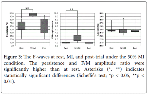
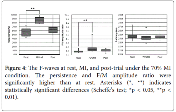
The changes in F-waves during and after MI under 10%, 30%, 50%, and 70% MVC. Persistence during MI under the four MI conditions was significantly increased than at Rest (Scheffe’s test; 10% MI vs. Rest, 70% MI vs. Rest, **p < 0.01; 30% MI vs. Rest, 50% MI vs. Rest, *p < 0.05) (Figures 1-4).
Figure 1: The F-waves at rest, MI, and post-trial under the 10% MI condition. The persistence and F/M amplitude ratio during motor imagery (MI) under maximal voluntary contraction of 10% (10% MI) were significantly higher than at rest. Asterisks (*, **) indicates statistically significant differences (Scheffe’s test; * < 0.05, **p < 0.01).
Persistence immediately after MI (at Post) were significantly decreased compared with at MI (Scheffe’s test; 10% MI vs. Post, 30% MI vs. Post, 70% MI vs. Post, *p < 0.05) (Figures 1 and 2). Persistence at Post was tended to be decreased compared with at 50% MI (Scheffe’s test; p = 0.067) (Figure 3).
The F/M amplitude ratio during MI under the three MI conditions was significantly increased than at Rest (Scheffe’s test; 10% MI vs. Rest, 50% MI vs. Rest, **p < 0.01; 30% MI vs. Rest, *p < 0.05) (Figures 1-3). The F/M amplitude ratio during 70% MI was tended to be increased than at Rest (Scheffe’s test; p = 0.082) (Figure 4). The F/M amplitude ratio immediately after 50% MI was significantly decreased compared at 50% MI (Scheffe’s test; *p < 0.05) (Figure 3).
Alternatively, no significant differences in latency were observed among three trials (Rest, MI, Post) under the four MI conditions (Figures 1-4).
Differences in the changes of F-waves during MI under the four muscle contraction strengths.
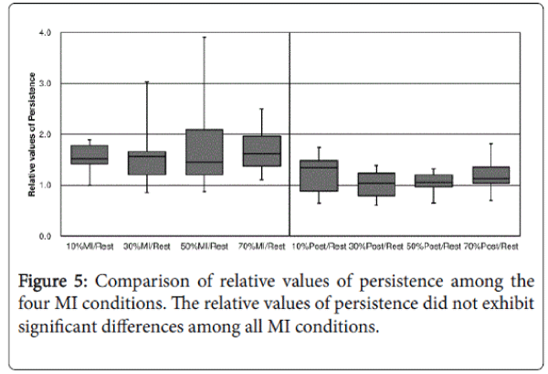
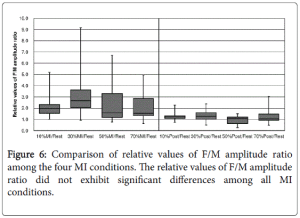
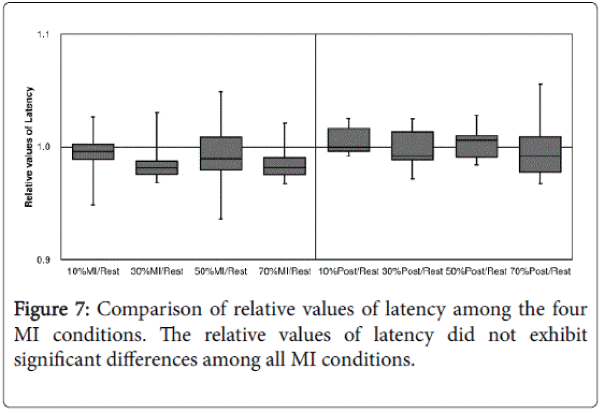
The relative values of persistence, F/M amplitude ratio, and latency did not exhibit significant differences among the four MI conditions (Figures 5-7).
Discussion
In this study, both the persistence and F/M amplitude ratio during MI under various imagined contraction strengths (10% MI, 30% MI, 50% MI, and 70% MI) were significantly increased than that at Rest.
Although, no significant differences in the relative values of persistence, F/M amplitude ratio, and latency were observed among the four MI conditions. These results indicate that MI of muscle contraction can increase the spinal motor neurons excitability, but the imagined muscle contraction strengths were not involved in the changes of spinal motor neurons excitability, at least from 10% to 70% MVC.
MI under 10%, 30%, 50%, and 70% MVC can increase the spinal motor neurons excitability. The spinal motor neurons excitability during MI was significantly greater than at rest for all MI conditions, suggesting that MI activates (excites) descending pathways to spinal motor neurons innervating the thenar muscles. The previous studies have demonstrated activation of multiple cortical and subcortical regions contribute to the motor preparation and planning during MI [5-8].
The cerebral cortex activity during MI presumably increases the spinal motor neurons excitability via descending pathways. In addition, the subjects performed MI while holding the sensor of a pinch meter. Therefore, tactile and proprioceptive inputs may have influenced the response. Mizuguchi et al. [18,19] reported that the MEP amplitude during MI was larger when a ball was squeezed than when no ball was held. In contrast, somatosensory evoked potentials (SEPs) in response to median nerve stimulation were not modulated by MI or by holding the ball. Suzuki et al. [10] analyzed the changes in spinal motor neurons excitability between with and without holding sensor MI tasks.
The F-wave during MI with holding sensor was grater facilitated than without holding sensor. The haptic and proprioceptive perception also contributes to the increase in spinal motor neurons excitability together with MI-activated pathways.
Differences in the imagined muscle contraction strengths during MI do not influence the changes in spinal motor neurons excitability. Our previous study suggested that the facilitation magnitude of spinal motor neurons excitability during MI under three MVC conditions (i.e. 10%, 30%, 50% MVC) were similar [14]. The novel result compared with our previous results is as follows, there were not observed significant increase in the spinal motor neurons excitability during MI under 70% MVC compared with other imagined muscle contraction strengths. Hence, it suggested that differences in the imagined muscle contraction strength did not influence the magnitude of the change in spinal motor neurons excitability.
Similarly, in previous study, Hale et al. [12] reported that the amplitude of the soleus H-reflex were increased during ankle plantar flexion MI under 40%, 60%, 80%, and 100% MVC. However, no significant differences were observed in the amplitude of the H-reflex among four MI conditions. About higher imagined muscle contraction strength also not progressively enhance spinal motor neuron excitability, one possibility is the contribution of neural mechanisms that inhibit actual movement and muscle contraction during MI. Park and Li [20] reported that the corticospinal excitability during MI of finger movement under 10%, 20%, 30%, 40%, 50%, and 60% MVC was higher than that rest, with no differences among all MI conditions. They also suggested that the imagined muscle contraction strengths cannot influence the magnitude of the change in corticospinal excitability.
Similarly, an event-related potential study found that the magnitude of primary motor cortex activity during MI did not correlate with the imagined contraction strengths but supplementary motor area (SMA) and premotor cortex (PM) activity during MI did [21]. The SMA and PM have the various important roles. In detail, these two areas are more activated for larger force generation [22,23], and participated in motor preparation, planning, and motor inhibition [24,25]. Therefore, the SMA and PM may inhibit the actual muscle activity depending on the muscle contraction strength. These inputs from the SMA and PM may suppress any additional excitability conferred by MI with high imagined contraction strength. Furthermore, the spinal motor neurons excitability during MI is thought to be affected by central nervous system via the corticospinal and extrapyramidal tract. Thus, the degree of the changes of spinal motor neurons excitability during MI under different imagined muscle contraction strengths may be modulated by both excitatory and inhibitory inputs from the central nervous system.
In clinical use of MI of isometric thenar muscles activity
Functional reorganization of the central nervous system may occur after brain and spinal cord injury. After brain and spinal cord injury, motor cortex excitability reduced due to various factors, including the damage of neural substrates, loss of sensory inputs, and disuse of the affected limb [26]. The significant decrease was shown in both size and number of corticospinal neurons. Consequently, the corticospinal excitability would be decreased [27].
Therefore, we considered that facilitating the excitability of central and spinal neural level could be necessary for improvement motor function. MI can increase the MEPs amplitude in patients with post stroke [28] and spinal cord injury [29], and the F-waves in post-stroke [30]. The present results implicated that MI of isometric thenar muscles activity under slight MVC (i.e. 10% MVC) can substantially facilitate the spinal motor neurons excitability. Therefore, MI under the slight MVC might be enough to improve motor function in clinical setting. Further research will be required about the influence of imagined muscle contraction strength on actual performance.
Conclusion
The present study aimed to investigate the influence of imagined muscle contraction strengths on the changes in spinal motor neurons excitability in more detail. Especially, the novelty of this study compared with our previous study [10,14] is to investigate the changes in spinal motor neurons excitability during MI under larger imagined muscle contraction strength than 50% MVC. Here, we assessed the changes of spinal motor neurons excitability during MI under 10%, 30%, 50%, and 70% by the F-wave. The present results demonstrated that MI of thenar muscles activity under 10%, 30%, 50%, and 70% MVC can facilitate the spinal motor neurons excitability. However, the imagined muscle contraction strengths did not influence the changes of spinal motor neurons excitability.
Acknowledgments
None declared.
References
- Guillot A, Rienzo FD, Maclntyre T, Moran A, Collet C (2012) Imagining is not doing but involves specific motor commands: a review of experimental data related to motor inhibition. Front Hum Neurosci 6: 247.
- Yue G, Cole KJ (1992) Strength increases from the motor program: comparison of training with maximal voluntary and imagined muscle contractions.J Neurophysiol 67: 1114-1123.
- Guillot A, Tolleron C, Collet C (2010) Does motor imagery enhance stretching and flexibility?J Sports Sci 28: 291-298.
- Jackson PL, Lafleur MF, Malouin F, Richards C, Doyon J (2001) Potential role of mental practice using motor imagery in neurologic rehabilitation.Arch Phys Med Rehabil 82: 1133-1141.
- Stephan KM, Fink GR, Passingham RE, Silbersweig D, Ceballos-Baumann AO, et al. (1995) Functional anatomy of the mental representation of upper extremity movements in healthy subjects.J Neurophysiol 73: 373-386.
- Luft AR, Skalej M, Stefanou A, Klose U, Voigt K (1998) Comparing motion- and imagery-related activation in the human cerebellum: a functional MRI study.Hum Brain Mapp 6: 105-113.
- Lotze M, Montoya P, Erb M, Hülsmann E, Flor H, et al. (1999) Activation of cortical and cerebellar motor areas during executed and imagined hand movements: an fMRI study.J CognNeurosci 11: 491-501.
- Matsuda T, Watanabe S, Kuruma H, Murakami Y, Watanabe R, et al. (2011) Neural correlates of chopsticks exercise for the non-dominant hand: comparison among the movement, images and imitations–A functional MRI study. Rigakuryoho Kagaku 26: 117-122.
- Kasai T, Kawai S, Kawanishi M, Yahagi S (1997) Evidence for facilitation of motor evoked potentials (MEPs) induced by motor imagery.Brain Res 744: 147-150.
- Suzuki T, Bunno Y, Onigata C, Tani M, Uragami S (2013) Excitability of spinal neural function during several motor imagery tasks involving isometric opponenspollicis activity. NeuroRehabilitation 33: 171-176.
- Oishi K, Kimura M, Yasukawa M, Yoneda T, Maeshima T (1994) Amplitude reduction of H-reflex during mental movement simulation in elite athletes. Behav Brain Res 62: 55-61.
- Hale BS, Raglin JS, Koceja DM (2003) Effect of mental imagery of a motor task on the Hoffmann reflex.Behav Brain Res 142: 81-87.
- Cowley PM, Clark BC, Ploutz-Snyder LL (2008) Kinesthetic motor imagery and spinal excitability: the effect of contraction intensity and spatial localization.ClinNeurophysiol 119: 1849-1856.
- Bunno Y, Yurugi Y, Onigata C, Suzuki T, Iwatsuki H (2014) Influence of motor imagery of isometric opponenspollicis activity on the excitability of spinal motor neurons: a comparison using different muscle contraction strengths. J PhysTherSci 26: 1069-1073.
- Suzuki T, Saitoh E (2000) Recommendations for the practice of the evoked EMG: H-reflex and F-wave–Guidelines of the International Federation of Clinical Neurophysiology. Rigakuryoho Kagaku 15: 187-192.
- Mesrati F, Vecchierini MF (2004) F-waves: neurophysiology and clinical value.NeurophysiolClin 34: 217-243.
- Fisher MA (2007) F-waves--physiology and clinical uses.ScientificWorldJournal 7: 144-160.
- Mizuguchi N, Sakamoto M, Muraoka T, Kanosue K (2009) Influence of touching an object on corticospinal excitability during motor imagery.Exp Brain Res 196: 529-535.
- Mizuguchi N, Sakamoto M, Muraoka T, Nakagawa K, Kanazawa S, et al. (2011) The modulation of corticospinal excitability during motor imagery of actions with objects.PLoS One 6: e26006.
- Park WH, Li S (2011) No graded responses of finger muscles to TMS during motor imagery of isometric finger forces.NeurosciLett 494: 255-259.
- Romero DH, Lacourse MG, Lawrence KE, Schandler S, Cohen MJ (2000) Event-related potentials as a function of movement parameter variations during motor imagery and isometric action. Behav Brain Res 117: 83-96.
- Wright DJ, Holmes PS, Smith D (2011) Using the movement-related cortical potential to study motor skill learning.J Mot Behav 43: 193-201.
- Oda S, Shibata M, Moritani T (1996) Force-dependent changes in movement-related cortical potentials.J ElectromyogrKinesiol 6: 247-252.
- Watanabe J, Sugiura M, Sato K, Sato Y, Maeda Y, et al. (2002) The human prefrontal and parietal association cortices are involved in NO-GO performances: an event-related fMRI study.Neuroimage 17: 1207-1216.
- Nakata H, Sakamoto K, Ferretti A, Gianni Perrucci M, Del Gratta C, et al. (2008) Somato-motor inhibitory processing in humans: an event-related functional MRI study.Neuroimage 39: 1858-1866.
- Liepert J, Bauder H, Wolfgang HR, Miltner WH, Taub E, et al. (2000) Treatment-induced cortical reorganization after stroke in humans.Stroke 31: 1210-1216.
- Wrigley PJ, Gustin SM, Macey PM, Nash PG, Gandevia SC, et al. (2009) Anatomical changes in human motor cortex and motor pathways following complete thoracic spinal cord injury.Cereb Cortex 19: 224-232.
- Cicinelli P, Marconi B, Zaccagnini M, Pasqualetti P, Filippi MM, et al. (2006) Imagery-induced cortical excitability changes in stroke: a transcranial magnetic stimulation study. Cereb Cortex 16: 247-253.
- Cramer SC, Nelles G, Benson RR, Kaplan JD, Parker RA, et al. (1997) A functional MRI study of subjects recovered from hemiparetic stroke.Stroke 28: 2518-2527.
- Naseri M, Petramfar P, Ashraf A (2015) Effect of Motor Imagery on the F-Wave Parameters in Hemiparetic Stroke Survivors.Ann Rehabil Med 39: 401-408.
Relevant Topics
- Electrical stimulation
- High Intensity Exercise
- Muscle Movements
- Musculoskeletal Physical Therapy
- Musculoskeletal Physiotherapy
- Neurophysiotherapy
- Neuroplasticity
- Neuropsychiatric drugs
- Physical Activity
- Physical Fitness
- Physical Medicine
- Physical Therapy
- Precision Rehabilitation
- Scapular Mobilization
- Sleep Disorders
- Sports and Physical Activity
- Sports Physical Therapy
Recommended Journals
Article Tools
Article Usage
- Total views: 11035
- [From(publication date):
specialissue-2016 - Apr 03, 2025] - Breakdown by view type
- HTML page views : 10176
- PDF downloads : 859

