The Hernia of Rieux: A Rare Cause of Digestive Obstruction
Received: 03-Jun-2023 / Manuscript No. roa-23-103173 / Editor assigned: 05-Jun-2023 / PreQC No. roa-23-103173 (PQ) / Reviewed: 19-Jun-2023 / QC No. roa-23-103173 / Revised: 22-Jun-2023 / Manuscript No. roa-23-103173 (R) / Published Date: 29-Jun-2023 DOI: 10.4172/2167-7964.1000455
Keywords
Occlusion; Internal hernia; Peri caecal; CT
Case History
A 59-year-old patient was admitted to the emergency room with a cessation of matter and gas, vomiting and abdominal pain, dating back 6 days. His history did not reveal any particular history. The clinical examination showed fever at 38.2°, hemodynamic and respiratory stability with abdominal distension. His biological workup revealed inflammatory syndrome and hyperleukocytosis.
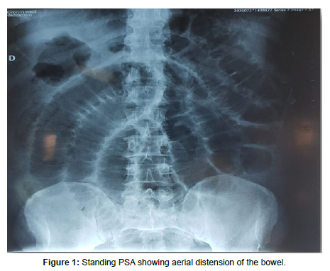
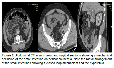
Thus, a radiograph of the abdomen without preparation was performed (Figure 1) showing aerial distension of the bowel, followed by an abdominal CT scan (Figure 2) showing a mechanical occlusion of the small intestine on pericaecal hernia.
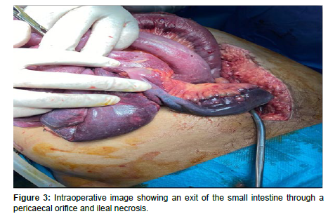
The patient was then operated in emergency and the operative exploration confirmed the diagnosis of occlusion on pericaecal internal hernia (Figure 3).
Internal hernia intestinal (IHI) obstruction represents 5% of the causes of acute intestinal obstruction [1]. It corresponds to hernias forming where hollow organs are incarcerated via intra-abdominal or retroperitoneal orifices. A variety of internal hernias are described, depending on the site and the orifice involved. We distinguish hernias formed through normal or paranormal orifices of the peritoneum (Winslow’s hiatus, para duodenal, pericaecal and inter sigmoidal spaces) from hernias through abnormal orifices of the peritoneum (trans mesenteric, trans epiploic, trans mesosigmoidal, broad and hepato gastric ligaments) or iatrogenic (small bowel surgery, gastric bypass) [2]. IHI occurs when the right Toldt’s fascia is detached, resulting in the formation of a hernia sac. The particularity of this sac is that it is formed in contact with the posterior or lateral walls of the ascending colon that can be stretched and compressed by the herniated loops [2,3].
The usual clinical presentation of internal hernias is an occlusive syndrome in a patient with a history of spontaneously resolving subocclusive episodes without any particular context.
Imaging through abdominal CT scan allows suggesting the diagnosis thus indicating a surgical intervention which will confirm the diagnosis secondarily. It allows to confirm the presence of an occlusion, its mechanical character, its type but also to look for signs of gravity. The CT appearance of pericaecal internal hernias is a closed loop occlusion recognized in axial section by the radial arrangement of the more or less distended coves. Depending on the size and location of the hernia sac, three main types of pericaecal hernia are described, namely pericaecal hernia in the external (retrocaecal), internal (retro ileocaecal) and ileo-appendicular (posterior to the appendix and the last appendix) positions appendix and the last ileal loop). In this case, the CT scan found distension of some of the bowel coves with hydroaeric level and radial arrangement with a sac in the right iliac fossa behind the coecum suggesting a pericaecal internal hernia in its retro ileocaecal form (Figure 2).
The management of this type of condition remains urgent, and is done by surgery, consisting of disinterment of the herniated loop, its treatment and then closure of the breach. Surgical exploration confirmed our initial diagnosis by showing a sac in the right iliac fossa containing some necrotic loops (Figure 3).
The immediate postoperative course was simple. In the absence of an early diagnosis, the evolution is towards strangulation, hence the interest of performing an imaging at the slightest doubt for a better prognosis.
References
- Ouedraogo S, Ouedraogo S, Kambire JL, Zida M, Sanou A (2017) Occlusion secondary to congenital internal transmesenteric hernia: about 2 cases. Pan Afr Med J 27: 131.
- Mathias J, Phi I, Brout O, Ganne PA, Laurent V, et al. (2014) Hernies internes. EMC 33-015.
- Armstrong O, Hamel A, Hamel O, Rogez JM, Robert R, et al. (2005) Hernies internes: anatomie chirurgicale. Morphologie 89: 195.
Indexed at, Google Scholar, Crossref
Citation: Rostoum S, Zhim M, Naggar A, Haddad SE, Allali N (2023) The Hernia of Rieux: A Rare Cause of Digestive Obstruction. OMICS J Radiol 12: 455. DOI: 10.4172/2167-7964.1000455
Copyright: © 2023 Rostoum S, et al. This is an open-access article distributed under the terms of the Creative Commons Attribution License, which permits unrestricted use, distribution, and reproduction in any medium, provided the original author and source are credited.
Share This Article
Open Access Journals
Article Tools
Article Usage
- Total views: 1298
- [From(publication date): 0-2023 - Mar 29, 2025]
- Breakdown by view type
- HTML page views: 1079
- PDF downloads: 219


