The Focal Hepatic Hot Spot Sign
Received: 08-Jul-2021 / Accepted Date: 16-Aug-2021 / Published Date: 23-Aug-2021 DOI: 10.4172/2167-7964.1000335
Keywords: Focal; Hepatic Hot spot sign
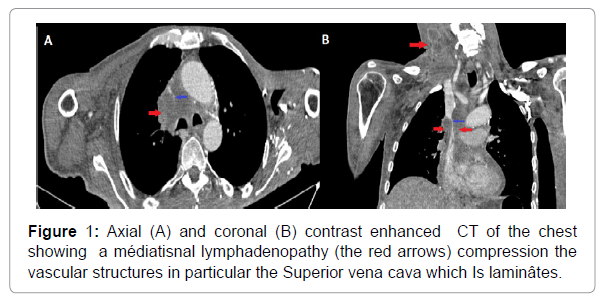
The focal hepatic hot spot sign is due to a Porto systemic venous shunt between the SVC and the portal vein. it is created by areas of focal increased blood flow that result from this shunt. This sign is found mainly in Budd-Chiara syndrome, the causes of which are multiple, notably thorax neoplasms.
Clinical image
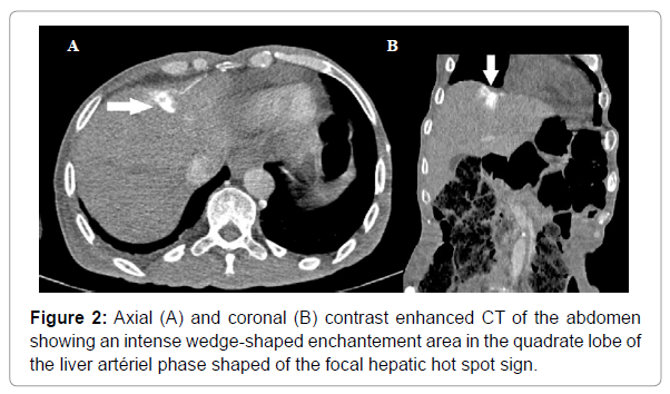
A 65-year-old man followed for a left pulmonary adenocarcinoma with lymph node metastasis treated by chemotherapy, the control scanner showed an increase in mediastina lymphadenopathy compressing the mediastina vascular structures, in particular the superior vena cava Figure 1, and the abdomen-CT showing an area of intense, homogeneous wedge-shaped enhancement in the square lobe of the liver Figure 2 [1-3].
Discussion
The focal hepatic hot spot sign was first described in 1983 by Ishikawa. it is due to a portosystemic venous shunt between the SVC and the portal vein. The hot spot is created by areas of focal increased blood flow that result from this shunt. This sign is found mainly in Budd-Chiari syndrome, the causes of which are multiple, notably thorax neoplasms such as pulmonary carcinoma and lymphoma, Vasculo-Behcet disease, fibro sing mediastinitis and lactic aneurysm, the differential diagnosis is made mainly with haemangioma, hepatocellular carcinoma, focal nodular hyperplasia located in segment IV and which can mimic the focal hepatic hot spot sign.
References
- Hoang VT, Nhu Quynh Vo, Trinh CT, (2020) The focal hepatic hot spot sign with Lung cancer in computed tomographie. Respira Case Rep 8 :(8)
- Dickson AM. (2005) The focal hepatic hot spot sign. Radiology, 237Â :(2)Â :647- 648
- Phoophiboon V, Tantiprawan J, Vanakiatkul H, Wongkarnjana A (2020). Systemic to pulmonary venus shunt and the focal hepatic hot spot sign from SVC obstruction in Behcet’s disease. . BMJ Case Rep 13 :(2) : e234017
Citation: Drissi A, Hatim E, Sahar M, Jerguigue H, Latib R (2021) Osteopoikilosis is a Disorder Diagnosed only by Radiology. OMICS J Radiol 10: 335. DOI: 10.4172/2167-7964.1000335
Copyright: © 2021 Drissi A, et al. This is an open-access article distributed under the terms of the Creative Commons Attribution License, which permits unrestricted use, distribution, and reproduction in any medium, provided the original author and source are credited.
Select your language of interest to view the total content in your interested language
Share This Article
Open Access Journals
Article Tools
Article Usage
- Total views: 3207
- [From(publication date): 0-2021 - Dec 23, 2025]
- Breakdown by view type
- HTML page views: 2420
- PDF downloads: 787


