The Enzyme Antioxidant Defence System in Libyodrilus Violaceus in Response to Crude Oil Pollution in the Niger Delta Region of Nigeria
Received: 03-Nov-2023 / Manuscript No. EPCC-23-116396 / Editor assigned: 06-Nov-2023 / PreQC No. EPCC-23-116396 / Reviewed: 20-Nov-2023 / QC No. EPCC-23-116396 / Revised: 23-Nov-2023 / Manuscript No. EPCC-23-116396 / Accepted Date: 30-Nov-2023 / Published Date: 30-Nov-2023
Abstract
Crude oil exploration inevitably contributes to environmental pollution owing to endless combustion that contributes to climate change and its effects. Crude oil toxicity is a matter of global concern and has been a major concern in the oilrich Niger Delta region of Nigeria. Earthworms have been scientifically proven to be worthy sentinel for ecotoxicological studies and their toxicity studies have been proven to be highly significant. The present study supports an array of past studies that sub-lethal concentrations of crude oil and its fractions were able to down-regulate the activities of some key antioxidant enzymes. In this short-term study, antioxidant enzymes including Superoxide dismutase (SOD), catalase (CAT), glutathione peroxidase (GPX), and glutathione reductase (GRX) were assessed in earthworms exposed to various concentrations of crude oil fractions and its fractions. Antioxidant enzyme alterations visible in this study mean that soil organisms could suffer untold system degradation damage and even mortality in crude oil-contaminated environments.
Keywords
Crude oil; Superoxide dismutase; Catalase; Glutathione peroxidase; Glutathione reductase; Lybiodrilus Violeceus
Introduction
Crude oil contamination caused by crude oil spillages is rampant in the Niger Delta region of Nigeria because it is the main region for crude oil extraction, transportation and distribution in the Country [1- 6]. Ecotoxicological studies have shown that crude oil contamination could cause injurious effects and even death of living organisms because it contains harmful compounds such as heavy metals, BTEX, and Polycyclic Aromatic Hydrocarbons (PAH), that get into the ecosystem and into these species, generating free radicals and Reactive Oxygen Species (ROS) [4,5,7-9]. Free radicals and ROS are known to be able to cause damage to both proteins, lipids, DNA and cell membranes that eventually affect the living organisms that are exposed to these toxicants. The enzyme antioxidant defence system is an important aspect of the complex antioxidant defence system that protects the living organism from the adverse effect of free radicals and ROS that can cause oxidative damage. Crude oil toxicity causes oxidative stress, disrupting normal cellular processes that can lead to adverse health effects such as tissue damage, cell death and various clinical conditions [4-6]. The antioxidant defence system is disrupted and overwhelmed in crude oil contamination owing to free radicals and ROS generation [10]. Scientific research geared at understanding the effect of crude oil toxicity on the enzyme antioxidant system will help highlight the possible mechanisms of crude oil toxicity in living organisms and also help inform remediation strategies that could help minimise ecological and human health consequences of crude oil pollution.
Methodology
This research was carried out in the Department of Biochemistry, College of Life Sciences, University of Benin, Benin City, Edo State and in Igbinedion University, Okada, Edo state. This study was conducted from September, 2009 to May, 2015.
Materials
Test samples
Bonny Light Crude Oil samples used were obtained from Shell Petroleum Development Company.
Already contaminated soil samples from three Crude Oil spillage sites located in Obrikom village, Ebocha village and Mgbede village in Ogba \ Ndoni \ Egbema local Government Area in Rivers State were collected for this research study.
The samples were taken from the topsoil (0-15 cm) and subsoil from about 15-20 cm depth from surface level.
The total numbers of samples from the contaminated sites were six. All samples collected were put in screw capped containers and taken to the laboratory for processing.
Apparatus used
Top loading weighing balance, sensitive weighing scale, centrifuge, Refrigerator, Water bath, separating funnel, magnetic stirrer, magnetic Hot plate, retort stand, oven, plastic bowls, test tubes, conical flask, beaker, magnifying glass, surgical blade, spectrophotometer, plain storage bottle, surgical glove, mortar and pestle, nets, Test-tubes, Vials for storing extracts (2 ml), Glass funnel, Separating funnel, 100 ml volumetric flask, 100 ml clean and dried conical flask, 100 ml sintered glass funnel, Clamp and stand, Refrigerator (specially designated for storage of standards and extracts at 4.0°C.
Test animals
The test specimen (Libyodrilus violaceus) were collected by handpicking from a moist subsurface soil in Okada town, Benin city, Edo state Nigeria, The earthworms were collected from the same site in order to reduce variability in biotype.
Identification and characterisation of earthworm
Samples of earthworms were sent to the department of Zoology, faculty of Animal Science, University of Benin, Edo State for identification and characterisation. The specimen was identified to be Libyodrilus violaceus.
Acclimatization of earthworm
The earthworms were held in an open culture bowls for 10 days prior to the start of the experiment. Each acclimatization bowl contained 1000 g of habitat soil. At the end of the 10th day, the earthworms were removed from the acclimatization bowl, rinsed with clean water and weighed before being introduced into the various test bowls.
Preparation of earthworm bed
1000 g of habitat soil was added into 10 well labelled open plastic bowls. 10 g of finely meshed clean paper as source of cellulose was added into each of the 10 bowls. The soils in the different bowls were mixed with different concentration and fractions of crude oil. 20 earthworms were individually weighed and introduced into each of the different bowls.
Contaminant/pollutant
Bonny light crude oil Crude oil was the pollutant used. Crude oil sample was obtained from Shell Petroleum Development Company, Sapele, Delta state, Nigeria.
Methods
Fractionation of crude oil
The Bonny light crude oil was fractionated by the method of Anderson et al, 1974 [11]. The crude oil and distilled water were introduced into a 1000 ml conical flask at a ratio of 1:2 (20 ml of crude oil to 40 ml of distilled water). A magnetic stirrer was introduced into the mixture and the mixture was placed on a magnetic hot plate and allowed to stir for 48 hrs. The mixture after stirring separated into two distinct fractions; a dark dense phase (water insoluble fraction) and a very light brown watery phase (water soluble fraction). This was done to mimic the separation of the spilt crude oil in to land and water bodies in the event of a spillage.
Collection of crude oil fractions
A 2000 ml separating funnel was allowed to stand erect on a retort stand. The separated mixture was introduced into the separating funnel and was allowed to stand for 24 hrs. The two distinct fractions were separated by carefully allowing them pass through the tap and each fraction was collected in a different conical flask. The conical flasks were covered with aluminium foil sheets and kept at room temperature until commencement of experiment.
Preparation of the different test concentrations of crude oil
Concentrations of crude oil used for these sublethal studies were selected based on the 96 h exposure LC50 value (10.33 ml/kg) for bonny light crude oil exposed to earthworms, Eudrilus eugeniae [10].
0.1% concentration of the WSF, WIF and WC oil fraction were prepared by adding 1 ml of the crude oil or its fractions to 1000 g of the habitat (control) soil and mixed with 999 ml of distilled water.
0.2% concentration of WSF, WIF and WC oil fraction was prepared by adding 2 ml of the crude oil or its fractions to 1000 g of the habitat (control) soil and mixed with 998 ml of distilled water.
0.3% concentration of crude oil fraction was prepared by adding 3 ml of the crude oil or its fractions to 1000 g of the habitat (control) soil and mixed with 997 ml of distilled water.
Preparation of assay samples
The earthworms were weighed using a sensitive weighing balance before termination of experiment. The Clitellum and Post Clitellum of the various earthworms were excised, weighed and homogenized using 2 ml of ice cold 0.9% normal saline solution. The homogenate were centrifuged at 4000 rpm for 10 mins and the clear supernatant aspirated and the residue recovered for relevant biochemical estimations by standard methods. The supernatant were put in plain air tight experimental tubes and stored in the refrigerator until ready for analysis. The clitellum and post clitellum of 100 earthworms for lipid study were analysed using the same method of lipid extraction modified by Bligh Dyer, (1959) [12], the clitellum and post clitellum were homogenized using a mixture of chloroform and methanol in the ratio 1:2. The homogenates were also centrifuged at 4000 rpm for 10 mins, the clear supernatant aspirated and the residue recovered for relevant biochemical estimations by standard methods.
In vivo antioxidative activity
Estimation of superoxide dismutase (sod) activity
Principle: Adrenaline auto-oxidizes rapidly in aqueous solution to adrenochrome, whose absorbance can be determined at 420 nm using a spectrophotometer. The auto-oxidation of adrenaline depends on the presence of superoxide anions. The enzyme SOD inhibits the autooxidation of adrenaline by catalyzing the breakdown of superoxide anions. The degree of inhibition is thus a reflection of the activity of SOD, and is determined at one unit of the enzyme activity [13].
Procedure: An aliquot of 0.4 ml dilute supernatant of the sample was added to 5 ml of 0.05 M carbonate buffer (pH 10.2) to equilibrate in the spectrophotometer and the reaction started by the addition of 0.6 ml of freshly prepared 0.3 mM epinephrine as the substrate to the buffersupernatant mixture which was mixed by inversion. The reference cuvette contained 5 ml of the buffer, 0.6 ml of the epinephrine and 0.4 ml of distilled water. The increase in absorbance at 480 nm was due to the adrenochrome formed which was monitored every 30 seconds for 120 seconds. The unit of SOD activity was given as the amount of SOD necessary to cause 50% inhibition of the oxidation of epinephrine to adrenochrome 60 seconds.

ABStest=Oxidation of adrenochrome in the presence of SOD
ABSRef=Oxidation of adrenochrome in the absence of SOD
The enzyme activity can be calculated as follows:

Where y=mg of tissue in the volume of the sample used. Unit of SOD activity=Amount of SOD giving 50% inhibition. Estimation of catalase (CAT) activity Principle: Catalase is present in nearly all animal cells, plant and bacteria and acts to prevent accumulation of noxious H202 which was converted to O2 and H2O

At high concentrations of low molecular weight alcohols or formaldehyde and low peroxide concentration, catalase exhibits peroxidative activity and can utilize organic hydroperoxides with small aliphatic substituents such as hydrogen peroxide peracetic acid as substrate [14].
Procedure: Aliquots of the homogenate supernatant (0.5 ml) were added to ice-cold test tubes while the blank contained 0.5 ml of distilled water. The reaction was initiated by adding sequentially, at fixed intervals, 5 ml of 30 MmH2O2 and was mixed thoroughly by inversion. After exactly 3 minutes, the reaction was stopped sequentially by adding 1 ml of 6 M H2SO4 and was mixed quickly by inversion. The test sample and the blank were taken one at a time, and 7 ml of 0.01 M potassium permanganate was added, mixed twice by inversion and absorbance read between 30 - 60 seconds at 480 nm. The spectrophotometric standard was prepared by adding 7 ml of 0.01 M potassium permanganate to a mixture of 5.5 ml 0.05 M phosphate buffer pH 7.0 and 1 ml 6 M sulphuric acid solution. The spectrophotometer was zeroed with distilled water.
Calculations: The decomposition of hydrogen peroxide by catalase follows first-order kinetics given by the equation:

First-order rate constant=K
Absorbance of blank=AB
Absorbance of standard=Astd
Absorbance of test=AT
S0=Astd - AB
S3=Astd - AT
S0=[S] at zero time
S3=[S] at t=3 minutes
Assay of glutathione peroxidase (GPx)
GPx activity was measured according to the continuous Spectrophotometric Rate Determination method [15].
Principle: This method is based on the oxidation of pyrogallol to purpurogallin by peroxidase at 20°C. Oxidation of pyrogallol to purpurogallin by peroxidase at 20°C. The amount of purpurogallin formed was taken as an activity unit and expressed as unit/mg protein
Reagent used
Reagent I: 100 mM Potassium Phosphate buffer, pH 6.0 at 20°C; prepare 100 ml in deionized water using potassium phosphate,monobasic, Anhydrous. Adjust pH to 6.0 at 20°C with 1.0 M KOH.
Reagent II: 0.50% (w/w) Hydrogen Peroxide solution (H2O2),prepare 50 ml in deionized water using Hydrogen Peroxide, 30% (w/w) solution. Prepare fresh.
Reagent III: 5% (w/v) Pyrogallol solution; prepare 10 ml in deionized water using Pyrogallol. Prepare fresh and keep from light. Obtain the Δ A420nm/20 seconds using the maximum linear rate for both the Test and Blank.

S=seconds
3=Volume (in millilitres) of assay
Df=Dilution factor
12=Extinction coefficient of 1 mg/ml of purpuragallin at 420 nm 0.1=Volume (in millilitres) of sample used.
Assay of glutathione reductase (GRx)
Reagent I: Elman’s (DTNB); 0.0198 g of 2 – nitrobenzoic acid and 1.0 g of sodium citrate were dissolved in 100 ml of distilled water.
Reagent II: 5% Trichloroacetic acid (TCA); 20 g of TCA was dissolved in 400 ml of distilled water.
Reagent III: Phosphate buffer

Statistical analysis
Data collected were subjected to statistical analysis using the SPSS version 20. Results obtained were expressed as mean ± SEM. One-way Analysis of variance (ANOVA) was also used to compare the means of the parameters measured and where significant differences were observed at 95% confidence level, Duncan’s New Multiple Range test [16] was used to separate the means.
Results
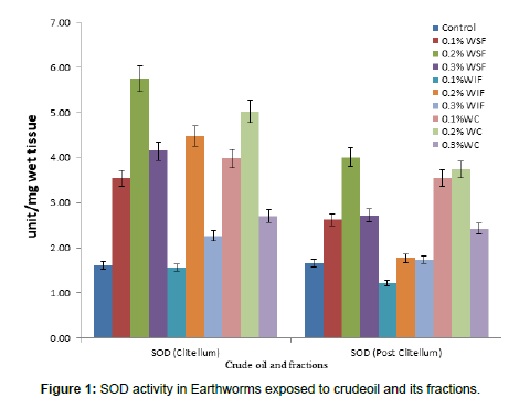
Ant oxidative status of Earthworms (Libyodrilus violaceus) exposed to crude oil and its fractions.SOD activity in the Clitellum and post clitellum of Earthworms exposed to different concentrations of crude oil and its fractions. It was generally observed that there was a significant increase (p<0.05) in all test subjects exposed to the toxicants when compared to control subjects except the group exposed to 0.1%WIF. However, within the test groups, the 0.2% WSF produced the highest effect on SOD activity in the clitellum of Earthworms exposed to the pollutant while the lowest effect was seen in the post clitellum of the test group exposed to 0.1% WIF. A bell-shaped increase was observed in the clitellum parts of the Earthworms exposed to all the concentrations of the pollutant except those exposed to the 0.1% WIF where the lowest SOD activity was observed (Table 1).
| Clitellum | Post Clitellum | |
|---|---|---|
| Groups | (unit/mg wet tissue) | (unit/mg wet tissue) |
| Control | 1.61 ± 0.29e | 1.67 ± 0.19e |
| 0.1% WSF | 3.54 ± 0.55bc | 2.63 ± 0.51b |
| 0.2% WSF | 5.75 ± 0.73a | 4.01 ± 0.92a |
| 0.3% WSF | 4.15 ± 0.40ab | 2.72 ± 0.13bc |
| 0.1%WIF | 1.57 ± 0.19e | 1.22 ± 0.98d |
| 0.2% WIF | 4.48 ± 0.84ab | 1.77 ± 0.21cd |
| 0.3% WIF | 2.27 ± 0.33dd | 1.74 ± 0.20cd |
| 0.1%WC | 3.98 ±0.66bc | 3.55 ±0.31b |
| 0.2% WC | 5.03 ± 0.64ab | 3.74 ± 0.60b |
| 0.3%WC | 2.71 ± 0.31d | 2.42 ± 0.32c |
Table 1: SOD activity in Earthworms exposed to crude oil and its fractions.
Values represent mean ± standard error of mean (SEM). Values represent mean ± SEM; n=10; WSF=Water soluble fraction, WIF=Water insoluble fraction, WC=whole crude.
Means of the same column followed by different lettered superscripts differ significantly (P<0.05)
Means of the same column with the same overlapping superscripts are statistically similar or show no significant difference (p>0.05) (Figure 1).
Catalase (CAT) activity in the Clitellum and post clitellum of Earthworms exposed to different concentrations of crude oil and its fractions
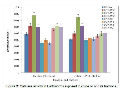
The catalase activity of clitellum and Post clitellum parts of Earthworms exposed to different doses of crude oil and its fractions are presented in Figure 2. It was observed that there was a significant increase (p<0.05) in the activities of catalase in the clitellum and Post clitellum of subjects exposed to all doses of the WSF toxicant when compared to the control subjects. There was also a significant increase (p<0.05) in the activities of catalase in the clitellum of subjects exposed to the WC when compared to the control subjects. There was an insignificant increase (p>0.05) in the Post clitellum of Earthworms exposed to WC. In this study, there was an insignificant reduction (p>0.05) in the activities of catalase in the clitellum of the subjects exposed to the WIF when compared to the control subjects. There was no significant difference (p>0.05) in the Post clitellum of subjects exposed to the WIF of the pollutant when compared to the Post clitellum of the control subjects. The highest catalase activity was seen in the clitellum of the subjects exposed to the 0.2% WSF while the lowest catalase activity was observed in the clitellum of subjects exposed to the 0.1% WIF.
Catalase (CAT) activity in the Clitellum and Post clitellum of earthworms exposed to different concentrations of crude oil and its fractions (Table 2).
Crude oil /fraction |
Clitellum | Post Clitellum |
|---|---|---|
| (µM/mg wet tissue) | (µM/mg wet tissue) | |
| Control | 0.059 ± 1.45 × 10⁻³ de | 0.051 ± 2.72 × 10⁻³ d |
| 0.1% WSF | 0.072 ± 2.18 × 10⁻³ b | 0.060 ± 1.83 × 10⁻³ c |
| 0.2% WSF | 0.088 ± 5.04 × 10⁻³ a | 0.085 ± 7.29 × 10⁻³ a |
| 0.3% WSF | 0.070 ± 2.73 × 10⁻³ b | 0.072 ± 2.94 × 10⁻³ b |
| 0.1%WIF | 0.046 ± 1.62 × 10⁻³ d | 0.050 ± 1.68 × 10⁻³ d |
| 0.2% WIF | 0.050 ± 1.80 × 10⁻³ e | 0.053 ± 4.91 × 10⁻³ d |
| 0.3% WIF | 0.045 ± 5.82 × 10⁻³ d | 0.052 ± 3.38 × 10⁻³ d |
| 0.1%WC | 0.068 ± 2.98 × 10⁻³ c | 0.056 ± 3.34 × 10⁻³ cd |
| 0.2% WC | 0.072 ± 2.94 × 10⁻³ b | 0.060 ± 1.83 × 10⁻³ cd |
| 0.3%WC | 0.070 ± 3.31 × 10⁻³ b | 0.061 ± 1.90 × 10⁻³ d |
Table 2: Catalase (CAT) activity in the Clitellum and Post clitellum of earthworms exposed to different concentrations of crude oil and its fractions.
Values represent mean ± standard error of mean (SEM). Values represent mean ± SEM; n=10; WSF=Water soluble fraction, WIF=Water insoluble fraction, WC=whole crude.
Means of the same column followed by different lettered superscripts differ significantly (P<0.05)
Means of the same column with the same overlapping superscripts are statistically similar or show no significant difference (p>0.05) (Figure 2).
Glutathione peroxidase (GPX) activity in the Clitellum and Post clitellum of Earthworms exposed to different concentrations of crude oil and its fractions
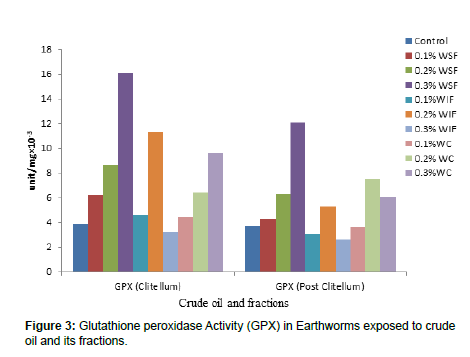
The Glutathione peroxidase (GPX) activity of clitellum and Post clitellum parts of Earthworms exposed to different doses of crude oil and its fractions are presented in Table 3 and Figure 3. It was observed that there was a dose dependent significant increase (p<0.05) in the activities of GPX in the clitellum and Post clitellum of subjects exposed to all doses of the WSF toxicant when compared to the control subjects. There was also a significant increase (p<0.05) in the activities of GPX in the clitellum and Post clitellum of subjects exposed to the WC except those exposed to the 0.1% WC when compared to the control subjects. There was no significant difference (p>0.05) in the Post clitellum of Earthworms exposed to 0.1%WC, 0.1% WIF, 0.3% WIF when compared to control. In this result it was also observed that there was no significant difference in the clitellum of subjects exposed to the 0.3% WIF when compared to the control subjects. There was an insignificant reduction (p>0.05) in the activities of GPX in the Post clitellum of the subjects exposed to the 0.3%WIF when compared to the control subjects. While the subjects exposed to 0.1,0.2 and 0.3% WSF and 0.1,0.2 and 0.3% showed a dose depended increase in the activity. The highest GPX activity was seen in the clitellum of the subjects exposed to the 0.3% WSF while the lowest GPX activity was observed in the Post clitellum of subjects exposed to the 0.3% WIF. Glutathione peroxidase (GPX) activity in the Clitellum and post clitellum of earthworms exposed to different concentrations of crude oil and its fractions (Table 3).
| Crude oil /fraction | Clitellum (unit/mg×10-3) | Post Clitellum |
|---|---|---|
| (unit/mg×10-3 ) | ||
| Control | 3.9 ± 0.348 e | 3.7 ± 0.300e |
| 0.1% WSF | 6.2 ± 1.937cd | 4.3 ± 1.001cd |
| 0.2% WSF | 8.6 ± 0.846bc | 6.3 ± 1.513bc |
| 0.3% WSF | 16.1 ± 2.383a | 12.1± 1.792a |
| 0.1%WIF | 4.6 ± 1.002d | 3.1 ± 0.314e |
| 0.2% WIF | 11.3 ± 2.033b | 5.3 ± 0.716d |
| 0.3% WIF | 3.2 ± 0.359e | 2.6 ± 0.306e |
| 0.1%WC | 4.4 ± 0.427d | 3.6 ± 0.340e |
| 0.2% WC | 6.4 ± 1.087d | 7.5 ± 0.749b |
| 0.3%WC | 9.6 ± 1.360cd | 6.1 ± 1.169bc |
Table 3: Glutathione peroxidase (GPX) activity in the Clitellum and post clitellum of earthworms exposed to different concentrations of crude oil and its fractions.
Values represent mean ± standard error of mean (SEM). Values represent mean ± SEM; n=10; WSF=Water soluble fraction, WIF=Water insoluble fraction, WC=whole crude.
Means of the same column followed by different lettered superscripts differ significantly (P<0.05)
Means of the same column with the same overlapping superscripts are statistically similar or show no significant difference (p>0.05) (Figure 3).
Glutathione reductase (GRX) activity in the Clitellum and Post clitellum of Earthworms exposed to different concentrations of crude oil and its fractions.
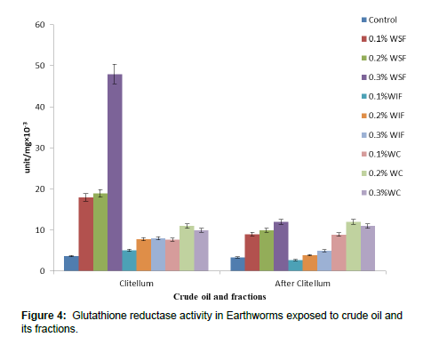
The Glutathione reductase (GRX) activity of clitellum and Post clitellum parts of Earthworms exposed to different doses of crude oil and its fractions are presented in Figure 4. It was observed that there was a dose dependent significant increase (p<0.05) in the activities of GRX in the clitellum and Post clitellum of all subjects exposed to all doses of the toxicant except the Post clitellum of the subjects exposed to 0.1% and 0.2% WIF when compared to the control subjects. It was observed that the GRX activities were significantly low (p<0.05) in the Earthworms exposed to 0.1%WIF when compared to the control subjects. Worthy of note in this result is that the activities of GRX were generally higher in the clitellum of all subjects when compared to its activity in the Post clitellum. The highest GRX activity was seen in the clitellum of the subjects exposed to the 0.3% WSF while the lowest GRX activity was observed in the Post clitellum of subjects exposed to the 0.1% WIF (Table 4).
Crude oil |
Clitellum | Post Clitellum |
|---|---|---|
| /fraction | (unit/mg×10-3 ) | (unit/mg×10-3 ) |
| Control | 3.7 ± 0.39b | 3.4 ± 0.77ab |
| 0.1% WSF | 18 ± 1.216cd | 9 ± 1.028c |
| 0.2% WSF | 19 ± 1.783cd | 10 ± 0.946c |
| 0.3% WSF | 48 ± 0.504d | 12 ± 0.79c |
| 0.1%WIF | 5.1 ± 0.51c | 2.7 ± 1.67a |
| 0.2% WIF | 7.8 ± 1.28c | 3.9 ± 0.21b |
| 0.3% WIF | 8 ± 1.067c | 5 ± 0.490c |
| 0.1%WC | 7.6 ± 0.65c | 8.9 ± 1.84c |
| 0.2% WC | 11± 0.988c | 12 ± 1.38c |
| 0.3%WC | 10 ± 1.783c | 11± 0.988c |
Table 4: Glutathione reductase (GRX) activity in the Clitellum and post clitellum of earthworms exposed to different concentrations of crude oil and its fractions.
Values represent mean ± standard error of mean (SEM). Values represent mean ± SEM; n=10; WSF=Water soluble fraction, WIF=Water insoluble fraction, WC=whole crude.
Means of the same column followed by different lettered superscripts differ significantly (P<0.05)
Means of the same column with the same overlapping superscripts are statistically similar or show no significant difference (p>0.05) (Figure 4).
Discussion
Effect of crude oil and its fractions on in vivo antioxidative status of clitellum and post clitellum of earthworms
As a defence against reactive free radicals, the body produces antioxidant enzymes like Superoxide dismutase SOD, catalase (CAT) glutathione peroxidase (GPX) and glutathione reductase (GRX) which are some of the main antioxidant enzymes. Superoxide dismutase metabolizes the superoxide anion (O- 2) into molecular oxygen and
H2O2, this is then deactivated by catalase, thus preventing oxidative damage. GRX also plays an important role in cellular protection by reducing glutathione in the oxidized form (GSSG) to the reduced form (GSH). The effect of crude oil on antioxidant enzymes in animals is one of mixed results. A study by Eriyamremu et al., (2008) [17] reveal that SOD and GRX activities are lowered in the head region of tadpoles but increased in the tail region when exposed to different concentrations of crude oil and its fractions [18] demonstrated that the exposure of earthworms (Eisenia fetida) to toluene, ethylbenzene and xylene (chemicals present in the crude oil) induced a bell-shaped change in SOD activities with a tendency of inducement firstly and then inhibition with increasing concentrations of the pollutants. In the present study SOD and CAT activities showed a similar trend in all exposed animals similar to that observed by Liu et al.,., (2010) [18]. A hyperbolic increase (inducement and further inhibition with increase in concentration) was observed in SOD activity in all animals when compared to the control earthworms except in those exposed to the 0.1% WIF. These observations did not however correlate completely with work done by Otitoloju and Olagoke, (2011) [19] who reported a significant reduction in SOD and CAT activities only in the liver and gills of fishes (Clarias gariepinus) exposed to crude oil, xylene toluene and benzene after a 28 day exposure. The duration of exposure to the toxicant, the type and species of test animals might be the reason for these varied observations. The consensus however seem to support a decrease in antioxidant enzyme activities under stress conditions. Several studies have shown these antioxidant enzymes to be capable biomarkers for assessing effect of pollutants in terrestrial ecosystem at early stages.
Superoxide dismutase is a first order antioxidant enzyme but also a metalloenzyme which requires zinc for its activity. Cadmium can substitute zinc in enzymes where the metal is required [20] and it may be an essential part for the way cadmium bring about a reduction in SOD activity. These possible effects of cadmium contribute to increased lipid peroxidation and may in part account for the observed high levels of malondialdehyde in the crude oil treated animals. It therefore follows that these observed decreases in these enzymes would decrease the ability of the tissues to scavenge free radicals. This would have profound effects on the animal as it would compromise the protection offered by these enzymes (SOD and CAT) against free radical damage and problems that may be associated with free radicals.
Glutathione reductase (GRX) and glutathione peroxidase (GPX) activities in the clitellum followed the same trend. Dose dependent perturbations were observed in all exposed animals when compared to the control, except those exposed to the WIF. Observed significant increase could be due to an imbalance between the production of free radicals and the antioxidant defence systems owing to the effect of the toxicant. These findings are in accordance with the earlier findings of Sulata et al., 2008 [21]; who reported a dose dependent increase in the activities of GPX and GR during the early phase (14th day) of the exposure to lead (pb).
Conclusion
The observed increases in the activities of the antioxidant enzymes recorded in the earthworms exposed to the WSF and WC are in consonant with observed results from the in vitro studies that revealed higher concentrations of toxic constituents in these two fractions. The observed positive activities of the antioxidant enzymes in the animals exposed to the WIF must have been due to it possessing the least concentrations of the toxic constituents and higher concentrations of the total hydrocarbon content (THC) as shown in, the in vitro studies. This study further provides scientific evidence that earthworms influence the degradation of THC in the polluted soil positively. The result of this study provides scientific data that earthworm is a useful stress indicator to identify and assess recovery of crude oil contaminated soils.
References
- Hozumi T, Tsutsumi H , Kono M (2000) Bioremediation on the shore after an oil spill from the Nakhodka in the sea of Japan. Mar Pollut Bull40: 308-314.
- Ezemonye LIN (2004) Comparative studies of macroinvertebrates community structure in two river-catchment areas (Warri and Forcados rivers) in delta state, Nigeria. Afr Sci 5: 181-192.
- UNEP (2011) Environmental assessment of Ogoniland. UNEP Nairobi : 1–262.
- Erifeta G, Eriyamremu GE, Omage K, Njoya HK (2017) Alterations in the bio-membrane of Libyodrilus violaceus following exposure to crude oil and its fractions.Int j biochem res20: 1-11.
- Erifeta GO, Njoya HK, Josiah SJ, Nwangwu SC, Osagiede PE, et al. (2019) Physicochemical characterisation of crude oil and its correlation with bioaccumulation of heavy metals in earthworm (Libyodrilus violaceus). IJRSI 6: 370-375.
- Georgina O Erifeta (2023) Biomolecule (Protein) Monitoring and Histopathological Changes in Earthworms (Libyodrilus violaceous). Sci Environm6: 169.
- Albers PH (1995) Petroleum and individual polycyclic aromatic hydrocarbons. In: Handbook of Ecotoxicology, Hoffman DJ, Rattner BA, Burton CA, Cairns J (eds.), Boca Raton, Lewis Publishers: 330-355.
- McLaughlin MJ, Hamon RE, McLaren, RG Speir TW, Rogers SL, et al. (2000) A bioavailability-based rationale for controlling metal and metalloid contamination of agricultural land in Australia and New Zealand. Soil Res 38: 1037-1086.
- Asagba, Samuel , Eriyamremu G (2007) Oral cadmium exposure alters haematological and liver function parameters of rats fed a Nigerian-like diet. J Nutr & Environ Med .
- Otitoloju, Adebayo A, (2005) Stress indicators in earthworms Eudriluseugeniae inhabiting a crude oil contaminated ecosystem. acta satech 2: 1-5.
- Anderson J Neff, Jerry Cox, BA Tatem, H Hightower G (1974) Characteristics of Dispersions and Water-Soluble Extracts of Crude and Refined Oils and Their Toxicity to Estuarine Crustaceans and Fish. Mar Biol 27: 75-88.
- Bligh EG, Dyer W J (1959) A rapid method of total lipid extraction and purification. Can J Biochem Physiol 37: 911–917.
- Misra HP, Fridovic I (1972) The role of superoxide anion in the autoxidation of epinephrine and a simple assay for superoxide dismutase. JBC 247: 3170-3175.
- Cohen A, Gagnon M , Nugegoda D (2005) Alterations of Metabolic Enzymes in Australian Bass, Macquaria novemaculeata, After Exposure to Petroleum Hydrocarbons. AECT 49: 200–205.
- Maehly AC, Chance B (1954) The assay of catalases and peroxidases. Methods of biochemical analysis 1:357–424.
- IBM Corp, (2013) IBM SPSS Statistics for Windows, version 22.0. Armonk.
- Eriyamremu GE, VE Osagie, SE Omoregie, and CO Omofoma (2008) Alterations in glutathione reductase, superoxide dismutase and lipid peroxidation of tadpoles (Xenopus laevis) exposed to Bonny Light crude oil and its fractions. Ecotoxicol Environ Saf 71 : 284-290.
- Liu Y, Zhou Q, Xie X, Lin D, Dong L (2010) Oxidative stress and DNA damage in the earthworm Eisenia fetida induced by toluene, ethylbenzene and xylene. Ecotoxicol 19:1551-1559.
- Otitoloju A , Olagoke O (2011) Lipid peroxidation and antioxidant defense enzymes in Clarias gariepinus as useful biomarkers for monitoring exposure to polycyclic aromatic hydrocarbons.Environ Monit Assess182: 205-213.
- Predki PF, Sarkar BI Budhendra(1992) Effect of replacement of “zinc finger” zinc on estrogen receptor DNA interactions.JBC 267: 5842-5846.
- Sulata M, Roy S, Chaudhury S, Bhattacharya S (2008) Antioxidant responses of the earthworm Lampito mauritii exposed to Pb and Zn contaminated soil.Environ Pollut 151:1-7.
Indexed at, Google Scholar, Crossref
Indexed at, Google Scholar, Crossref
Indexed at, Google Scholar, Crossref
Indexed at, Google Scholar, Crossref
Indexed at, Google Scholar, Crossref
Indexed at, Google Scholar, Crossref
Indexed at, Google Scholar, Crossref
Indexed at, Google Scholar, Crossref
Indexed at, Google Scholar, Crossref
Indexed at, Google Scholar, Crossref
Indexed at, Google Scholar, Crossref
Indexed at, Google Scholar, Crossref
Indexed at, Google Scholar, Crossref
Indexed at, Google Scholar, Crossref
Indexed at, Google Scholar, Crossref
Indexed at, Google Scholar, Crossref
Indexed at, Google Scholar, Crossref
Citation: Georgina EO, Kingsley EE, Augustine AO, Diepreye EV, Ozego IA, etal. (2023) The Enzyme Antioxidant Defence System in Libyodrilus Violaceus inResponse to Crude Oil Pollution in the Niger Delta Region of Nigeria. Environ PollutClimate Change 7: 357.
Copyright: © 2023 Georgina EO, et al . This is an open-access article distributedunder the terms of the Creative Commons Attribution License, which permitsunrestricted use, distribution, and reproduction in any medium, provided theoriginal author and source are credited.
Select your language of interest to view the total content in your interested language
Share This Article
Recommended Journals
Open Access Journals
Article Usage
- Total views: 1544
- [From(publication date): 0-2023 - Oct 19, 2025]
- Breakdown by view type
- HTML page views: 1234
- PDF downloads: 310


