Research Article Open Access
The Effects of Different Force Directions and Resistance Levels during Unilateral Resistive Static Contraction of the Lower Trunk Muscles on the Ipsilateral Soleus H-reflex in the Side-lying Position
Arai Mitsuo1*, Shiratani Tomoko2 and Kuruma Hironobu2
1Division of Physical Therapy, Tokyo Metropolitan University, Japan
2Division of Physical Therapy, Sonoda Hospital, Japan
- *Corresponding Author:
- Arai Mitsuo
Ph.D, Division of Physical Therapy, Faculty of Health Sciences
Tokyo Metropolitan University, 7-2-10, Higashioku
Arakawa-ku, Tokyo, 116-8551, Japan
Tel: +81-3-3819-1211
Fax: +81-3-3819-1406
E-mail: arai-mitsuo@tmu.ac.jp
Received date: April 28, 2016; Accepted date: May 11, 2016; Published date: May 20, 2016
Citation: Mitsuo A, Tomoko S, Hironobu K (2016) The Effects of Different Force Directions and Resistance Levels during Unilateral Resistive Static Contraction of the Lower Trunk Muscles on the Ipsilateral Soleus H-reflex in the Side-lying Position. J Nov Physiother 6:290. doi:10.4172/2165-7025.1000290
Copyright: © 2016 Mitsuo A, et al. This is an open-access article distributed under the terms of the Creative Commons Attribution License, which permits unrestricted use, distribution, and reproduction in any medium, provided the original author and source are credited.
Visit for more related articles at Journal of Novel Physiotherapies
Abstract
The objective of this study was to compare the effects of resistive static contraction of the pelvic depressor (RSCPD) with different direction–strength combinations on the H-reflex of the ipsilateral soleus. The participants were 16 normal subjects with a mean (SD) age of 21.6 (0.8) years. The subjects performed RSCPD under four distinct direction–strength combinations (straight-weak, straight-strong, diagonal-weak, and diagonal-strong) in a random order. Three-way analysis of variance of the H/Mmax ratio and Scheffé's post-hoc test revealed that the RSCPD caused an initial reflexive facilitatory phase on the H-reflex of the soleus during RSCPD followed by subsequent gradual inhibitory phases after completion of the RSCPD, excluding the interval 80–100 s after RSCPD. Compared with diagonal-weak RSCPD, neutral-strong RSCPD also significantly influenced the facilitatory effects on the H-reflex of the soleus, reflecting facilitation of the reflex excitability of the motor neurons.
Keywords
PNF; Remote aftereffect; Active range of motion; Resistive static contraction; Pelvic depressors; H-reflex
Introduction
Where it can be difficult to use direct approaches to improve the active and passive range of motion of severely restricted joints (AROM and PROM, respectively) because of pain or weakness of the agonist muscle, indirect approaches can be useful in clinical practice. Contractions are not restricted to the target muscle, and thus, activities occur in both the ipsilateral and contralateral (non-target) muscles during strong unilateral contraction [1].
When direct approaches attempting to improve the maximal AROM and strengthen the agonist muscles of restricted joints become difficult, due to pain or weakness of the agonist or antagonist muscles, indirect neurorehabilitation therapy, including specific static contractions (SCs), can be useful in improving function [2].
Facilitation of trunk control can also be used to influence the extremities [3]. PNF is one technique that can be used during treatment, which involves manual resistance to direct pelvic motion of the posterior depressors [3].
As an indirect approach, resistive static contraction of the posterior depressors (RSCPD) using a PNF pattern in the mid-range of pelvic motion, to induce unilateral resistive SC of the lower trunk muscles while side-lying, increases the flexibility of remote joints, such as the upper shoulder and knee, without stretching as remote aftereffect [4].
The neurophysiological remote ascending effects of RSCPD on the H-reflex of the flexor carpi radialis result in reflexive inhibition during RSCPD, followed by gradual excitation after completion of the RSCPD [5].
The impact of resistive exercise on remote joints depends on both the degree of strength and the position during SC, as reported previously in our study of the upper extremities [2]. RSCPD has also been reported to exhibit remote ascending effects leading to improvement of the hand-behind-back range of motion in patients with rotator cuff tears [6] and improving the range of motion in patients with restricted wrist flexion range of motion [2]. As a descending remote effect, RSCPD significantly improved the AROM and PROM of knee extension in patients with orthopedic diseases compared with that by sustained stretch of knee flexors [4,7].
In addition to the degree of activity of the strength of force, the direction of force during RSCPD may determine indirect neurophysiological remote descending effects of RSCPD.
However, the descending neurophysiological effects of both the strength and direction of force during RSCPD are unknown.
The amplitude of the H reflex provides a fairly good estimate of the strength of the reflexly recruited motor units [8]. RSCPD-induced inhibition or facilitation of the H-reflex of the soleus provides supporting evidence of the descending neurophysiological effects of RSCPD.
Therefore, the objective of this study was to compare the effects of different unilateral RSCPD types, i.e., distinct direction–strength combinations, on the H-reflex of the ipsilateral soleus leading to descending neurophysiological effects.
Relevance
The application of a specific type of RSCPD may be effective in the indirect treatment of the ipsilateral lower extremity, which cannot be exercised directly. Inhibition or facilitation of the H-reflex of the soleus by a specific type of RSCPD will provide evidence supporting the induction of descending neurophysiological effects in the ipsilateral lower extremity.
Methods
Participants
Nine female and seven male subjects, aged 20–23 years (mean, 21.6; standard deviation (SD), 0.8 years) with no history of neurological illness, volunteered to participate in this study. The subjects were volunteer students of University in Tokyo Metropolitan University in Japan. They have no training in the pelvic elevation and depression. The exclusion criteria included injuries to the extremities or back that required medical attention within the last year. All subjects signed informed consent forms that were approved by the Ethics Committee of Tokyo Metropolitan University, prior to enrolment in this study.
Each subject was asked to identify their preferred hand for writing, in order to determine their dominant upper extremity. Based on this criterion, all subjects were right-handed.
They have no training in the pelvic elevation and depression.
Experimental design
Four different RSCPD types featuring different force directions (straight and diagonal), as shown in Figure 1, and resistant levels (10% of the maximal voluntary contraction (MNC), weak; 20% of MVC, strong) were randomly performed by each subject.
The MVC strength of the RSCPD force was measured using a hand-held dynamometer (Mobie MT-100, SAKAI Medical Co., Ltd., Tokyo, Japan).
The effect of order was controlled by randomly assigning numbers to each subject from a random number table, in order to determine the order of each of the RSCPD conditions i.e., the direction–strength combinations (straight-weak, straight-strong, diagonal-weak, and diagonal-strong). Each exercise was performed for 20 s.
The following verbal exercise cues were used: (1) when measuring the maximal force, “Push the plate of the dynamometer as much as you can” and (2) when performing the RSCPD exercise protocol, “Please keep your position and pelvic steady.”
Subjects laid on their left side with both legs flexed in left side-lying position and maintained their body segments against the traction force.
Each subject was sufficiently well-trained in the SC methods prior to the start of the study, so that they would be able to perform each activity independently. Each exercise was performed for 20s, with a resting period between exercises.
Two types of resistive SC exercises that also lasted for 20 s were performed by each subject.
During each exercise, subjects were positioned in a left side-lying position on the bed, with their hips and knees flexed at 60°. Both arms of each subject were straightened in front at shoulder height.
Each exercise was performed during the measurement of upper (right) soleus H-waves in the side-lying position with both legs flexed.
A corset with attachments for a cable that were connected to a pulling ring, to which the cable may be attached, were used to pull from the pelvis to apply the weight of the load. Each subject actively resisted the traction force applied by the corset acting diagonally or straight upward without movement to induce SC of the lower trunk muscles. The traction force line of the straight direction was from the upper (right) greater trochanter to the upper acromion process.
The traction force line of the diagonal direction was 30° against the straight line passing through the right ischial tuberosity.
The subjects maintained their lower trunk against the traction force.
H-reflex stimulation
The peak-to-peak amplitude and latencies were measured from the onset of stimulation to the initial positive deflection of the evoked H-reflex and M-wave. For comparisons, the amplitude ratio of H/Mmax was calculated.
The H-reflex of the upper (right) soleus was measured at rest, during and after each resistive exercise. The soleus H- reflex was evoked by stimulating the posterior tibial nerve via a monopolar electrode (1-ms rectangular pulse) in the popliteal fossa using a constant-current stimulator (Neuropack μ MEB9100, Nihon Kohden Corp., Tokyo, Japan).
H-reflexes with small M-waves were elicited below the cubital fossa over the belly of the soleus.
The signal was amplified with a bandpass filter, with a passband of 20 Hz to 3 kHz, using the evoked potential measuring system. Skin care was maintained to ensure that the impedance was less than 2 kΩ at the recording site.
The soleus H- reflex was elicited via the stimulation of the tibial nerve using an AgCl cathode in the popliteal fossa and a 9-mm diameter anode placed over the lateral malleolus. Electromyographic signals were recorded from the soleus with standard nonpolarizable Ag-AgCl surface disk electrodes (outer diameter, 9 mm). Electrical stimuli, with a rectangular pulse (1-ms duration), were delivered at 1 Hz using a stimulator. The current was increased from 0 in 0.1-mA increments until the maximal amplitude of the H-reflex with a small M-wave was obtained. When the H-reflex markedly increased, demonstrating ankle plantar flexion with no pure eversion or eversion, it was considered to mainly originate from the soleus.
The number of additional motoneurons recruited by a constant excitatory conditioning stimulus in a monosynaptic test reflex has been reported to be highly dependent on the size of the test reflex itself (Crone, 1990). The M-wave size was maintained throughout the experiment at approximately 4%–8% of the Mmax (maximal M-wave amplitude) in order to ensure that displacement of the stimulation electrode did not occur, and that reflex recruitment gain during the stimulus did not contribute to the observed effects [9,10].
Parameters
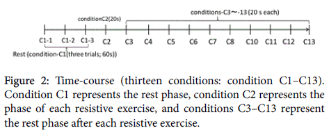
H-reflexes and M-waves (1 Hz) were elicited sequentially without interruption for 320 s. The 320 s study duration was divided into 13 conditions (condition C1, 80 s; conditions C2–C13, 20 s each. C1 (four 20 s trials) represented the resting phase, C2 represented the resistive exercise phase, and C3–C13 represented the resting phases after the resistive exercise, as shown in Figures 1 and 2. The intensity of the induction of H-reflexes with small M-waves via the tibial nerve was determined in C1. The initial stimulus intensity was maintained constant for each subject throughout all of the subsequent experimental trials.
Parameter of excitability
The H-reflex amplitude during and after each resistive exercise (C1– C13) was standardized to the corresponding Mmax H-reflex (the H/ Mmax ratio).
Statistical analysis
All statistical analyses were performed using SPSS ver. 21.0 for Windows (IBM Corp., Somers, NY).
The three-way analysis of variance (ANOVA) for the H/Mmax ratio with Scheffé’s post-hoc tests was used to determine the effects of individuals, time course (thirteen conditions: conditions C1–C13), and RSCPD type (four levels; straight-weak, straight-strong, diagonal-weak, and diagonal-strong). P-values < 0.05 were considered significant.
Results
To assess the reliability of the measures of the H-reflexes of the soleus (peak-to-peak amplitude), three trials were conducted under C1 and analyzed using ANOVA, in order to derive the interclass coefficients (ICCs). The ICC [1,3] was 0.98 for the H-reflexes of the soleus, which indicated a significant degree of consistency in C1.
The means and SDs of the H/Mmax ratio for different time courses are presented in Table 1.
| Time course | Mean | SD |
|---|---|---|
| C1 | 0.42 | 0.38 |
| C2 | 0.52 | 0.53 |
| C3 | 0.42 | 0.4 |
| C4 | 0.39 | 0.35 |
| C5 | 0.38 | 0.34 |
| C6 | 0.38 | 0.34 |
| C7 | 0.44 | 0.47 |
| C8 | 0.41 | 0.39 |
| C9 | 0.42 | 0.4 |
| C10 | 0.41 | 0.38 |
| C11 | 0.41 | 0.39 |
| C12 | 0.4 | 0.39 |
| C13 | 0.41 | 0.38 |
| Condition C1 represents the rest phase, condition C2 represents the phase of each resistive exercise, and conditions C3–C13 represent the rest phase after each resistive exercise. | ||
Table 1: Means and standard deviations of the H/Mmax ratio of each time course
The means and SDs of the H/Mmax ratio of each direction–strength combination are presented in Table 2.
| RSCPD | Mean | SD |
|---|---|---|
| diagonal-weak | 0.4 | 0.37 |
| straight-weak | 0.42 | 0.4 |
| diagonal-strong | 0.42 | 0.39 |
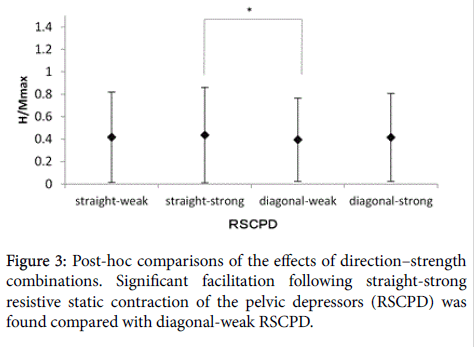
| straight-strong | 0.44 | 0.42 |
Table 2: Means and standard deviations of the H/Mmax ratio of each direction–strength combination.
The three-way ANOVA revealed significant main effects of direction–strength combinations [F (3,765) = 4.47, P = 0.00], time course [F (12,765) = 6.63, P = 0.00], individuals [F (15,765) = 624.75, P = 0.00, partial eta squared = 0.93; observed power=1.00], no significant interactions between time course and direction–strength combinations [F (36,765) = 0.59, P = 0.97].
The results of three-way ANOVA suggested that RSCPD-induced descending neurophysiological effects on the H-reflex of the soleus depend on both the direction–strength combinations of RSCPD and the time course, but no significant relationship was noted between the direction–strength combinations of RSCPDs and time course because of non-significant interactions.
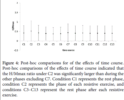
Post-hoc comparisons of the time course effects indicated that the H/Mmax ratio under C2 (during RSCPD) was significantly greater than that during the other phases, with the exception of excluding C7 (Table 1, Figures 3 and 4). In addition, neutral-strong RSCPD elicited significant facilitatory effects on the H-reflex of the soleus compared with those produced by diagonal-weak RSCPD (Table 2 and Figure 3).
Figure 4: Post-hoc comparisons for of the effects of time course. Post-hoc comparisons of the effects of time course indicated that the H/Mmax ratio under C2 was significantly larger than during the other phases excluding C7. Condition C1 represents the rest phase, condition C2 represents the phase of each resistive exercise, and conditions C3–C13 represent the rest phase after each resistive exercise.
Discussion
Post-hoc comparisons of the effects of time course indicated that the H/Mmax ratio under C2 (during RSCPD) was significantly larger than that during the other rest phases excluding C7 (the 80–100 s phase after RSCPD), indicating a temporary facilitatory effect after the inhibitory effects following RSCPD. While the ascending remote rebound-effects on the FCR H-reflex induced by RSCPD elicited an initial reflexive inhibitory phase during RSCPD, followed by a gradual facilitatory phase after RSCPD [6], the RSCPD on the H-reflex of the soleus elicited an initial reflexive facilitatory phase during RSCPD followed by gradual inhibitory phases after RSCPD, excluding C7.
After a vigorous session of concentric-eccentric triceps surae exercise, as a direct approach (eight sets of 10 repetitions), the amplitudes of the soleus and lateral gastrocnemius H-reflexes were attenuated for 10–60 s [11]. When the plantar flexors were active at 10% of the MVC as a direct approach, the H/Mmax ratio also decreased significantly and only returned to baseline levels after 1 min [12]. The reduction in H/Mmax was further attenuated and was more pronounced in the lateral gastrocnemius compared with the soleus muscle [12].
In contrast to the direct approach, the indirect approach employed in this study revealed that significant inhibition occurred immediately after RSCPD, and the values gradually returned to baseline levels, after which a temporary facilitatory phase under C7, and a subsequent inhibitory phase from C8 to C13 were observed (Figure 4).
Neutral-strong RSCPD influences the facilitatory effects on the H-reflex of the soleus, reflecting the facilitation of the reflex excitability of the motor neurons compared with diagonal-weak RSCPD. The straight line from the upper (right) ischial tuberosity of the pelvis to the lower acromion process in this study was equivalent to the lines in our previous methods of RSCPD [2,5,6].
Conversely, diagonal-weak RSCPD produced significant inhibitory effects on the H-reflex of the soleus compared with the neutral-strong RSCPD.
The descending neurophysiological effects of RSCPD on the H-reflex of the soleus included movement direction and load-dependent activity. The induction of descending neurophysiological effects for facilitation or inhibition requires the consideration of the force direction and resistance level.
It was suggested that there is a central interaction between load-related afferent input from the periphery and descending signals [13]. Elucidating load-dependent adaptations in monopedal stance, an increase in loading revealed a significant increase in the H/Mmax ratio; thereby, a reduction of the loading level led to a decrease of the H/Mmax ratio [14]. These receptor signals might arise from Golgi tendon organs and be conducted via type Ib afferents to the spinal locomotor generator [13], indicating that the strength of SCs of the lower trunk muscles, such as RSCPD, may influence the muscles of the lower extremity via Ib afferents.
The increase of the amplitude of the soleus H-reflex as a descending neurophysiological effect during RSCPD may also reflect increased motor unit response and underlie enhanced muscle contractility [9], which leads to improved AROM of the lower extremities. A decrease of the amplitude of the soleus H-reflex following RSCPD may also reduce muscle compliance leading to improvement in the PROM of the lower extremities. If the objective of the therapy is to increase PROM, weak-diagonal RSCPD may be an efficient method for inducing inhibition. Conversely, if the aim of the therapy was to increase AROM, strong-straight RSCPD may be an efficient method for inducing facilitation.
As to remote descending effects, remote descending facilitation of the quadriceps motor nucleus was induced following voluntary contraction of the upper limb muscles (wrist extensors) as a Jendrássik maneuver (JM) [15]. JM effects should not be limited to the monosynaptic reflex pathway but extend to the entire response elicited in the stretched muscle [16].
JM operates by gating a long loop, possibly transcortical pathway [16]. RSCPD may be also influenced by brain activities. Further research is needed to identify the neurophysiological remote aftereffects of the RSCPD. The central nervous system plays a prominent role in muscle stiffness [17]. Establishing the evidence base for identifying efficient methods for improving ROM of the remote joints requires further investigation using modalities such as fMRI.
Conclusion
Compared with diagonal-weak RSCPD, neutral-strong RSCPD exerts a facilitatory effect on the H-reflex of the soleus, which reflects facilitation of the reflex excitability of the motor neurons.
Induction of descending neurophysiological effects for facilitation or inhibition requires consideration of the force direction and resistance level.
References
- Post M, Bayrak S, Kernell D, Zijdewind I (2008) Contralateral muscle activity and fatigue in the human first dorsal interosseous muscle. J Appl Physiol (1985) 105: 70-82.
- Arai M, Shiratani T (2015) Effect of remote after-effects of resistive static contraction of the pelvic depressors on improvement of restricted wrist flexion range of motion in patients with restricted wrist flexion range of motion. J Bodyw Mov Ther 19: 442-446.
- Trueblood PR, Walker JM, Perry J, Gronley JK (1989) Pelvic exercise and gait in hemiplegia. Phys Ther 69: 18-26.
- Shiratani T, Arai M, Masumoto K, Akagi S, Shimizu A, et al. (2013) Effects of a resistive static contraction of the pelvic depressors technique on the passive range of motion of the knee joints in patients with lower-extremity orthopedic problems. PNF Res 13: 8-17.
- Arai M, Shiratani T (2012) The remote after-effects of a resistive static contraction of the pelvic depressors on the improvement of active hand-behind-back range of motion in patients with symptomatic rotator cuff tears. Biomed Res 23: 415-419.
- Arai M, Shiratani T, Shimizu ME, Shimizu H, Tanaka Y, et al. (2012) Neurophysiological study of remote rebound-effect of resistive static contraction of lower trunk on the flexor carpi radialis H-reflex. Current Neurobiol 3: 25-29.
- Shiratani T, Arai M, Shimizu ME, Nitta O, Masumoto K, et al. (2014) Effects of a resistive static contraction of the pelvic depressors technique on the active range of motion of the knee joints in patients with lower-extremity orthopedic conditions. PNF Res 14: 1-10.
- Maffiuletti NA, Martin A, Van Hoecke J, Schieppati M (2000) The relative contribution to the plantar-flexor torque of the soleus motor units activated by the H reflex and M response in humans. Neurosci Lett 288:127-130.
- Knikou M (2008) The H-reflex as a probe: pathways and pitfalls. J Neurosci Methods 171: 1-12.
- Crone C, Hultborn H, Mazières L, Morin C, Nielsen J, et al. (1990) Sensitivity of monosynaptic test reflexes to facilitation and inhibition as a function of the test reflex size: a study in man and the cat. Exp Brain Res 81: 35-45.
- Trimble MH, Harp SS (1998) Postexercise potentiation of the H-reflex in humans. Med Sci Sports Exerc 30: 933-941.
- Xenofondos A, Patikas D, Koceja DM, Behdad T, Bassa E, et al. (2015) Post-activation potentiation: The neural effects of post-activation depression. Muscle Nerve 52: 252-259.
- Dietz V, Muller R, Colombo G (2002) Locomotor activity in spinal man: significance of afferent input from joint and load receptors. Brain 125: 2626-2634.
- Ritzmann R, Freyler K, Weltin E, Krause A, Gollhofer A (2015) Load Dependency of Postural Control--Kinematic and Neuromuscular Changes in Response to over and under Load Conditions. PLoS One 10: e0128400.
- Toulouse P, Delwaide PJ (1980) Reflex facilitation by remote contraction: topographic aspects. Arch Phys Med Rehabil 61: 511-516.
- Nardone A, Schieppati M (2008) Inhibitory effect of the Jendrassik maneuver on the stretch reflex. Neuroscience 156: 607-617.
- Milner TE, Cloutier C, Leger AB, Franklin DW (1995) Inability to activate muscles maximally during cocontraction and the effect on joint stiffness. Exp Brain Res 107: 293-305.
Relevant Topics
- Electrical stimulation
- High Intensity Exercise
- Muscle Movements
- Musculoskeletal Physical Therapy
- Musculoskeletal Physiotherapy
- Neurophysiotherapy
- Neuroplasticity
- Neuropsychiatric drugs
- Physical Activity
- Physical Fitness
- Physical Medicine
- Physical Therapy
- Precision Rehabilitation
- Scapular Mobilization
- Sleep Disorders
- Sports and Physical Activity
- Sports Physical Therapy
Recommended Journals
Article Tools
Article Usage
- Total views: 11712
- [From(publication date):
June-2016 - Apr 02, 2025] - Breakdown by view type
- HTML page views : 10763
- PDF downloads : 949


