Research Article Open Access
The Effect of EMD Application for Intentional Replantation of Periodontally Involved Teeth in Dogs
Akira Saito1 and Emiko Saito2*
1Department of Oral Rehabilitation, Division of Oral Functional Science, Hokkaido University Graduate School of Dental Medicine, Sapporo, Japan
2Department of Periodontology and Endodontology, Division of Oral Health Science, Hokkaido University Graduate School of Dental Medicine, Sapporo, Japan
- *Corresponding Author:
- Emiko Saito, DDS, PhD
Department of Periodontology and Endodontology
Division of Oral Health Science
Hokkaido University Graduate School of Dental Medicine Kita-13
Nishi-7, Kita-ku, Sapporo 060-8586, Japan
Tel: +81 11 706 4266
Fax: +81 11 706 4334
E-mail: water@den.hokudai.ac.jp
Received date: April 04, 2014; Accepted date: May 30, 2014; Published date: June 06, 2014
Citation: Saito A, Saito E (2014) The Effect of EMD Application for Intentional Replantation of Periodontally Involved Teeth in Dogs. J Interdiscipl Med Dent Sci 2:125. doi:10.4172/2376-032X.1000125
Copyright: © 2014 Saito A, et al. This is an open-access article distributed under the terms of the Creative Commons Attribution License, which permits unrestricted use, distribution, and reproduction in any medium, provided the original author and source are credited.
Visit for more related articles at JBR Journal of Interdisciplinary Medicine and Dental Science
Abstract
Background: Intentional replantation of periodontally involved teeth has been reported to result in unfavorable healing like root resorption and ankylosis. However, many recent clinical reports using enamel matrix derivative (EMD) showed a good outcome based on clinical and radiographic examination. However, histological findings are lacking. The purpose of this study was to evaluate healing after intentional replantation with EMD in a periodontally involved teeth model.
Methods: A total of 20 incisors from seven beagle dogs were used. The periodontal ligament and cementum 5 mm from the coronal part of the roots were removed, whereas those in the apical part were preserved. Ten teeth of the experimental group were transplanted following application of EMD to the root surface. Ten teeth from the control group were transplanted without application. Eight weeks after transplantation, periodontal healing was analyzed.
Results: Surface root resorption in the experimental group was significantly greater than in the control. New cementum formation was observed near the apical end of the planed root of the EMD group. Replacement resorption of the EMD group was significantly less than in the control. There was no significant difference in inflammatory resorption between groups.
Conclusion: The combination use of EMD in intentional replantation resulted in new cementum formation on the root planed surface and inhibited root resorption and ankylosis. However, root resorption occurred at the coronal part in areas where the surface was root planed.
Keywords
Intentional replantation; Root resorption; Ankylosis; Enamel matrix derivative; Periodontal; Animal model; Cementum
Introduction
Intentional replantation is performed in cases requiring apicoectomy, root sealing of teeth that are difficult to cure by root canal therapy [1-5], filling at the perforation site [6] and the adhesion of root fracture [7-11] outside the oral cavity. Teeth which are out of occlusion due to missing antagonists, especially third molars, are implanted to another place in the jaw intentionally to improve oral function by intentional tooth implantation [12].
Intentional replantation was reported mostly by studies which used clinical and radiographic evaluation to investigate the success rate and a high survival rate of 50% [13] to 95% [14] was reported. The most common causes of failure are root resorption and ankylosis caused by damage/lacking of periodontal ligament [15-18]. As patients requiring intentional tooth replantation are mostly elderly people as compared to those with avulsed teeth, the teeth would often be periodontally involved.
Some clinical studies reported a good outcome when using a combination of intentional replantation and Enamel Matrix Derivative (EMD) for severe periodontally involved teeth [14,19]. It is suggested that the remaining periodontal ligament cells have the potential to populate on the root planed surface and enhance connective attachment using EMD [20].
However, histometric evaluations in clinical and animal models are lacking and it is not known whether periodontal ligament cells populating the root-planed surface after application of EMD would induce formation of connective tissue attachment.The purpose of this study was to evaluate healing after intentional replantation with EMD in a periodontally involved tooth model.
Materials and Methods
Preparation and transplantation of teeth
Twenty maxillary incisors of seven beagle dogs (male, 1 year old; mean weight, 11 kg) were used. This study protocol (No. 08-0257) followed the guidelines for care and use of laboratory animals of the Graduate School of Medicine, Hokkaido University. The dogs received plaque control, consisting of twice-weekly brushing and application of 0.5% chlorhexidine gluconate solution, in order to establish healthy gingival conditions prior to surgical procedures.
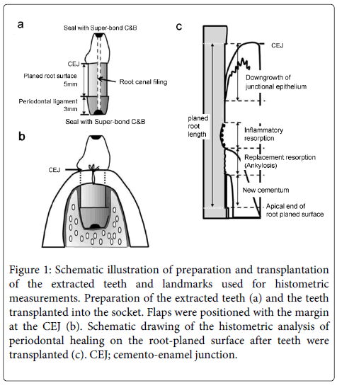
The surgical procedures were performed under anesthesia by an intramuscular injection of medetomidine hydrochloride (5 μg/kg, Domitor®; Meiji Seika, Tokyo, Japan) and ketamine hydrochloride (2.9 mg/kg, Ketaral 50®; Sankyo, Tokyo, Japan) and local infiltration (xylocaine 2% with 1:80,000 epinephrine, Xylocaine®; DENTSPLY SANKIN, Tokyo, Japan). We used teeth that were not adjacent in this study in order to fix to adjacent teeth after implantation. The incisors were carefully extracted with forceps and then immersed in sterile phosphate buffer saline (PBS). Approximately half of the crown was excised and the length of the root was adjusted to 8 mm using a bur and the contents of the root canal were removed with Kerr type file and Peeso reamer. The root canal was filled with gutta-percha and ZOE-free sealer (Canals®N; Showa Yakuhin, Tokyo, Japan). The root-end cavities were prepared and applied aqueous solution of 10% citric acid and 3% ferric chloride (activator Green®, Sun Medical Co. Ltd., Shiga, Japan) with a Benda® Brush (Centrix, Shelton, CT, USA) for five seconds and then filled with 4-META/MMA-TBB resin (Super-bond® C&B, Sun Medical Co. Ltd., Shiga, Japan). Periodontal ligament and cementum were removed 5 mm from the coronal part of the roots and the root surfaces were planned. The periodontal ligament in the apical part of the root was retained (Figure 1a). At this point, the teeth were randomly assigned to one of two treatment groups based on a random computer-generated list.
Figure 1: Schematic illustration of preparation and transplantation of the extracted teeth and landmarks used for histometric measurements. Preparation of the extracted teeth (a) and the teeth transplanted into the socket. Flaps were positioned with the margin at the CEJ (b). Schematic drawing of the histometric analysis of periodontal healing on the root-planed surface after teeth were transplanted (c). CEJ; cemento-enamel junction.
In 10 teeth in the EMD group, the root-planed surface was conditioned with 24% EDTA (pH 7.4) for 2 min and rinsed with PBS. Then, 0.5 ml of EMD was applied over the entire root-planed surface. Then, the teeth were placed into the sockets and the gingival margin was positioned with the margin at the cemento-enamel junction (CEJ) of the replanted teeth (Figure 1b). The teeth were adjusted to occlude and fixed to adjacent teeth with Super-bond C&B.
Ten teeth in the control group were similarly prepared including root conditioning and replanted without application of EMD.
Plaque control included once-weekly brushing and application of 0.5% chlorhexidine gluconate solution throughout the healing period.
Histological processing and histometric analysis
The dogs were sacrificed 8 weeks after replantation. Block sections were dissected, fixed in 10% buffered formalin, decalcified in Plank-Rychlo solution, trimmed, dehydrated, and embedded in paraffin. Serial sections of 3-4 μm thickness were prepared in the bucco-lingual plane. Sections were stained with hematoxylin and eosin.
Each section was carefully measured once by a single examiner, who was blind to the status of the block. Three sections (500 mm apart) representing the central part of the root was selected for measurements. The following measurements were performed on the root-planed surface of the teeth by light microscopy (Figure 1c).
Planed root length: distance between CEJ and apical extent of the root-planed surface; 2) Down growth of junctional epithelium: distance from CEJ to apical margin of the junctional epithelium; 3) New cementum: longitudinal length of regenerated cementum or cementum-like deposit on the root with or without resorption lacunae; 4) Inflammatory resorption: resorption lacunae on the root surface with inflammatory cells in the area; and 5) Replacement resorption (Ankylosis): the periodontal ligament is replaced by bone. The alveolar bone is in contact with cementum or dentin. Values were expressed in percentages of planed root length.
Data Analysis
The mean and standard deviation for each measurement were calculated for each tooth from selected sections. Differences between two groups were statistically analyzed using the Mann-Whitney U test.
Results
Clinical healing after transplantation of teeth was uneventful with minimum indications of inflammation throughout the experimental period.
Histological observations
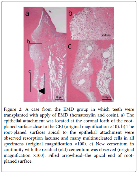
EMD group - The epithelial attachment was located in the coronal fourth of root planed surface close to the CEJ (Figure 2). Connective tissue was observed at the apical part of the epithelium and the root planed surface with resorption lacunae containing many multinucleated cells were observed in all specimens. New cementum formation was limited to the apical part of the root planed surface in all specimens. However, new cementum was observed on root surface with shallow root resorption lacunae. Ankylosis was observed in two specimens.
Figure 2: A case from the EMD group in which teeth were transplanted with apply of EMD (hematoxylin and eosin). a) The epithelial attachment was located at the coronal forth of the rootplaned surface close to the CEJ (original magnification ×10). b) The root-planed surfaces apical to the epithelial attachment were observed resorption lacunae and many multinucleated cells in all specimens (original magnification ×100). c) New cementum in continuity with the residual (old) cementum was observed (original magnification ×100). Filled arrowhead=the apical end of rootplaned surface.
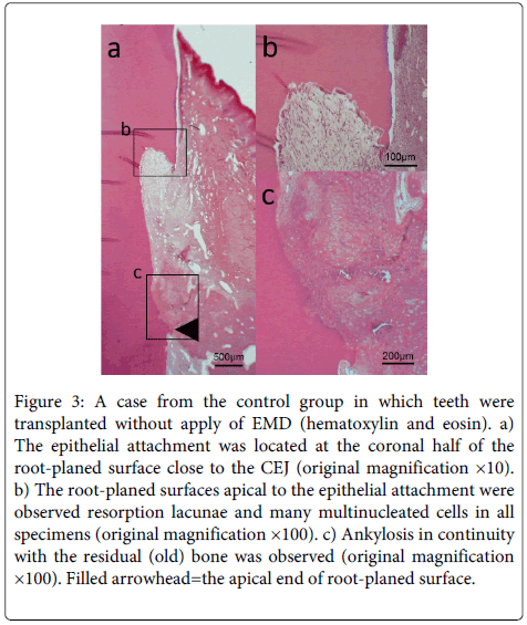
Control group - The epithelial attachment was located at the coronal half of root planed surface close to the CEJ (Figure 3). Connective tissue was observed at the apical part of the epithelial attachment and the root planed surface with many resorption lacunae containing many multinucleated cells was observed in all specimens. Ankylosis was noted at the apical part of the root planed surface in eight specimens.
Figure 3: A case from the control group in which teeth were transplanted without apply of EMD (hematoxylin and eosin). a) The epithelial attachment was located at the coronal half of the root-planed surface close to the CEJ (original magnification ×10). b) The root-planed surfaces apical to the epithelial attachment were observed resorption lacunae and many multinucleated cells in all specimens (original magnification ×100). c) Ankylosis in continuity with the residual (old) bone was observed (original magnification ×100). Filled arrowhead=the apical end of root-planed surface.
Histometric Analysis
New cementum formation in the EMD group was 30.41 ± 6.46%, significantly greater than that in the control group (P<0.05) (Table 1). Replacement resorption (ankylosis) was 2.93 ± 5.73% in the EMD group, significantly less than that in the control group (P<0.05). Downgrowth of junctional epithelium in the EMD group were less than that in the control group, but was not statistically significant.
| parameter | EMD group (n = 10) | Control group (n = 10) | P value |
|---|---|---|---|
| Downgrowth of junctional epithelium (%) | 28.36±8.96 | 55.44±27.9 | NS |
| New cementum (%) | 30.41±6.46 | 0.36±0.49 | <0.05 |
| Inflammatory resorption (%) | 23.6±7.2 | 14.28±12.69 | NS |
| Replacement resorption (%) | 2.93±5.73 | 24.56±19.59 | <0.05 |
Table 1: Histometric Analysis of Periodontal Healing. SD=standard deviation; n=number of sites; P value by Mann-Whitney U test. (Group means ± SD in percentages).
Discussion
Nyman et al. [21] reported that root resorption and ankylosis occurred on the root-planed surface during healing after replantation with periodontally involved teeth in 1985. Periodontal ligament cells covering the denuded root surface are required for normal healing of implanted and replanted teeth. Saito et al. observed connective tissue attachment on the denuded root surface of teeth covered with proliferating cultured periodontal ligament [22] and Zhou et al. reported similar findings when using periodontal ligament sheet [23].
Several studies have reported that EMD stimulated proliferation of periodontal ligament in vitro [20,24,25]. Recently, a clinical study reported the use of severe periodontally involved teeth for intentional replantation and showed the effect of EMD on periodontal repair [26,27]. However, most of these findings were based on radiographic and clinical examination and histological analysis had not been performed. In this study, we used periodontally involved teeth that had 5 mm of the root surface planed from the coronal part of the roots, to examine the effect and behavior of EMD on intentional replantation of periodontally involved teeth with partially remaining healthy periodontal ligament. New cementum formation was observed about 1.5 mm from the coronal part in the EMD group, significantly greater than that in the control group. This result was similar to that of a study that examined the effect of EMD in a periodontitis model with vertical bone defect [28-31]. These suggested that EMD could promote migration, proliferation and differentiation of undifferentiated periodontal ligament cells onto the root planed surface adjacent to remaining healthy periodontal ligament and could enhance new cementum formation.
Regarding inflammatory root resorption, there was no significant difference between the two groups. Yagi et al. [32] have reported that amelogenins inhibited root resorption by decreasing the number of osteoclasts. Amelogenin is the main component of EMD. However, EMD did not definitely show inflammatory root resorption of the implantation teeth model in which the periodontal ligament was removed [33-36]. On the other hand, several studies using animal models have reported that root application and root filling of EMD were effective against root resorption [19,37-39]. Therefore, EMD might be effective under limited conditions. However, the periodontitis model in this study could not show the potency of EMD. Root resorption was observed on root planed surface at the coronal portion in both groups and the site was thought to be where periodontal ligament cell could not migrate and proliferate. Root resorption could be inhibited if periodontal ligament cells covered all over the root surface. Therefore, it was important that a circumference could be ensured where the periodontal ligament did not inhibit migration and proliferation. For example, the combination of EMD and GTR might be effective in a clinical setting.
Ankylosis in the EMD group was significantly less than that in the control group. This result was similar to that of Iqbal et al., who used a replantation model with damaged periodontal ligament or no periodontal ligament [19,34,35]. However, Arau´jo et al. reported that ankylosis in EMD application group was larger than that of the no EMD application group [33]. They submerged the replantation tooth and continued to submerge it until the end of the observation period. In general, ankylosis occurred over a wide area without occlusion in an appropriate time after replantation [39,40]. Jianq et al. reported that EMD enhanced proliferation of osteoblasts in vitro and reduced characteristics of osteoblasts such as ALP activity, type I collagen, ALP, runt-related protein 2, osteocalcin, bone sialoprotein and osteopontin, and reversed the ability of cell attachment [41]. Therefore, EMD was thought to inhibit ankylosis in a clinical model that brings teeth into occlusion after replantation.
This study suggested that intentional replantation with EMD application induced cementum formation by periodontal ligament cells that had migrated, proliferated and differentiated from the adjacent remaining periodontal ligament to the root-planed surface by EMD. Periodontal ligament cells inhibited ankylosis and root resorption; however, root resorption occurred at the coronal portion in areas where the surfaces were extensively root planed. If the attachment between root planed surface and connective tissue attachment could be quantified, this technique may have the potential to become a foreseeable therapy. Furthermore, creating an environment where migration and proliferation of the periodontal ligament by EMDOGAIN is not inhibited might help to expand indications.
Conclusion
This study demonstrated the combination use of EMD as intentional replantation was formed new cementum on the root planed surface and inhibited root resorption and ankylosis, and on the other hand, root resorption occurred at the coronal portion in the case that root planed surface became extensive.
Acknowledgement
The authors declare that they have no conflict of interests. This study was supported by Grants-in-Aid for Encouragement of Scientific Research (22592307 and 25463211) from the Ministry of Education, Science, Sports and Culture of Japan.
References
- Sherman P Jr (1968) Intentional replantation of teeth in dogs and monkeys. J Dent Res 47: 1066-1071.
- Kaufman AY (1982) Intentional replantation of a maxillary molar. A 4-year follow-up. Oral Surg Oral Med Oral Pathol 54: 686-688.
- Nosonowitz DM, Stanley HR (1984) Intentional replantation to prevent predictable endodontic failures. Oral Surg Oral Med Oral Pathol 57: 423-432.
- Goel BR, Satish C, Suresh C, Goel S (1983) Clinical evaluation of gold foil as an apical sealing material for replantation. Oral Surg Oral Med Oral Pathol 55: 514-518.
- Herrera H, Leonardo MR, Herrera H, Miralda L, Bezerra da Silva RA (2006) Intentional replantation of a mandibular molar: case report and 14-year follow-up. Oral Surg Oral Med Oral Pathol Oral RadiolEndod 102: e85-87.
- Tang PM, Chan CP, Huang SK, Huang CC (1996) Intentional replantation for iatrogenic perforation of the furcation: a case report. Quintessence Int 27: 691-696.
- Kawai K, Masaka N (2002) Vertical root fracture treated by bonding fragments and rotational replantation. Dent Traumatol 18: 42-45.
- Hayashi M, Kinomoto Y, Takeshige F, Ebisu S (2004) Prognosis of intentional replantation of vertically fractured roots reconstructed with dentin-bonded resin. J Endod 30: 145-148.
- Kudou Y, Kubota M (2003) Replantation with intentional rotation of a complete vertically fractured root using adhesive resin cement. Dent Traumatol 19: 115-117.
- Hayashi M, Kinomoto Y, Miura M, Sato I, Takeshige F, et al. (2002) Short-term evaluation of intentional replantation of vertically fractured roots reconstructed with dentin-bonded resin. J Endod 28: 120-124.
- Selden HS (1996) Repair of incomplete vertical root fractures in endodontically treated teeth--in vivo trials. J Endod 22: 426-429.
- Grossman LI (1982) Intentional replantation of teeth: a clinical evaluation. J Am Dent Assoc 104: 633-639.
- Grossman LI, Ship II (1970) Survival rate of replanted teeth. Oral Surg Oral Med Oral Pathol 29: 899-906.
- Filippi A, Pohl Y, von Arx T (2002) Treatment of replacement resorption with Emdogain--a prospective clinical study. Dent Traumatol 18: 138-143.
- Emmertsen E, Andreasen JO (1966) Replantation of extracted molars. A radiographic and histological study. ActaOdontolScand 24: 327-346.
- Asgary S, AlimMarvasti L2, Kolahdouzan A3 (2014) Indications and case series of intentional replantation of teeth. Iran Endod J 9: 71-78.
- Lee EU, Lim HC1, Lee JS1, Jung UW1, Kim US2, et al. (2014) Delayed intentional replantation of periodontally hopeless teeth: a retrospective study. J Periodontal Implant Sci 44: 13-19.
- Andreasen JO, Kristerson L (1981) The effect of limited dryinnremovalof the periodontal ligament after replantation of mature permanentincisors in monkeys. ActaOdontolScand 39: 1-19.
- Kenny DJ, Barrett EJ, Johnston DH, Sigal MJ, Tenenbaum HC (2000) Clinical management of avulsed permanent incisors using Emdogain: initial report of an investigation. J Can Dent Assoc 66: 21.
- Cattaneo V, Rota C, Silvestri M, Piacentini C, Forlino A, et al. (2003) Effect of enamel matrix derivative on human periodontal fibroblasts: proliferation, morphology and root surface colonization. An in vitro study. J Periodontal Res 38: 568-574.
- Nyman S, Houston F, Sarhed G, Lindhe J, Karring T (1985) Healing following reimplantation of teeth subjected to root planing and citric acid treatment. J ClinPeriodontol 12: 294-305.
- Saito A, Saito E, Yoshimura Y, Takahashi D, Handa R, et al. (2011) Attachment formation after transplantation of teeth cultured with enamel matrix derivative in dogs. J Periodontol 82: 1462-1468.
- Zhou Y, Li Y, Mao L, Peng H (2012) Periodontal healing by periodontal ligament cell sheets in a teeth replantation model. Arch Oral Biol 57: 169-176.
- Gestrelius S, Andersson C, Lidström D, Hammarström L, Somerman M (1997) In vitro studies on periodontal ligament cells and enamel matrix derivative. J ClinPeriodontol 24: 685-692.
- Van der Pauw MT, Van Den Bos T, Everts V, Beertsen W (2000) Enamel matrix-derived protein stimulates attachment of periodontal ligament fibroblast and enhances alkaline phosphatase activity and transforming growth factor 1 release of periodontal ligament and gingival fibroblasts. J Periodontol 71: 31-43.
- Baltacioglu E, Tasdemir T, Yuva P, Celik D, Sukuroglu E (2011) Intentional replantation of periodontally hopeless teeth using a combination of enamel matrix derivative and demineralized freeze-dried bone allograft. Int J Periodontics Restorative Dent 31: 75-81.
- Sugai K, Sato S, Suzuki K, Ito K (2008) Intentional reim plantation of a tooth with severe periodontal involvement using enamel matrix derivative in combination with guided tissue regeneration and bone grafting: a case report. Int J Periodontics Restorative Dent 28: 89-94.
- Heijl L, Heden G, Svärdström G, Ostgren A (1997) Enamel matrix derivative (EMDOGAIN) in the treatment of intrabony periodontal defects. J ClinPeriodontol 24: 705-714.
- Froum SJ, Weinberg MA, Rosenberg E, Tarnow D (2001) A comparative study utilizing open flap debridement with and without enamel matrix derivative in the treatment of periodontal intrabony defects: a 12-month re-entry study. J Periodontol 72: 25-34.
- Pontoriero R, Wennström J, Lindhe J (1999) The use of barrier membranes and enamel matrix proteins in the treatment of angular bone defects. A prospective controlled clinical study. J ClinPeriodontol 26: 833-840.
- Tonetti MS, Lang NP, Cortellini P, Suvan JE, Adriaens P, et al. (2002) Enamel matrix proteins in the regenerative therapy of deep intrabony defects. J ClinPeriodontol 29: 317-325.
- Yagi Y, Suda N, Yamakoshi Y, Baba O, Moriyama K (2009) In vivo application of amelogenin suppresses root resorption. J Dent Res 88: 176-181.
- Araújo M, Hayacibara R, Sonohara M, Cardaropoli G, Lindhe J (2003) Effect of enamel matrix proteins (Emdogain') on healing after re-implantation of "periodontally compromised" roots. An experimental study in the dog. J ClinPeriodontol 30: 855-861.
- Lam K, Sae-Lim V (2004) The effect of Emdogain gel on periodontal healing in replanted monkeys' teeth. Oral Surg Oral Med Oral Pathol Oral RadiolEndod 97: 100-107.
- Molina GO, Brentegani LG (2005) Use of enamel matrix protein derivative before dental reimplantation: a histometric analysis. Implant Dent 14: 267-273.
- Guzmán-Martínez N, Silva-Herzog FD, Méndez GV, Martín-Pérez S, Cerda-Cristerna BI, et al. (2009) The effect of Emdogain and 24% EDTA root conditioning on periodontal healing of replanted dog's teeth. Dent Traumatol 25: 43-50.
- Iqbal MK, Bamaas N (2001) Effect of enamel matrix derivative (EMDOGAIN) upon periodontal healing after replantation of permanent incisors in beagle dogs. Dent Traumatol 17: 36-45.
- Filippi A, Pohl Y, von Arx T (2001) Treatment of replacement resorption with Emdogain--preliminary results after 10 months. Dent Traumatol 17: 134-138.
- Chen CC, Kanno Z, Soma K (2005) Occlusal forces promote periodontal healing of transplanted teeth with enhanced nitric oxide synthesis. J Med Dent Sci 52: 59-64.
- Mine K, Kanno Z, Muramoto T, Soma K (2005) Occlusal forces promote periodontal healing of transplanted teeth and prevent dentoalveolarankylosis: an experimental study in rats. Angle Orthod 75: 637-644.
- Jiang SY, Shu R, Song ZC, Xie YF (2011) Effects of enamel matrix proteins on proliferation, differentiation and attachment of human alveolar osteoblasts. Cell Prolif 44: 372-379.
Relevant Topics
- Cementogenesis
- Coronal Fractures
- Dental Debonding
- Dental Fear
- Dental Implant
- Dental Malocclusion
- Dental Pulp Capping
- Dental Radiography
- Dental Science
- Dental Surgery
- Dental Trauma
- Dentistry
- Emergency Dental Care
- Forensic Dentistry
- Laser Dentistry
- Leukoplakia
- Occlusion
- Oral Cancer
- Oral Precancer
- Osseointegration
- Pulpotomy
- Tooth Replantation
Recommended Journals
Article Tools
Article Usage
- Total views: 16102
- [From(publication date):
June-2014 - Jul 18, 2025] - Breakdown by view type
- HTML page views : 11343
- PDF downloads : 4759


