The Dental Application of Cone Beam CT Imaging
Received: 01-Nov-2022 / Manuscript No. roa-22-82181 / Editor assigned: 03-Nov-2022 / PreQC No. roa-22-82181 (PQ) / Reviewed: 17-Nov-2022 / QC No. roa-22-82181 / Revised: 23-Nov-2022 / Manuscript No. roa-22-82181 (R) / Published Date: 30-Nov-2022 DOI: 10.4172/2167-7964.1000413
Image Article
The use of new imaging technologies in dentistry for diagnosis and treatment evaluation is gaining popularity [1]. Cross-sectional 3D computed tomography (CT) offers a more accurate visualization of the craniofacial structures than the 2D method [2,3] and naturally avoids superimposition and issues caused by amplification.
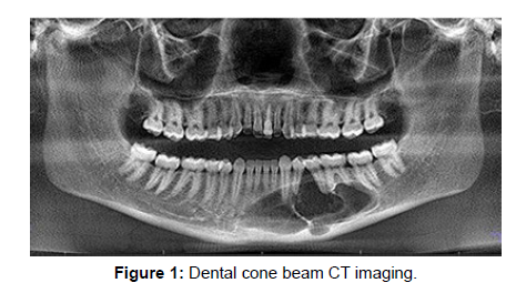
Medical CT (MDCT) was used in dentistry to take pictures right from the start. Concentrations on MDCT demonstrated the accuracy and dependability of 3D CT evaluations of dental procedures [4].Despite these advantages, MDCT uses a significantly higher effective dose than conventional radiographs that make use of dental and maxillofacial imaging. Because of this, it is unrestricted in its application for routine assessments, particularly those related to growth and cephalometric research. In addition, MDCT is an expensive method, and dental specialists and small clinics do not effectively open scanners for maxillofacial imaging (Figure 1).
Another method for maxillofacial imaging, known as cone beam computed tomography (CBCT), was first described in a paper by Mozzo P, et al. [5]. It was only recently proposed. A CBCT check uses a substitute sort of getting that the practice MDCTs. Because the x-ray source produces a cone-shaped beam, it is possible to capture the image in a single shot rather than individually capturing the cut as in MDCT. This method of imaging has the following advantages significantly less radiation than MDCT, the possibility of individual overlap-free reconstructions, and the import and export of DICOM data for various applications. In addition, this imaging technology enables three-dimensional (3D) imaging and data, providing a picture of the craniofacial and dental designs needed for orthodontics and maxillofacial careful applications.
References
- Kumar V, Ludlow J, SoaresCevidanes LH, Mol A (2008) In vivo comparison of conventional and cone beam CT synthesized cephalograms. Angle Orthod 78: 873-879.
- Kumar V, Ludlow JB, Mol A, Cevidanes L (2007) Comparison of conventional and cone beam CT synthesized Cephalograms. Dentomaxillofac Radiol 36: 263-269.
- Sandler PJ (1988) Reproducibility of cephalometric measurements. Br J Orthod 15: 105-110.
- Swennen GR, Schutyser F, Barth EL, De Groeve P, De Mey A (2006) A new method of 3-D cephalometry Part I: the anatomic Cartesian 3-D reference system. J Craniofac Surg 17: 314-325.
- Mozzo P, Procacci C, Tacconi A, Martini PT, Andreis IA (1998) A new volumetric CT machine for dental imaging based on the cone-beam technique: preliminary results. Eur Radiol 8: 1558-1564.
Indexed at, Google Scholar, Crossref
Indexed at, Google Scholar, Crossref
Indexed at, Google Scholar, Crossref
Indexed at, Google Scholar, Crossref
Citation: Patel P (2022) The Dental Application of Cone Beam CT Imaging. OMICS J Radiol 11: 413. DOI: 10.4172/2167-7964.1000413
Copyright: © 2022 Patel P. This is an open-access article distributed under the terms of the Creative Commons Attribution License, which permits unrestricted use, distribution, and reproduction in any medium, provided the original author and source are credited.
Share This Article
Open Access Journals
Article Tools
Article Usage
- Total views: 1708
- [From(publication date): 0-2022 - Mar 29, 2025]
- Breakdown by view type
- HTML page views: 1376
- PDF downloads: 332

