The Crossing Duct Sign of Pancras Divisum
Received: 04-Mar-2024 / Manuscript No. roa-24-130692 / Editor assigned: 06-Mar-2024 / PreQC No. roa-24-130692 / Reviewed: 20-Mar-2024 / QC No. roa-24-130692 / Revised: 25-Mar-2024 / Manuscript No. roa-24-130692 / Published Date: 29-Apr-2024
Pancreas divisum is the most common congenital malformation of the pancreatic ductal system, it has been reported to affect approximately 4-14% of the general population [1].
It results from failure of fusion of dorsal and ventral pancreatic duct during the early weeks of embryonic life (6-8 weeks). As a result, the dorsal duct (Santorini duct) drains most of the pancreatic parenchyma through the minor papilla, whereas the ventral duct (duct of Wirsung) drains a portion of the pancreatic head and uncinate process, through the major papilla [2].
Most patients with pancreas divisum are asymptomatic; however, it has been considered as a predisposing factor for chronic abdominal pain and recurrent idiopathic pancreatitis [3].
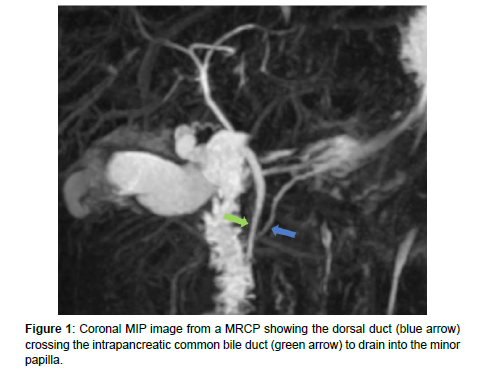
Magnetic resonance cholangiopancreatography (MRCP) is the modality of choice for diagnosis. It shows the typical crossing duct sign (Figure 1) which refers to the joining of the main pancreatic duct (dorsal) into the accessory papilla, crossing over the common bile duct (CBD), which joins with the ventral pancreatic duct into the duodenum at the major papilla.
The treatment of pancreas divisum is typically reserved for symptomatic patients [4]. Endoscopic sphincterotomy is considered the first-line treatment.
References
- Kim HJ, Kim MH, Lee SK, Seo DW, Kim YT, et al. (2022) Normal structure, variations, and anomalies of the pancreaticobiliary ducts of Koreans: a nationwide cooperative prospective study. Gast rointest Endosc 55: 889-896.
- Guirat A, Abid M, Amar MB, Rebai W, Beyrouti MI (2009) Pancréas divisum. La Presse Médicale 38: 1353–1359.
- Mortelé K, Rocha T, Streeter J, Taylor A (2006) Multimodality Imaging of Pancreatic and Biliary Congenital Anomalies. Radiographics 26: 715-731.
- Morgan D, Logan K, Baron T, Koehler R, Smith J (1999) Pancreas Divisum: Implications for Diagnostic and Therapeutic Pancreatography. AJR Am J Roentgenol 173: 193-198.
Indexed at, Google Scholar, Crossref
Indexed at, Google Scholar, Crossref
Indexed at, Google Scholar, Crossref
Citation: Sara E, Imrani K, Faraj C, Ez-zaky S, Billah NM, et al. (2024) TheCrossing Duct Sign of Pancras Divisum. OMICS J Radiol 13: 547.
Copyright: © 2024 Sara E, et al. This is an open-access article distributed underthe terms of the Creative Commons Attribution License, which permits unrestricteduse, distribution, and reproduction in any medium, provided the original author andsource are credited.
Share This Article
Open Access Journals
Article Usage
- Total views: 680
- [From(publication date): 0-2024 - Mar 29, 2025]
- Breakdown by view type
- HTML page views: 499
- PDF downloads: 181

