The Concept of Computer Assisted Implantology: The Accuracy Outcomes!
Received: 19-Sep-2016 / Accepted Date: 04-Oct-2016 / Published Date: 11-Oct-2016 DOI: 10.4172/2161-119X.1000270
253429Introduction
Implantology has made a huge progress in recent years, not only through the development of implants (design, surface finish, connections...) but even more thanks to CAD/CAM.
But how the concept of CAI allows a surgical- prosthetic therapeutic approach different from a so-called "conventional"?
Well In the conventional method; the steps for implant planning are designed to confront an ideal prosthetic project with the patient's anatomy. They help the practitioner to make the right decisions when developing the treatment plan. Depending on the edentulous patient and requests, some criteria are more important than others, but, basically, the general process is the same [1,2].
The digital technology gives us the necessary information to be able to simulate the implant on the computer in advance. This increases our patients’ safety!
The main of this publication is to specify the value of a virtual surgical-prosthetic planning a project with CAD/CAM.
Materials and Methods
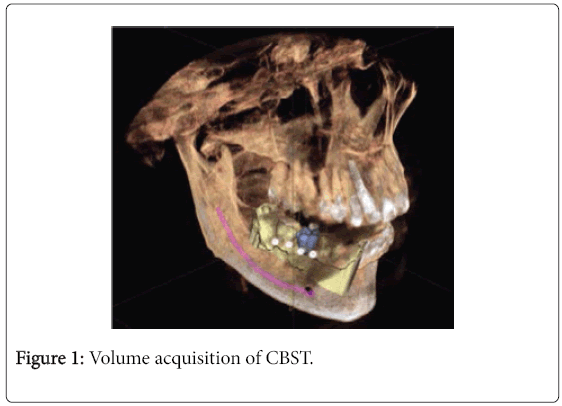
Coupled the CEREC system to CBCT: That’s allow three dimensional acquisition type CBST [1,2].
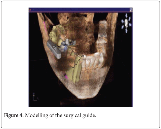
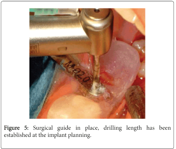
The second application of CAI resides in the ability to accurately guide the surgeon's drill through a "surgical guide". 3D planning is made by using a radiological guide and an X-ray scanner. 3D planning software sends computer data to sophisticated equipment that builds a surgical guide by stereolithography [3].
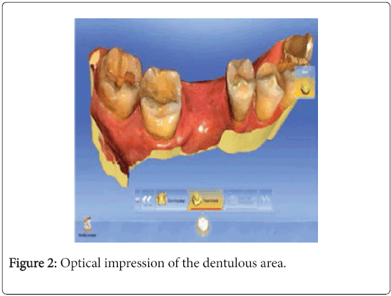
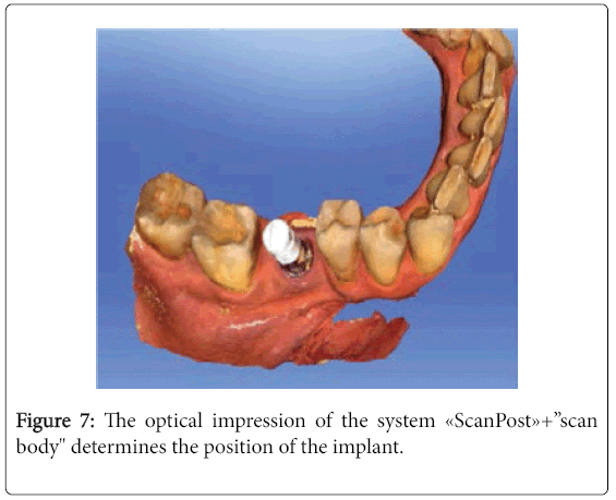
The Second step we have to take is an optical impression by using camera with coating by dioxide titane consequently a virtual three dimensional model is done [4,5].
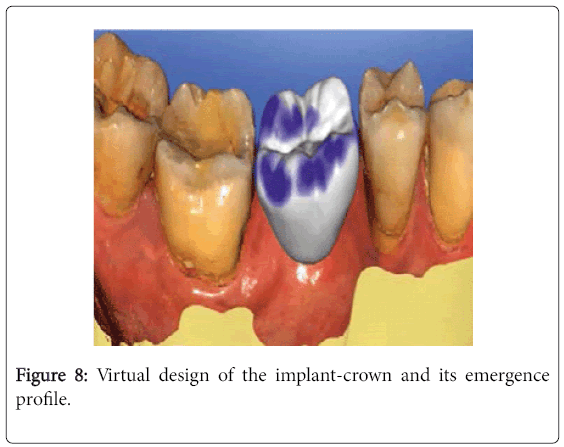
We use CEREC software to plan our prosthetic with superposition data from the CBCT and CEREC.
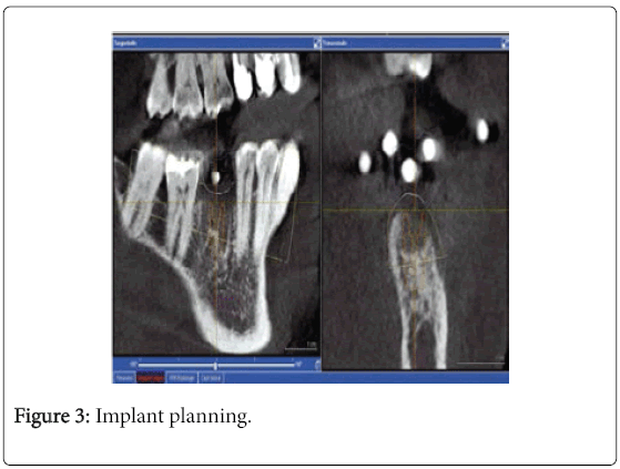
Implant planning through software Galiloes Galaxis. The software alerts us if the correct positioning of implant parameters is not met. The practitioner performs well planning and sees if whether or not a bone planning pre-or per -implant transversely or vertically (bone graft, guided bone regeneration or «sinus lift") [5].
Whether digital or conventional therapy, radiological guide has the same specifications. He must:
• Be rigid,
• Be radiopaque,
• Be stable,
• Not to issue artifacts
• Allow objectify the height of the soft tissue,
• Objectify the ideal axis of the implants
3D software overcomes the lack of a radiological guide in creating a virtually prosthetic project and then doing the implants [6].
It allows the practitioner, through the Cerec software to visualize bone volumes in 3D that will receive the implants and the draft.
Clinical Case
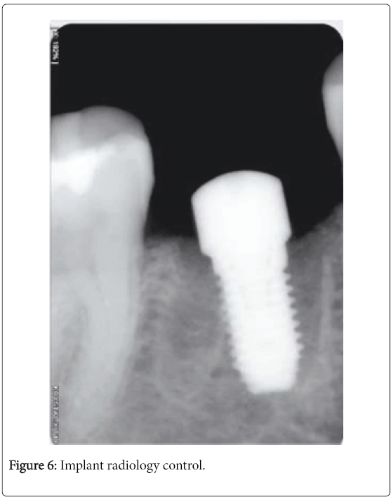
The figures shows the future implants and prosthetic project (Figures 1-8).
Results and Discussion
This concept allows communication and educational tools with software, the practitioner can explain to the patient throughout the treatment by showing the implant sites directly, instead that will make the future implant and the prosthetic project.
CEREC system coupling to CBCT helps to determine the implant site, but also the choice of type of implant, the design and positioning in 3D.
The prosthetic project is modelled virtually (digital wax up) and the surgical guide is designed by (CAD) thus the temporary prosthetic is designed before surgery.
The optical impression makes the maximum reduce of dependence materials (less impression materials, less steps, no disinfection and shipping) [7].
With the conventional method, it takes between 24 and 48 h to manufacture a surgical guide. This is due to numerous laboratory steps (casting plaster, processing of the radiological guide, processing resins, finishing, polishing). With the numerical method, it takes only 15 min to design the guide and only a few minutes for it to be manufactured by the machine drill [8,9].
The study by Johansson et al. [10] showed stabilities prosthetic and implant at 1 year. For them, results are promising for the planned implants digitally, immediately loaded with fixed prosthesis type bridge Procera (NobelBiocare) Prefabricated for rebuilding an edentulous maxilla. The authors observed in a 1 year implant survival rate of 99.4% on 312 implants placed in 52 patients. Marginal bone loss average after a year of follow up was 1.3 mm.
This new implant approach ensures speed, accuracy with many limitations: lack of access to post sites, the surgical guide depends on the type of tooth loss (dental, mucous, bone) with its risk of fracture [9,10].
Errors in the acquisition and processing of radiological images , on average less than 0.5 mm , mistakes when performing the surgical guide, generally around 0.1 to 0.2 mm for the guides made by CAM.
Apical deviation of the position of the implant between the virtual planning and actually obtained implant position ranging between 0.6 mm and 1.2 mm.
Of course this method has his limits such as: High cost.
Conclusion
With implantology, modern dentistry has experienced a breakthrough. This new option, now widely recognized, can treat all types of edentulous. The implant therapy should respect the concept of minimally invasive treatment. In addition, the aesthetic and functional requirements are a central part of the specifications of the treatment because we must not forget that the implant is in the service of the prosthesis.
The Concepts have evolved and today CAI seems to be emerging as a sub discipline through the many contributions and interest it conveys: It could well sign the evolution of a new era.
References
- Delphine l (2014) Thèseuniversité de Nantes N°2014-TOU3-3038: Planificationimplantaire par CFAO à partir d’un projetprothétique et chirurgieassistée par ordinateur: Etat des lieux, avancées et perspectives. Université Toulouse III-Paul Sabatier.
- D'haese J, Van De Velde T, Komiyama A, Hultin M, De Bruyn H (2012) Accuracy and complications using computer-designed stereolithographic surgical guides for oral rehabilitation by means of dental implants: A review of the literature. Clin Implant Dent Relat Res 14: 321-335.
- Ender A, Mehl A (2011) Full arch scans: Conventional versus digital impressions-an in-vitro study. Int J Comput Dent 14: 11-21.
- Amoyel F (2010) Implantologieassistée par ordinateur:Traitement d’un édentementcompletmaxillaire à l’aide du systémeNobelGuide: Le FilDentaire N°53 Avril 2010.
- Telara GL (2012) N ouveaux concepts d’implontologieguidée par ordinateur,PartieI:Forage en temps réel et phase d’implant.
- Georgel S (2012) La chirurgieimplantaireguidéeassistée par ordinateur. Thèseuniversité de Nantcy Metz N°3975.
- (2015) Thèseuniversité de Nantes 2015 N°022: Apport de l’empreinteoptique pour la réalisation des restaurations supra-implantaire.
- Johansson B, Friberg B, Nilson H (2009) Digitally planned, immediately loaded implants with prefabricated prostheses in the reconstruction of edentulous maxillae:A 1-year prospective multicenter study. ClinImpl Dent Relat Res3: 194-200.
Citation: Zenati L, Boukais H, Bouarfa A (2016) The Concept of Computer Assisted Implantology: The Accuracy Outcomes! Otolaryngol (Sunnyvale) 6:270. DOI: 10.4172/2161-119X.1000270
Copyright: © 2016 Zenati L, et al. This is an open-access article distributed under the terms of the Creative Commons Attribution License, which permits unrestricted use, distribution, and reproduction in any medium, provided the original author and source are credited.
Share This Article
Recommended Journals
Open Access Journals
Article Tools
Article Usage
- Total views: 11017
- [From(publication date): 10-2016 - Apr 01, 2025]
- Breakdown by view type
- HTML page views: 10198
- PDF downloads: 819

