The Cluster Sign
Received: 04-Mar-2024 / Manuscript No. roa-24-130691 / Editor assigned: 06-Mar-2024 / PreQC No. roa-24-130691 / Reviewed: 20-Mar-2024 / QC No. roa-24-130691 / Revised: 25-Mar-2024 / Manuscript No. roa-24-130691 / Published Date: 29-Apr-2024
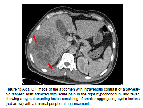
The cluster sign is a radiological finding associated with pyogenic liver abscesses that better appreciated on contrast-enhanced CT and MRI images. It was first described by Jeffrey et al. in 1988 [1].
On CT scan (Figure 1), the cluster sign manifests as a hypoattenuating lesion consisting of smaller aggregating cystic formations, which demonstrate peripheral enhancement on post-contrast imaging. On MRI, the smaller cystic formations cluster together, appearing hypointense on T1-weighted images and hyperintense on T2-weighted images, with rim enhancement noted on post-gadolinium images.
The main differential diagnoses of the liver cluster sign include cystic metastasis and biliary cystadenocarcinoma [2].
References
- Jeffrey RB, Tolentino CS, Chang FC, Federle MP (1988) CT of small pyogenic hepatic abscesses: the cluster sign. Am J Roentgenol 151: 487-489.
- Halvorsen RA, Korobkin M, Foster WL, Silverman PM, Thompson WM (1984) The variable CT appearance of hepatic abscesses. Am J Roentgenol 142: 941-946.
Indexed at, Google Scholar, Crossref
Citation: Sara E, Imrani K, Faraj C, Bahlouli N, Billah NM, et al. (2024) The ClusterSign. OMICS J Radiol 13: 546.
Copyright: © 2024 Sara E, et al. This is an open-access article distributed underthe terms of the Creative Commons Attribution License, which permits unrestricteduse, distribution, and reproduction in any medium, provided the original author andsource are credited.
Share This Article
Open Access Journals
Article Usage
- Total views: 642
- [From(publication date): 0-2024 - Mar 29, 2025]
- Breakdown by view type
- HTML page views: 458
- PDF downloads: 184

