Case Report Open Access
The Cardiac Mass; Is it A Thrombus, Tumor or Vegetation? Take it in the Context of the Disease
Adam M Au*Specialty in Internal Medicine, Plantation General Hospital, 401 Nw 42nd Ave, Plantation, FL 33317, Florida, United States
- *Corresponding Author:
- Adam M Au
Specialty in Internal Medicine
Plantation General Hospital
401 Nw 42nd Ave
Plantation, FL 33317
Florida, United States
Tel: 954-587-5010
Email: dradammau@gmail.com
Received date: June 16, 2016; Accepted date: August 29, 2016; Published date: September 02, 2016
Citation: Adam MA (2016) The Cardiac Mass; Is it A Thrombus, Tumor or Vegetation? Take it in the Context of the Disease. J Clin Diagn Res 4:128. doi:10.4172/2376-0311.1000128
Copyright: © 2016 Adam MA. This is an open-access article distributed under the terms of the Creative Commons Attribution License, which permits unrestricted use, distribution, and reproduction in any medium, provided the original author and source are credited.
Visit for more related articles at JBR Journal of Clinical Diagnosis and Research
Abstract
Background: Masses are common findings in echocardiography and cardiac imaging; largely confusing without surgical and pathological interventions for diagnosis. Method: Through case presentations and peer-reviewed publications, this paper elucidates a scientific methodology on how a clinician can arrive at a timely diagnosis by focusing on the respective properties of the mass on imaging. Results: Twenty-three cardiac masses and two imaging cases are delineated respectively to tumor, vegetation or emboli, as well as other findings. One of the masses is substantiated by histopathological analysis after additional assessment with transesophageal echocardiogram. Conclusion: With eminent symptoms and potentially perilous delay of treatment, a careful examination of cardiac masses provides numerous unique clues in helping the clinician expedite treatment.
Keywords
Valvular vegetation; Infective endocarditis; Cardiac mass; Thrombus; Valvular disease
The Cardiac Mass; Is it a Thrombus, Tumor or Vegetation ?
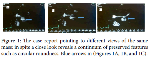
In every-day echocardiography, extra intracardiac structures namely in order of least to most common: tumor, vegetation, and thrombus are encountered and often easily confused without a pathological diagnosis. Sometimes sample anatomical specimens surgically excised are inevitably covered in blood products, and without histopathologic processes are difficult to diagnose (Figure 1A).
Hence to administer a timely treatment for in situ masses, it is necessary to wield a high pretest probability from the perspective of different acoustic windows obtained from transesophageal (TEE), transthoracic (TTE), multiplane imaging modalities as wells as from the patient’s clinical history. Using this approach, the table below unveils the most probable of the three of the named masses by fixating on their properties such as texture, and size variability [1].
Clinical Presentation
A 36 years of old female on multi-drug regimen for end-stage renal disease, insulin dependent diabetes mellitus, systemic arterial hypertension and a history of lung transplant presents with new-onset worsening palpitations [2], and feeling of episodic impending doom (Figure 1B). Initial work up with cardiac troponins are negative, electrocardiogram showed non-specific T wave abnormalities in the lateral precordial leads, while cardiac enzymes were elevated at 0.05 (Table 1). Physical examination shows a patient who is anxious, afebrile with faint 1/6 holosystolic murmur without radiation to axilla and a delayed plop [3]. Bedside echocardiogram in the emergency department was followed with a 2D echocardiogram (Figure 1C).
| Cardiac Masses | Comments on Features | Diagnosis |
|---|---|---|
| Left ventricular apical mass |
Apex of the LV a tapering regional cavity is predisposed to stasis. In association with anterior infarcts, there is a 10-40% incidence of thrombus reported. |
Thrombus |
| Ventricular regional wall mass |
Ventricular infarcts, aneurysms, dyskenesis, akinesis form endothelial injuries which by Virchow’s triad becomethrombogenic. |
Thrombus |
| Non-prosthetic aortic valve mass |
In pseudo aortic stenosis (reduced EF) and true aortic stenosis, stasis in the left ventrcle become precipitating factors for thrombogenicity |
Thrombus |
| Non - prosthetic valve mass on upstream side |
Vegetations are typically located on the upstream side of the valve, are usually irregular grotesque shaped and exhibit disordered motion not in pattern with the valve leaflets’ excursion. |
Vegetation |
| Mass with severe valvular regurgitation |
Unlike thrombi, most vegetations rarely cause stenosis. | Vegetation |
| Mass in cardiomyopathy | There is a 1.6-3.5% incidence of thromboembolic events in patients with CHF stage II-IV for which several studies indicate no benefit from anticoagulation. | Thrombus |
| Rheumatic valvular mass | M protein from Group A Streptococcuilicits an immune cascade leading to disruption of valvular endothelium and the valve basement membrane damage with erythrocyte rouleux formation. |
Thrombus |
| Mechanical prosthetic mitral valve mass |
There is a high occurrence of thrombus formation on mechanical valves,while thrombus on bioprosthetic valves are rare. |
Thrombus |
| Mechanical prosthetic aortic valve mass |
Thrombus are more likely on mechanical mitral valves, pannus formation occurs frequently on prosthetic aortic valves. Pannus are chronic fibrous tissue growth mostly flat and non-mobile and non-sessile. |
Pannus |
| Mass on early bioprosthetic valve |
Non-endothelized sewing rings and suture materials on the ring is adhesive to blood prodcts. |
Vegetation |
| Mass on AICD or Pacemaker lead |
Thoracotomy and device insertion predisposes to vegetation most of which attach to the electric lead. |
Thrombus |
| RA or LA appendage mass |
Morphologies of both appendages, have been associated with erythrocyte sludge formation and eventual thrombogenesis. |
Thrombus |
| Anti-phospholipid syndrome mass |
Libman Sacks verrucous non-bacterail thrombotic endocarditis are common in this herpercoagulableanticardiolipin syndrome |
|
| Pedunculated left atrial mass. |
Myxoma, the most common cardiac mass located in the LA. Thrombus can mimic myxoma even in anticoagulated patients. |
Myxoma |
| Line related mass | While you will suspect that line-related masses are infectious in etiology, on the contrary lines cause more thrombus. Certain factors such as: oscillating motion of the line, chemotherapeutic agents, and choice of specific line material can correlate withthrombogenesis. |
Thrombus |
| Echogenic mass in intravenous drug-user |
One typical example of right-sided masses. Bacteremia and endothelial mass or mass on valves are pointers to endocarditis Using the modified Dukes criteria in prosthetic valve is helpful in arriving at the diagnosis. |
Vegetation |
| Simultaneous biventricularapical obliterating masses | Cardiac involvement in hypereosinophilia affects both the left and right sides with fibrotic fibrin formation, with wall damage and ensuing thrombosis. |
Thrombus |
| Mass on papillary muscle |
Second most common cardiac benign tumors. Attachment usually contiguous with valve leaflet. Mostly found on the aortic valve possibly obstructing the outflow tract. |
Papillary Fibroleastma |
| Mass in a dilated LA. | Structural dilatation in the LA associated with poor forward flow is associated with thrombus formation. |
Thrombus |
| Mass in SLE | Libman-Sacks Vegetation are sterile growth on valvular structures in autoimmune lupus erythematosus. They, like other vegetations can be associated with severe regurgitation. |
Vegetation |
| Recent MI or CABG with mass |
In certain cases of hibernating myocardium post bypass graft, and ventricular infarcts there is a risk for blood stasis. |
Vegetation |
| Adjacent regional wall mass post valvular surgery |
Suturing and prosthetics valves cause artifacts. Shadowing artifacts can be mistaken for masses hence need formultiplane views and parameters for better identification. |
Artifact |
| Pulmonary vein mass | TTE is poor modality for detecting pulmonary emboli. Most masses seen in the proximity of the pulmonary valve should be seen in the context of the RVSP, and clinical symptoms such as hemoptysis for possible neoplastic migration or embolism |
Pulmonaryvalve remnant |
| Severe TV regurgitation | Patient with flushing, wheezing and diarrhea should raise suspicion for malignancy. The 5HIAA disease affects the TV first, except in septal defects without a closure – left heart valves involved. |
Carcinoid |
Table 1: This table is only a guide in addressing cardiac masses.
The ultimate diagnosis, however, depends on their bacteriologic and microscopic properties [4]. In all cases of suspected thrombus or pannus, vegetation should be excluded in the diagnosis. In addition, markers such as d-dimer, fibrin, prothrombin fragments, and serum levels of von Willebrand factor can be pointers to thrombus formation, while auto-immune markers are elevated in SLE [5]. Sometimes relentless search can end up to PCR in the diagnosis of marantic nodules such as in Hodgkin’s (Figure 2).
Lymphoma
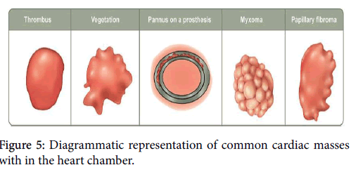
A careful examination of the images in the case demonstrates a well-defined rounded mass freely oscillating ipsilateral to the flow direction [6]. The 2D video images also show the mass coursing the motion of the valve. TEE and the pathologic diagnosis confirmed a thrombus (Figure 5).
Clinical Presentation of a Prosthetic Valve Lesion
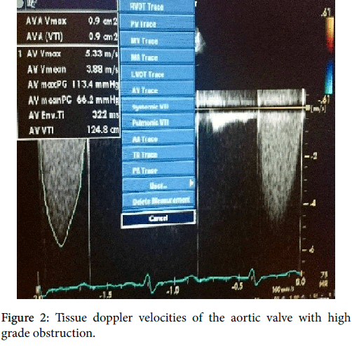
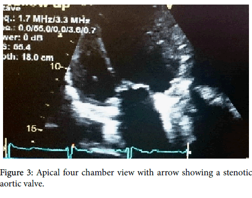
42 years old male referred for evaluation for valvular heart disease with history of valve replacement. Overall, the left ventricular systolic function is preserved with ejection fraction of 65-70%, with moderate to severe concentric left ventricular hypertrophy and a restrictive filling pattern [7]. The aortic valve showed obstructed mechanical prosthetic valve with a large mass noted causing severe aortic stenosis with peak/ mean pressure gradient 120 mmHg/70 mmHg. The estimated aortic valve area by the continuity equation 0.9 cm2 (Figure 3). The tricuspid valve had mild regurgitation with the right ventricular systolic pressure estimated at 65 mmHg [8].
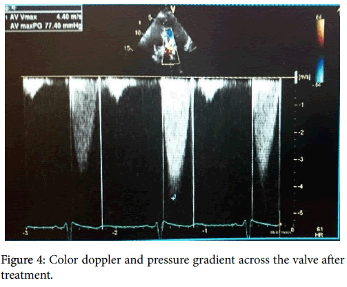
While prosthetic valves have some inherent degree of obstruction, a close look at this view reveals a lesion on the valve [9]. TEE confirmed the diagnosis. While transthoracic echocardiogram without contrast has 85% sensitivity in detecting intracavitary masses, the transesophageal procedure improves the diagnostic accuracy to about 95%, especially if the location is in the atrial appendage [10]. Patient was administered thrombolysis. Post treatment echocardiogram showed aortic valve stenosis with a better peak/mean pressure gradient of 77/40 mmHg with thrombus no longer seen and the right ventricular systolic pressure estimated at 35-40 mmHg (Figure 4). And normal proximal aortic diameter [11,12].
The mass in the second case was also a thrombus befitting its location on a mechanical aortic valve [13]. The clinician administered thrombolysis without surgical intervention or microscopic diagnosis based on most the criteria cited in the table above [14].
Conclusion
Cardiac masses either symptomatic or incidental are common findings in echocardiography and are problematic in precise diagnosis without intricate probing. While well-timed treatment is necessary, it is also absolutely essential to avoid administration of wrong treatment which could potentially be lethal to the patient. For a judicious diagnosis and treatment, characteristics such as regional location, mass morphology, clinical syndrome, and wave with the valve excursion are a few of the numerous clues to guide the clinician in the right direction.
References
- Chaudhry F, Grimm R, Kameswari M, Alicia A, Bruce L, et al. (2016) Guidelines for the Use of Echocardiography in the Evaluation of a Cardiac Source of Embolism: ASE Guideline.
- Alkindi F, Hamada A, Hajar R (2013) Thrombi in Different Clinical Scenarios. Heart Views 14: 101-105.
- Otto C (2009) Cardiac Masses and Potential Cardiac “Source of Embolus”. Textbook of Clinical Echocardiography: Expert Consult. Chapter 15. Saunders.
- Dunkman WB, Johnson GR,Carson PE, Bhat G, Farrell L, et al. (1993) Incidence of thromboembolism in congestive heart failure. The V-HeFT VA Cooperative Studies Group. Circulation 87: VI94-V101.
- Cunningham MW(2004) T cell mimicry in inflammatory heart disease. Molecular Immunology 40: 1121-1127.
- Griffin B, Rodriguez R, Carmela T, Paul C, Douglas J, et al. (Oct. 2015) Early Bioprosthetic Valve Failure: Mechanistic Insights via Correlation between Echocardography and Operative Findings. American Society of Echocardiography. Articles.
- Victor F, Place C De, Camus C,H Le Breton,C Leclercq, et al. (1999) Pacemaker Lead Infection: echocardiographic features, management and outcome. Heart 81: 82-87.
- Dhawan S, Tak T (2004 Oct) Left atrial mass: thrombus mimicking myxoma. Echocardiography 21:621-623.
- Fuchs S, Poliak A, Gilon D (1999) Central venous catheter mechanical irritation of the right atrial free wall: A cause for thrombus formation. Cardiology 91: 169-172.
- Pottecher T, Forrier M, Picardat P, Krause D, Bellocq JP, et al. (1984) Thrombogenicity of central venous catheters: Prospective study of polyethylene, silicone, and polyurethane catheters with phlebography or post-mortem examination. Eur J Anaesthesiol 1: 361-365.
- Alkindi F, Hamada A, Halar R (2013) Cardiac Thrombi in Different Clinical Scenarios. Heart Views 14: 101-105.
- Ogbogu P, Rosing DR, Horne MK (2007) Cardiovascular Manifestations of Hypereosinophilic Syndromes. Immunology and Allergy clinics of North America. 27:457-475.
- Chitwood W(2007) Cardiac Neoplasms: Current Diagnosis, Pathology, and Therapy. Journal of Cardiac Surgery 3: 119-154.
- Roldan C, Tolstrup K, Macias L (2015) LibmanSacks Endocarditis: Detection, Characterization, and Clinical Correlates by Three-Dimensional Transesophageal Echocardiography. American Society of Echocardiography 28: 770-779.
Relevant Topics
- Back Pain Diagnosis
- Cardiovascular Diagnosis
- Clinical Diagnosis
- Clinical Echocardiography
- COPD Diagnosis
- Diabetes Diagnosis
- Diagnosis Methods
- Diagnosis of cancer
- Diagnosis of CNS
- Diagnosis of Diabetes
- Diagnostic Products
- Diagnostics Market Analysis
- Heart diagnosis
- Immuno Diagnosis
- Infertility Diagnosis
- Medical Diagnostic Tools
- Preimplementation Genetic Diagnosis
- Prenatal Diagnostics
- Ultrasonography
Recommended Journals
Article Tools
Article Usage
- Total views: 35402
- [From(publication date):
December-2016 - Mar 30, 2025] - Breakdown by view type
- HTML page views : 33699
- PDF downloads : 1703

