Testicular Epidermoid Cyst: A Case Report.
Received: 04-Jun-2018 / Accepted Date: 14-Jun-2018 / Published Date: 21-Jun-2018 DOI: 10.4172/2161-0681.1000347
Keywords: Epidermoid cyst; Testis; Benign epithelial cysts
Case Presentation
70-year-old man was referred to hospital with the complaints of left testicular pain. In clinical examination, left testis was found as a palpable hard nodule. Right testis was completely normal. Patient had no past history and trauma. Evaluated laboratory tests showed no abnormal findings. Serum alpha-fetoprotein, beta-HCG and LDH were between normal range limits. Ultrasonography showed left testis larger than the right testis. Lesion was present in testicular parenchyma without invasion into the capsule.
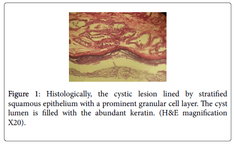
According to clinical and radiographic finding evaluation we diagnosed an initial testicular tumour primarily. The patient underwent orchiectomy of the testicular mass. Then, we performed a frozen section to rule out surgical margins malignancy. The resected mass was measured 5.2 × 7.5 × 1.0 cm. Cut section revealed a welldefined keratinizing cystic mass. Histologic findings revealed a benign cystic lesion lined by stratified squamous epithelium with a prominent granular cell layer. The cyst lumen is filled with the abundant keratin (Figure1). There was no evidence of malignancy. A final diagnosis of epidermoied cyst confirmed. The patient is under regular follow-up since last 10 months without any evidence of recurrence 10 months without any evidence of recurrence.
Discussion
Epidermal cysts are more common in men as in women [4-6]. Testicular epidermoid cyst is very rare .These benign lesions are about 3% of all testicular tumours [7]. The histopathologic features of testicular epidermoid cysts are similar with skin's epidermoid cysts [8]. Microscopically, epidermoid cysts revealed, a cystic lesion covered by stratified squamous epithelium with a prominent granular cell layer. The cyst lumen is filled with the abundant keratin, debris and desquamated cells [9]. Although the epithelial lining not to have malignant potential but malignant changes of basal cell carcinoma or squamous cell carcinoma from epidermal cysts have been reported [10]. Testicular sonographic findings in epidermal cysts described a sharply defined mass with a hyperechoic rim “onion ring” appearance of alternating hypoechogenicity layers of compacted keratin [8]. Magnetic resonance imaging (MRI) findings in these cysts revealed well-defined solid masses surrounded by a fibrous capsule [5]. This finding is similar to our case. Epidermoid cysts the prognosis is good. This cysts usually treated by conservative surgical excision and recurrence is uncommon [4].
Conclusion
Although testicular epidermal cysts are benign epithelial cysts and occurring of these lesions is very rare, malignant transformation has been reported in some cases. Therefore patient’s follow-up is required.
References
- Neville BW, Damm DD, Allen CM, Bouquot JE (2016) Oral and maxillofacial pathology. 4rd ed. Philadelphia: Saunder 357-370.
- Kondo T, Kawahara T, Matsumoto (2016) Epidermal Cyst in the Scrotum Successfully Treated while Preserving the Testis: A Case Report. Case Rep Oncol 9(1): 235–240.
- Dockerty M, Priestly JY (1942) Dermoid cysts of the testis. J Urol 48: 392–397.
- Çakıroglu B, Sönmez N, Sinanoğlu O (2015) Testicular epidermoid cyst. Afr J Paediatr Surg 12(1): 89–90.
- Pattamapaspong N, Muttarak M, Kitirattrakarn P (2014) Clinics in diagnostic imaging (149). Bilateral testicular epidermoid cysts. Georgian Med News 12(3): 14-7.
- Chen ST, Chiou HJ, Pan CC, Shen SH (2016) Epidermoid cyst of the testis: An atypical sonographic appearance. J Clin Ultrasound 44(7): 448-451.
- Bruni SG, Glanc P (2015) Bilateral Epidermoid Cysts of the Testes: A Characteristic Appearance on Ultrasonography. Ultrasound Q 31(3): 205-207.
- Lim J, Cho K (2015) Epidermoid cyst with unusual magnetic resonance characteristics and spinal extension. World J Surg Oncol 13: 240.
- Jeyaraj P, Sahoo NK (2015) An unusual case of a recurrent seborrheic/epidermal inclusion cyst of the maxillofacial region. J Maxillofac Oral Surg 14(1): 176–185.
- Prasad KK, Manjunath RD (2014) Multiple epidermal cysts of scrotum. Indian J Med Res 140: 318.
Citation: Ahmadi MH, Abdal K (2018) Testicular Epidermoid Cyst: A Case Report. J Clin Exp Pathol 8:347. DOI: 10.4172/2161-0681.1000347
Copyright: © 2018 Abdal K, et al. This is an open-access article distributed under the terms of the Creative Commons Attribution License, which permits unrestricted use, distribution, and reproduction in any medium, provided the original author and source are credited.
Share This Article
Recommended Journals
Open Access Journals
Article Tools
Article Usage
- Total views: 3623
- [From(publication date): 0-2008 - Feb 22, 2025]
- Breakdown by view type
- HTML page views: 2989
- PDF downloads: 634

