Case Report Open Access
Supplemental Permanent Mandibular Incisor- A Rare Case Report
Megha Gupta1* and Priya Subramaniam2
1Department of Preventive Dental Sciences, College of Dentistry, Jazan University,Jazan, Kingdom of Saudi Arabia
2Department of Pedodontics and Preventive Dentistry, The Oxford Dental College and Hospital, Bangalore, India
- Corresponding Author:
- Megha Gupta
Assistant Professor
Department of Preventive Dental Sciences
College of Dentistry, Jazan University
Jazan, Kingdom of Saudi Arabia
Tel: +96-6536856649
E-mail: meghaaguptaa@yahoo.com
Received Date: November 15, 2014; Accepted Date: December 18, 2014; Published Date: December 22, 2014
Citation: Megha Gupta and Priya Subramaniam (2015) Supplemental Permanent Mandibular Incisor- A Rare Case Report. J Interdiscipl Med Dent Sci 3:163. doi: 10.4172/2376-032X.1000163
Copyright: ©2015 Gupta M, et al. This is an open-access article distributed under the terms of the Creative Commons Attribution License, which permits unrestricted use, distribution, and reproduction in any medium, provided the original author and source are credited.
Visit for more related articles at JBR Journal of Interdisciplinary Medicine and Dental Science
Abstract
Anomaly in the number of teeth may manifest as a supernumerary tooth. Occurrence of a supernumerary tooth in the mandibular incisor region is uncommon. This report presents a rare case of five mandibular permanent incisors in a nine year old non-syndrome healthy male child. Clinical and radiographic examination revealed the presence of a supplemental (type of supernumerary) permanent incisor in the mandibular arch. Recognition of tooth anomalies is important during routine examination. Detection of supernumerary teeth is best achieved by thorough clinical and radiographic evaluation. Dentists should be aware of this condition when unusual crowding and displacement is seen in the mandibular incisor region. Early diagnosis may provide a better opportunity for optimal treatment outcome.
Keywords
Supernumerary teeth; Supplemental mandibular incisor teeth; Dental anomaly
Introduction
Supernumerary teeth may be defined as any teeth or tooth substance in excess of the usual configuration of twenty deciduous, and thirty-two permanent teeth [1]. They may occur as single or multiple, unilateral or bilateral and in one or both the jaws. Cases involving one or two supernumerary teeth affect the maxillary anterior region [2], whereas multiple supernumerary teeth are generally seen in the mandibular premolar region [3]. Single supernumerary teeth occur in 76-86 per cent cases, double supernumeraries in 12-23 percent cases and multiple supernumeraries in less than 1 per cent of cases [4].
Various studies have also reported the relative frequency of different supernumerary tooth. According to Luten [5], in the order of decreasing frequency: upper lateral incisors (50 per cent), mesiodens (36 per cent), upper central incisors (11 per cent), followed by bicuspids (3 per cent). Shapira and Kuftinec [6] state the order of decreasing frequency as: upper central incisors, molars (especially upper molars), premolars, followed by lateral incisors and canines.
The incidence of supernumerary permanent teeth is approximately 1-3% [1, 3], whereas that of supernumerary primary teeth is 0.3-1.7% of the population studied [7]. Males are affected more as reported by various studies [4,5,8]. Multiple supernumerary teeth are often associated with a syndrome. Common syndromes showing multiple supernumerary teeth include Gardiner’s syndrome, Cleidocranialdysostosis, cleft lip and palate, Apert syndrome, Crouzon disease, Down’s syndrome [9,10].
Supernumerary teeth can be found in any region of the dental arch. Classification of supernumerary teeth may be on the basis of location and form. Positional variations include mesiodens, para-molars, distomolars and para-premolars. There are four morphological types: conical or peg-shaped, tuberculate or invaginated, supplemental or incisiform, and odontome like [1]. Supplemental type of supernumerary teeth was present in our case.
The etiology of supernumerary teeth is not completely understood. One theory suggests that the supernumerary tooth is created as a result of a dichotomy of the tooth bud. The hyperactivity theory states that supernumeraries are formed as a result of local, independent, conditioned hyperactivity of the dental lamina [11]. Heredity may also play a role in the occurrence of this anomaly, as supernumeraries are more common in the relatives of affected children than in the general population. However, this does not follow a simple Mendelian pattern [1].
The effects of supernumerary teeth on the developing dentition vary. There may be no effect on the developing dentition whereas crowding may be evident due to an increased number of erupted teeth in a few cases. Failure of eruption of adjacent permanent teeth occurs in 30-60 per cent of the cases [2]. The supernumerary or the adjacent teeth may be displaced and ectopic eruption of either can also occur. Diastema, root resorption of adjacent teeth, malformation and /or loss of vitality of adjacent teeth can also be seen [8].
Treatment options include [1]:
1.Wait and watch in cases which have no effect on the developing dentition. This includes asymptomatic cases which do not hamper the eruption of teeth, and those with no active associated pathology
2.Removal of supernumerary tooth 3.Removal of supernumerary tooth and orthodontic treatment to re-establish sufficient space for the unerupted permanent tooth with or without surgical exposure of unerupted permanent tooth
Case Report
A nine year old normal and healthy boy reported to Department of Pedodontics and Preventive dentistry, with a chief complaint of sensitivity associated with trauma to an upper front tooth. The patient had a fall while playing and time lapse since the injury was 48 hours. His medical history was uneventful. Family history was non-contributory.
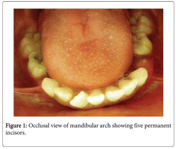
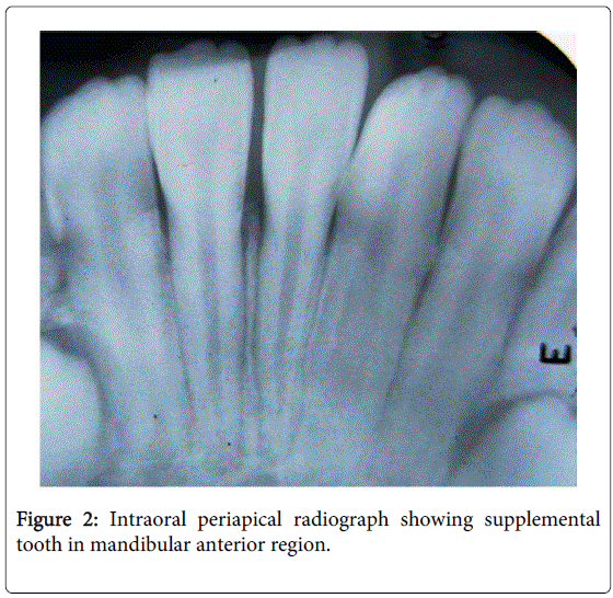
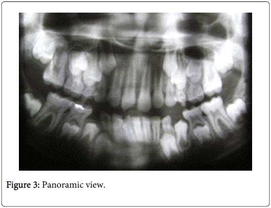
Extra oral examination revealed a convex profile and facial symmetry. Intra-oral examination showed marginal gingivitis in the maxillary anterior region. There was an Ellis’ Class II fracture of the maxillary permanent right central incisor. The patient had an increased over jet of 4mm and silver amalgam restoration of mandibular right primary second molar. In the mandibular anterior region, five permanent incisors were present. On the right side, there were three incisors, one of which was slightly rotated in a mesio-lingual version. All the incisors showed similar morphology with the presence of mammelons (Figure 1). Radiographic investigation confirmed the presence of a permanent supplemental tooth in the mandibular right incisor region. Roots of both central incisors showed apical closure. The supplemental incisor had an individual root with a single relatively wide root canal (Figure 2). An orthopantamograph was also taken to identify the presence of any additional and/or missing teeth (Figure 3).
The patient was referred to a pediatrician for detection of any other finding and/or syndrome. There was no abnormality reported. Dental treatment included esthetic restoration of the fractured maxillary permanent right central incisor with composite resin. The patient has been advised to be on a periodic follow-up.
Discussion
Supplemental teeth refer to a duplication of teeth in the normal series and are found at the end of a tooth series [1]. Very few cases of supplemental mandibular permanent incisors have been reported in literature [12-14]. Hyperplasia of tooth germs may occur in areas with wide intervals between tooth primordiums at the end of the dental lamina. Therefore, it does not easily occur in the mandibular anterior tooth, where there is a high inter-tooth primordium density. Therefore, occurrence of mandibular anterior supernumerary teeth is exceedingly low [15]. It is usually difficult to differentiate clinically between a normal tooth and supplemental tooth. In this case, rotation of the incisor associated with a wider root canal indicated it to be the supplemental incisor.
Erupted supplemental teeth most often cause crowding. The problem may be resolved by extracting the most displaced or deformed tooth. In this case, since there was no tooth-arch discrepancy, the supplemental incisor was not extracted. However, parents of the child were informed of presence of an additional tooth, and the need for subsequent treatment. In conclusion, a rare case of mandibular supplemental teeth is reported. Careful clinical and radiographic examination is essential for the detection of dental anomalies.
References
- Garvey MT, Barry HJ, Blake M (1999) Supernumerary teeth--an overview of classification, diagnosis and management.J Can Dent Assoc 65: 612-616.
- Mitchell L (1989) Supernumerary teeth.Dent Update 16: 65-66, 68-9.
- Yusof WZ (1990) Non-syndrome multiple supernumerary teeth: literature review.J Can Dent Assoc 56: 147-149.
- So LL (1990) Unusual supernumerary teeth.Angle Orthod 60: 289-292.
- Luten JR Jr (1967) The prevalence of supernumerary teeth in primary and mixed dentitions.J Dent Child 34: 346-353.
- Shapira Y, Kuftinec MM (1989) Multiple supernumerary teeth. Report of two cases.Am J Dent 2: 28-30.
- Taylor GS (1972) Characteristics of supernumerary teeth in the primary and permanent dentition.Dent Pract Dent Rec 22: 203-208.
- Högström A, Andersson L (1987) Complications related to surgical removal of anterior supernumerary teeth in children.ASDC J Dent Child 54: 341-343.
- Zhu JF, Marcushamer M, King DL, Henry RJ (1996) Supernumerary and congenitally absent teeth: a literature review.J ClinPediatr Dent 20: 87-95.
- Jensen BL, Kreiborg S (1990) Development of the dentition in cleidocranial dysplasia.J Oral Pathol Med 19: 89-93.
- Liu JF (1995) Characteristics of premaxillary supernumerary teeth: a survey of 112 cases.ASDC J Dent Child 62: 262-265.
- Sharma SA (2001) Mandibular midline supernumerary tooth: a case report.J Indian SocPedodPrev Dent 19: 143-144.
- Yokose T, Sakamoto T, Sueishi K, Yatabe K, Tsujino K, et al. (2006) Two cases with supernumerary teeth in lower incisor region.Bull Tokyo Dent Coll 47: 19-23.
- Yokose T, Sakamoto T, Sueishi K, Yatabe K, Tsujino K, et al. (2006) Two cases with supernumerary teeth in lower incisor region.Bull Tokyo Dent Coll 47: 19-23.
- Bhat M (2006) Supplemental mandibular central incisor.J Indian SocPedodPrev Dent 24 Suppl 1: S20-23.
Relevant Topics
- Cementogenesis
- Coronal Fractures
- Dental Debonding
- Dental Fear
- Dental Implant
- Dental Malocclusion
- Dental Pulp Capping
- Dental Radiography
- Dental Science
- Dental Surgery
- Dental Trauma
- Dentistry
- Emergency Dental Care
- Forensic Dentistry
- Laser Dentistry
- Leukoplakia
- Occlusion
- Oral Cancer
- Oral Precancer
- Osseointegration
- Pulpotomy
- Tooth Replantation
Recommended Journals
Article Tools
Article Usage
- Total views: 14576
- [From(publication date):
February-2015 - Jul 09, 2025] - Breakdown by view type
- HTML page views : 9954
- PDF downloads : 4622


