Make the best use of Scientific Research and information from our 700+ peer reviewed, Open Access Journals that operates with the help of 50,000+ Editorial Board Members and esteemed reviewers and 1000+ Scientific associations in Medical, Clinical, Pharmaceutical, Engineering, Technology and Management Fields.
Meet Inspiring Speakers and Experts at our 3000+ Global Conferenceseries Events with over 600+ Conferences, 1200+ Symposiums and 1200+ Workshops on Medical, Pharma, Engineering, Science, Technology and Business
Case Report Open Access
Superscan on both 99mTc-MDP and 153Samarium-EDTMP Bone Scans in a Patient with Breast Cancer
| Gonca G. Bural* and James M. Mountz | |
| Department of Radiology, Nuclear Medicine Division, University of Pittsburgh Medical Center, Pittsburgh, PA, USA | |
| Corresponding Author : | Gonca G. Bural Department of Radiology, Nuclear Medicine Division University of Pittsburgh Medical Center 200 Lothrop Street, Presbyterian Hospital Suite E-177 Pittsburgh, PA 15213, USA Tel: 412-647-0104 Fax: 412-647-2601 E-mail: buralgg@upmc.edu |
| Received February 05, 2012; Accepted April 02, 2012; Published April 08, 2012 | |
| Citation: Bural GG, Mountz JM (2012) Superscan on both 99mTc-MDP and 153Samarium- EDTMP Bone Scans in a Patient with Breast Cancer. OMICS J Radiology. 1:105. doi: 10.4172/2167-7964.1000105 | |
| Copyright: © 2012 Bural GG, et al. This is an open-access article distributed under the terms of the Creative Commons Attribution License, which permits unrestricted use, distribution, and reproduction in any medium, provided the original author and source are credited. | |
Visit for more related articles at Journal of Radiology
| Keywords |
| Super scan; Samarium bone scan; Breast cancer |
| Case Summary |
| A 64-year old woman with breast cancer was having severe exacerbating pain in her pelvic bones and lower extremities. She had a 99mTc-MDP bone scan which showed diffuse osseous metastases in a “superscan” pattern. |
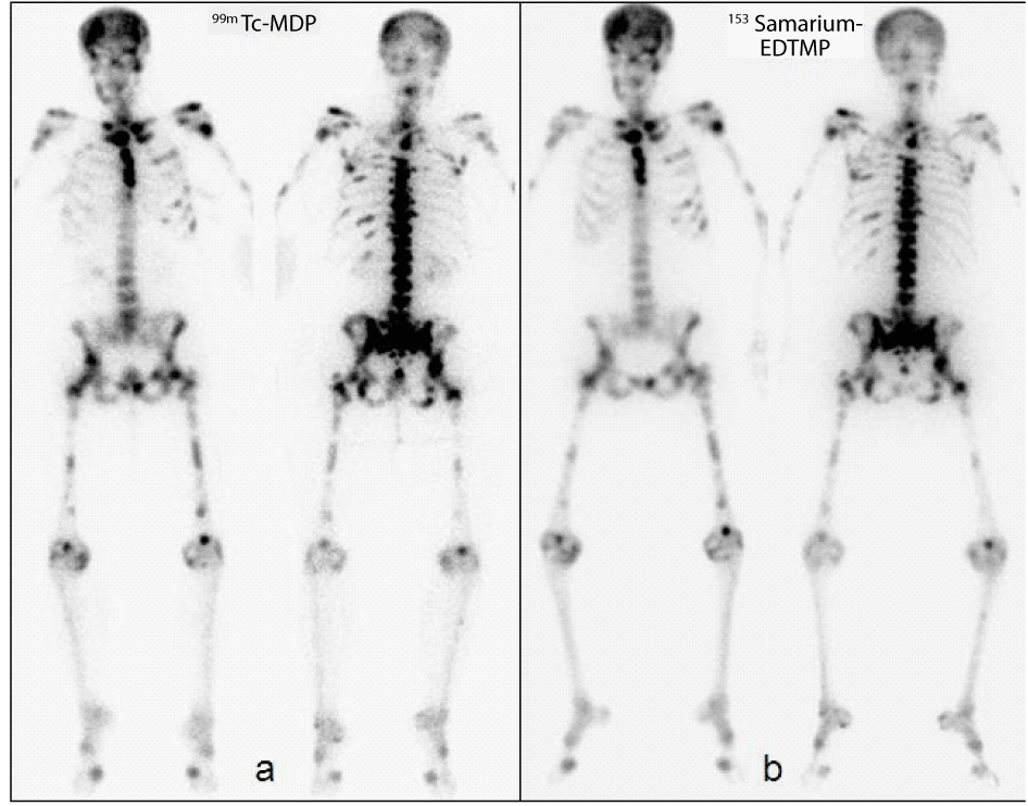
| Anterior and posterior whole body images of a 99mTc-MDP bone scan show diffuse metastatic disease involving the entire axial and appendicular skeleton, a non-visible left kidney, a very faint right kidney and minimal tracer accumulation in the bladder, representing a superscan (a). A superscan is defined as a bone scan which demonstrates markedly increased skeletal radioisotope uptake relative to soft tissues in association with absent or faint genito-urinary tract activity [1]. While a superscan is relatively uncommon, its recognition is important, as it is associated with a number of important underlying diseases [2,3] (Figure 1a). |
| One month later the patient received 4.22 GBq (114 mCi) 153Samarium- EDTMP for palliation of severe bone pain. Post treatment, whole body 153Samarium scan was also a “superscan” showing very similar osseous metastases pattern to the 99mTc-MDP bone scan. Anterior and posterior whole body images 24 hours after intravenous administration of 153Sm-EDTMP demonstrate diffuse osseous metastases in a very similar pattern with the prior bone scan. However on this scan both kidneys and the bladder were not visualized, consistent with a 153Sm-EDTMP superscan (b). 153Sm-EDTMP is used for relief of bone pain predominantly in breast cancer patients with painful osteoblastic skeletal metastases. 153Sm has a half life of 46.3 hours and has a 103 keV gamma emission, suitable for scintigraphic imaging. Superscans on the conventional bone scintigraphy have been described both in metastatic and metabolic bone diseases, with different patterns and appearances of radiotracer uptake [4-8] (Figure 1b). Presence of “superscan” both on conventional 99mTc-MDP bone scan and on the 153Samarium-EDTMP bone scan of the same patient with almost identical osseous metastatic pattern has yet not been reported. A superscan indicates the extensive presence of osseous metastases. A similar superscan pattern on both conventional and 153Samarium- EDTMP bone scans may indicate a better outcome for palliation of metastatic bone pain. All the lesions seen on conventional imaging will also be taking up the palliative bone treatment agent “153Samarium- EDTMP”, leading to efficient palliation of bone pain. |
| This case is unique with the presence of metastatic “superscan” both on conventional 99mTc-MDP bone scan and on the 153Samarium- EDTMP bone scan of the same patient. |
References |
|
Figures at a glance
 |
| Figure 1 |
Post your comment
Relevant Topics
- Abdominal Radiology
- AI in Radiology
- Breast Imaging
- Cardiovascular Radiology
- Chest Radiology
- Clinical Radiology
- CT Imaging
- Diagnostic Radiology
- Emergency Radiology
- Fluoroscopy Radiology
- General Radiology
- Genitourinary Radiology
- Interventional Radiology Techniques
- Mammography
- Minimal Invasive surgery
- Musculoskeletal Radiology
- Neuroradiology
- Neuroradiology Advances
- Oral and Maxillofacial Radiology
- Radiography
- Radiology Imaging
- Surgical Radiology
- Tele Radiology
- Therapeutic Radiology
Recommended Journals
Article Tools
Article Usage
- Total views: 8973
- [From(publication date):
May-2012 - Jul 11, 2025] - Breakdown by view type
- HTML page views : 4265
- PDF downloads : 4708
