Successful Management of Type I Rectal Prolapse in Crossbred Cattle: A Case Report
Received: 26-Apr-2022 / Manuscript No. jflp-22-61872 / Editor assigned: 02-May-2022 / PreQC No. jflp-22-61872 (PQ) / Reviewed: 16-May-2022 / QC No. jflp-22-61872 / Revised: 20-May-2022 / Manuscript No. jflp-22-61872 (R) / Accepted Date: 20-May-2022 / Published Date: 27-May-2022 DOI: 10.4172/2332-2608.1000344
Abstract
The current case report discussed about the complete successful management of rectal prolapse in a cattle. Two years old crossbred cattle was presented to a animal health camp held on burdhaman district in West Bengal (India) with a history of elongated cylindrical mass protruding through the anus. Clinical examination revealed normal appetite, pulse and respiratory rate with mild hyperthermia (102.8˚F) and congested mucous membrane. After diagnosed as type I (or incomplete) rectal prolapse the cattle was treated according therapy without perse-string suture. The epidural anaesthesia was done with 4 ml of 2% Lignocaine injection for proper restraining. Prolapsed mass washed with diluted solutions of Luke warm 2% potassium permanganate solution and after applying 50 gm of sugar granules on the mass, prolapse was manually replaced. In addition sugar, long-acting antibiotic injection enrofloxacin (Fortivir® @ 5 mg/kg B. Wt.) and injection meloxicam (Melonex® @ 0.2 mg/kg B. Wt.) was given intramuscularly. Reoccurrence of prolapse was not reported later and complete successful management of rectal prolapse was done.
Keywords: Type-I Rectal Prolapse; Management; Cattle
Keywords
Introduction
Rectal prolapse is protrusion of one or more layers of the rectum through the anus [1]. It may occur in animals of any age, breed, or sex [2]. In cattle, the condition may be a result of any disease that causes tenesmus [3] including diarrhoea, rectal neoplasia, severe enteritis and endoparasitic infestation [4], elevations in intra-abdominal pressure during parturition or episodes of coughing [5]. Based upon the involving layer of rectum, rectal prolapse divided into incomplete or type I where prolapse involves the rectal mucosa and sub mucosa only, appearing as a large soft tissue mass swelling at the rectum [6] and complete or type II where entire rectum stick out from anus. Excessive peristalsis due to periodic increase in muscular tone brings about pain of spasmodic nature [7]. When the prolapse cannot be corrected manually due to inflammation, ulceration and necrosis, surgical amputation or resection of exposed tissue may be required. In the present case increased peristalsis cause interruption of normal innervations of the external anal sphincter which resulted into type I prolapse and successfully corrected without any surgical intervention.
Case history and observation
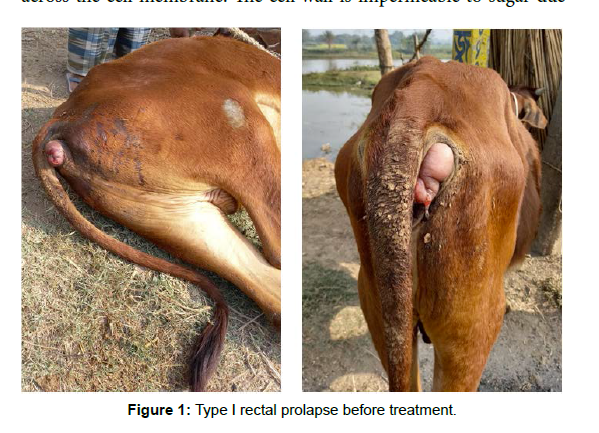
2 years old crossbred cattle was presented to a animal health camp held on Burdhaman district in West Bengal state (India) with a history of elongated cylindrical mass protruding through the rectum (Figure 1). Owner reported that the problem was occurred since 12 hours. On clinical examination it was found that there was congested mucous, membranes, mild hypothermia (102.8°F), pulse and respiratory rates were all within the reference values. Also, the animal has maintained a normal appetite. Closed examination of protruding elongated mass through the anus the case was diagnosed as type I (or incomplete) rectal prolapse.
Treatment and discussion
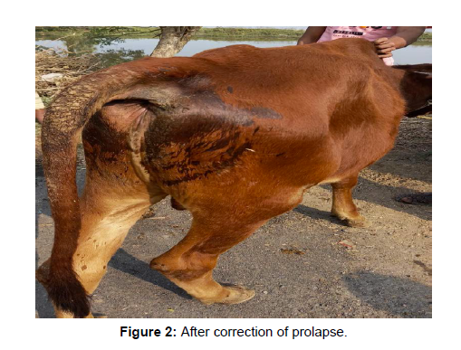
The prognosis depends on the severity of the case, degree of damage and contamination, duration of its existence or how quick it is attempted with suitable treatment. In the present case before correcting the prolapsed mass epidural anaesthesia was done by 4 ml of 2% Lignocaine injection in the first intercoccygeal space to restraint the animal properly. Prolapsed mass cleaned with diluted solution of Luke warm 2% potassium permanganate solution. After applying 50 gm sugar granules on the mass, the prolapse was manually replaced by gently force. Several studies [8, 9, 10] supports this indigenous method to correct the type I rectal prolapse manually. Sugar creates an environment where the solute concentration is greater on the exterior of the rectal tissues than inside of the cells that make up these tissues. As per laws of osmosis, water will passively move out of the rectal tissues and across the cell membrane. The cell wall is impermeable to sugar due to its stable and complex nature. This results in the shrinkage of cells and helps to reduce swelling. In addition to using sugar, long acting antibiotic injection enrofloxacin (Fortivir® @ 5 mg/kg B. Wt.) intramuscularly was given to prevent infections and injection meloxicam (Melonex® @ 0.2 mg/kg B. Wt.) intramuscularly was given to reduce discomfort due to pain. It was also advised to give easily digestible feed for avoiding the problems during defecation. As there was no history of diarrhoea or straining perse-string suture was not necessary in that case. The present case was managed successfully by this therapy (Figure 2) and no reoccurrence of prolapse was reported later.
Conclusion
Based upon the current study it can be concluded that indigenous method is effective for correction of type I or incomplete rectal prolapse without involving any suturing or surgical intervention.
Acknowledgement
The authors are thankful to Director, Block Livestock Development Officer (BLDO) of Burdhaman District for providing ample facilities to conduct the current work and Livestock Development Assistant (LDA) for working as helping hand.
Conflict of interest
The authors declare that there is no conflict of interest.
References
- Ettinger SJ, Feldman EC (1995) Textbook of veterinary internal medicine. 4th ed., W. B. Saunders, Philadelphia, 2: 1403-1404.
- Allu T, Rajapantula M, Killi L, Naidu G Venkata (2020) Clinical management of vaginal and rectal prolapse in crossbred Jersey cow- a case report. Int J Sci Environ Technol 9: 133-135.
- Alex Gallagher (2020) Rectal Prolapse in Animals.
- Turner TA, Fessler JF (1980) Rectal prolapse in the horse. J Am Vet Med Assoc 177: 1028-1032.
- Snyder JR, Pascoe JR and Williams JW (1985) Rectal prolapse and cystic calculus in a burro. J Am Vet Med Assoc 187: 421-422.
- Reed MS, Bayly MW, Sellon CD (2004) Ischaemic Disorders of the intestinal Tract. Equine Internal Medicine, Blikslager, AT and SL Jones (Eds.). Saunders Press, Missouri, pp: 919-920.
- Chakrabarti A (2006) Text book of clinical veterinary medicine. Kalyani Publication 6th Ed. pp: 211-213.
- Myer JO, Rothenberger DA (1991) Sugar in the reduction of incarcerated prolapsed bowel: report of two cases. Dis Colon Rectum 34: 416-418.
- Patel RK, Patel N, Singh N, Singh P (2016) Successful management of rectal prolapse in a mule (Equus mulus). J Livestock Sci 7: 35-37.
- Mandal S (2020) Indigenous method: A traditional method of treatment.
Indexed at, Google Scholar, Crossref
Citation: Halder B, Seikh B (2022) Successful Management of Type I Rectal Prolapse in Crossbred Cattle: A Case Report. J Fisheries Livest Prod 10: 344. DOI: 10.4172/2332-2608.1000344
Copyright: © 2022 Halder B, et al. This is an open-access article distributed under the terms of the Creative Commons Attribution License, which permits unrestricted use, distribution, and reproduction in any medium, provided the original author and source are credited.
Share This Article
Recommended Journals
Open Access Journals
Article Tools
Article Usage
- Total views: 3639
- [From(publication date): 0-2022 - Mar 29, 2025]
- Breakdown by view type
- HTML page views: 3160
- PDF downloads: 479


