Make the best use of Scientific Research and information from our 700+ peer reviewed, Open Access Journals that operates with the help of 50,000+ Editorial Board Members and esteemed reviewers and 1000+ Scientific associations in Medical, Clinical, Pharmaceutical, Engineering, Technology and Management Fields.
Meet Inspiring Speakers and Experts at our 3000+ Global Conferenceseries Events with over 600+ Conferences, 1200+ Symposiums and 1200+ Workshops on Medical, Pharma, Engineering, Science, Technology and Business
Case Report Open Access
Subdural Empyema in a 5 Month Old following E. coli Meningitis
| Alexander Blackwood R* | ||
| Department of Pediatrics and Communicable Diseases, University of Michigan Medical School, Ann Arbor, MI, USA Cornicelli | ||
| Corresponding Author : | Alexander Blackwood R Department of Pediatrics and Communicable Diseases University of Michigan Medical School D5101 Medical Professional Building/SPC 5718 1500 East Medical Center Drive Ann Arbor, MI 48109-5718, USA Tel: 734-763-2440 Fax: 734-232-3859 E-mail: rab@umich.edu |
|
| Received April 03, 2013; Accepted May 10, 2013; Published May 15, 2013 | ||
| Citation: Cornicelli, Blackwood M, Alexander Blackwood R (2013) Subdural Empyema in a 5 Month Old following E. coli Meningitis. J Infect Dis Ther 1:106. doi:10.4172/2332-0877.1000106 | ||
| Copyright: © 2013 Cornicelli, et al. This is an open-access article distributed under the terms of the Creative Commons Attribution License, which permits unrestricted use, distribution, and reproduction in any medium, provided the original author and source are credited. | ||
Related article at Pubmed Pubmed  Scholar Google Scholar Google |
||
Visit for more related articles at Journal of Infectious Diseases & Therapy
Abstract
Subdural empyema can be a rare complication of bacterial meningitis or an infected subdural hematoma. Reported here is a case of a 5 month old boy Ethiopian adoptee, with a suspected subdural hematoma that was discovered to have a subdural empyema culture positive for a multi-drug resistant E. coli. The child was managed successfully with three washout procedures and 8 weeks of meropenem. We present this case and a review of the literature on subdural empyema in pediatrics.
| Keywords | |
| Subdural empyema; E. coli meningitis | |
| Case | |
| A 5-month-old boy presented to the Emergency Department with new onset seizures fever and altered mental status. The child was a newly adopted boy from Ethiopia and his parents were in the process of bringing him to the United States. During the plane trip, the infant developed a fever (tactile), and had at least three episodes of seizure-like activity consisting of eye fluttering, lip smacking, and bilateral jerking of the upper extremities, with some eye rolling. Upon landing, the child was taken to a local emergency room, where he was noted to be postictal. The child received lorazepam, and was loaded with Phenobarbital, prior to transferring to Mott’s Children’s Hospital, University of Michigan, Ann Arbor, MI, USA. | |
| The past medical history is limited because the child was abandoned at a local orphanage at four weeks of age. However, the child had been admitted to a local hospital in Ethiopia seven times for recurrent pneumonia, having received several courses of ceftriaxone and gentamicin. There was no information regarding culture results or the indication for the antibiotic choices. However, the child reportedly responded to each antibiotic course. There was no history of prior seizure activity, but the child was known to be anemic and had been diagnosed with bilateral subdural fluid collections of unknown etiology (ultrasonogram and magnetic resonance imaging of the head, which were unavailable for review). Because of the growing head circumference, the parents anticipated having the hematomas drained following the child’s immigration. The parents had spent the past week at the orphanage participating in the care of the child, and noted no seizure activity or gross developmental abnormalities. In Ethiopia, the child had received Bacille Calmette–Guérin, (BCG), and was reportedly Human Immunodeficiency Virus (HIV) and intestinal parasite negative. | |
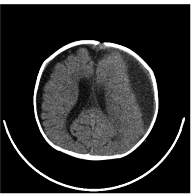
| On arrival to the hospital, physical exam was notable for temperature of 39.2°F, heart rate of 146 beats/min, BP 108/72 mm Hg, respiratory rate 58 breaths/min. His head was noted to be disproportionately large compared with the rest of his body (95th vs. 25th percentile), but without a bulging fontanelle. The pupils were equal round and reactive to light. Neurologically, the child responded to noxious stimuli in all extremities, left greater than right. Laboratory evaluation was significant for 16.9 k white blood cells/mm3 (61.2% neutrophils, 19.2% lymphocytes and 17.5% monocytes), hemoglobin of 7.5 g/ dl, platelet count of 1000 k platelets/mm3. Comprehensive metabolic panel was normal, except for mildly elevated transaminases (Alanine transaminase 492 IU/L, Aspartate transaminase 338 IU/L, Alkaline phosphatase 494 IU/L). Rapid HIV, blood, urine, and stool cultures were negative. A lumbar puncture revealed 23 white blood cells/mm3 (21% neutrophils), 1340 red blood cells/mm3, glucose 52 mg/dl and protein 82 mg/dl. Computerized tomography of the head revealed a large left-sided subdural hyperdense fluid collection, with mass effect on the adjacent brain. Roentgenogram of the chest revealed bilateral perihilar streaky opacities, with no focal opacification. The patient was loaded with Phenobarbital, and started empirically on vancomycin and ceftriaxone. The computerized tomography (CT) performed at the outside hospital was review revealing a large left-sided subdural fluid collection, measuring over 2 centimeters in thickness and a much smaller collection on the contralateral side (Figure 1). Neurosurgery was consulted and performed a left frontal frontoparietal craniotomy, after discovering purulent material and granulation tissue following bur hole placement and the subdural empyema was resected, no drains placed. Cultures from the brain specimen revealed E. coli, which was eventually noted to be producing an extended spectrum beta-lactamase (ESBL) (Table 1), and the antibiotics were switched to meropenem. Clinically, the child did reasonably well; however, the subdural fluid re-accumulated, requiring two additional neurosurgical drainage procedures. Cultures from repeat drainage procedures remained negative and the child received a total of 8 weeks of meropenem. | |
| An ELISA and Western blot for Human Immunodeficiency Virus (HIV) were positive, but the plasma quantitative RNA Polymerase Chain Reaction (PCR) was negative, indicating that the patient’s mother was HIV positive, but there was no perinatal transmission to the child. Stool for ova and parasites, as well as thick and thin smears for malaria were negative. A PPD (purified protein derivative) was positive at greater than 10 mm, but the child had reportedly received BCG (Bacille Calmette Guerin). A TB-Quantiferon gold was indeterminate and gastric aspirate cultures were ultimately negative for Mycobacterium tuberculosis, but in light of the positive PPD and infiltrate on the roentgenogram, the child was begun on treatment for presumed active tuberculosis (CDC guidelines). | |
| Discussion | |
| A subdural empyema is an infection located between the dura mater arachnoid mater, accounting for approximately 20% of the intracranial infections. While 95% are found in the cranium, they also occur along the spinal axis. While a subdural empyema is most frequently a complication from sinusitis, they can be associated with a paranasal or otogenic source [1,2]. They occur predominantly in adolescents and adults, but the can occur in neonates and infants. In infants and neonates, a subdural empyema is most frequently a complication from bacterial meningitis [1-4]. Interestingly, post-meningitic fluid collections are relatively common. However, subdural empyemas due to purulent meningitis are extremely rare, occurring in at most 1-2% of all cases of bacterial meningitis [5-7]. | |
| Since the advent of the vaccine against H. influenzae, the overwhelming majority of non-sterile cases of subdural empyemas are due to Streptococcal, Staphylococcal, anaerobic, or mixed bacteria [1-3]. In contrast, E. coli is found to be the causative organism in between 3-13% of all cases of subdural empyemas [1,2,4,7]. Subdural empyemas are potentially life threatening, their management requiring a combination of neurosurgical drainage and organism directed antibiotic therapy. Bur holes and external drains are often used to reduce the rate of empyema recurrence [8,9]. In total, there were 4 bur holes, but no drains placed in this patient. However, two additional washout procedures were required. | |
| The pathogenesis of this patient’s subdural empyema is unclear. Given the complexity of the presentation, the unknowns and highrisk past medical history, the patient may have acquired a subdural empyema through various ways. However, there are two leading possibilities. The first is that the patient experienced meningitis with an associated empyema that was inaccurately diagnosed, and managed as pneumonia and subdural hematomas. The meningitis and associated empyema was not adequately treated due to the antibiotic resistance of the organism, the duration of therapy, and the unavailability of neurosurgical intervention. The single causative agent of E. coli cultured from the subdural empyema, the extent of granulation tissue, disease burden, and the continual progression of his head circumference, further support this argument. The second possibility is that the patient had a subdural hematoma that subsequently became infected. While this phenomenon is rare, it has been reported in the literature [10-13]. Subdural hematomas in neonates and infants are usually the result of trauma and under normal circumstances, non-accidental trauma must be considered [14,15]. Hematomas occur through the rupture of cortical bridging veins and can become secondarily infected. Although we had no history of trauma, the patient’s background does not rule it out as a possibility. A similar event has been reported in an 8 month old, with a subdural hematoma infected with E. coli [11]. The negative finding on lumbar puncture and clinical presentation are consistent with either meningitis and subsequent empyema, or a subdural hematoma which was secondarily infected. The cell count and negative bacterial culture results in the CSF were not suggestive of incompletely treated or persistent meningitis. However, the level of organization of the empyema was suggestive of relatively longstanding process. | |
| The incidence of neonatal bacterial meningitis varies worldwide, with rates of 0.3/1000 live births in industrialized countries to 0.48 to 6.1/1000 live births in Africa and South Asia [16]. In the United States group B Streptococcus accounts for approximately 50% of the cases of neonatal meningitis, with E. coli representing an additional 20%. However, in underdeveloped countries, Klebsiella species and E coli account for a majority of the cases, followed by Staphylococcus aureus [17,18]. While ampicillin and gentamycin or cefotaxime are the primary antibiotic empirically used in the neonatal period, the emergence of cephalosporin-resistance is becoming more problematic worldwide, including in developing countries [19,20], as exhibited in this patient. Dexamethasone may be used in older infants and children, but it is not recommended in for neonatal meningitis [21]. Treatment courses for E. coli meningitis are generally 14-21 days, following documented sterilization of the CSF. However, in this case, there is no indication that the physicians in Ethiopia were aware of the bacterial meningitis, or that there was an ESBL producing organism which probably accounts for the inadequate duration of therapy. | |
| Conclusion | |
| This patient highlights some of the interesting challenges facing Pediatric Infectious Disease specialists globally. This patient was initially diagnosed with a subdural hematoma in the presence of intermittent fevers, thought to be secondary to recurrent pneumonia. Failure to irradiate the pneumonia using a beta-lactam raises the question of whether or not there is an ESBL producing organisms. While a subdural empyema may be difficult to diagnose and the management usually requires appropriate neurosurgical involvement, it must be part of the differential in a patient with a subdural hematoma and intermittent or persistent fevers. Additionally with an empyema there is a reasonable recurrence rate, so repeat washouts or drain placement may be required. | |
| References | |
References
- Banerjee AD, Pandey P, Devi BI, Sampath S, Chandramouli BA (2009) Pediatric supratentorial subdural empyemas: A retrospective analysis of 65 Cases. Pediatr Neurosurg 45: 11-18.
- Nathoo N, Nadvi SS, van Dellen JR, Gouws E (1999) Intracranial subdural empyemas in the era of computed tomography: a review of 699 cases.Neurosurgery 44: 529-535.
- Bockova J, Rigamonti D (2000) Intracranial empyema.Pediatr Infect Dis J 19: 735-737.
- Legrand M, Roujeau T, Meyer P, Carli P, Orliaguet G, et al.(2009) Paediatric intracranial empyema: differences according to age.Eur J Pediatr 168: 1235-1241.
- Han BK, Babcock DS, McAdams L (1985) Bacterial meningitis in infants: sonographic findings. Radiology 154: 645-650.
- Tunkel AR, Hartman BJ, Kaplan SL, Kaufman BA, Roos KL, et al. (2004) Practice guidelines for the management of bacterial meningitis. Clin Infect Dis 39: 1267-1284.
- Liu ZH, Chen NY, Tu PH, Lee ST, Wu CT (2010) The treatment and outcome of postmeningitic subdural empyema in infants. J Neurosurg Pediatr 6: 38-42.
- Miller ES, Dias PS, Uttley D. (1987) Management of subdural empyema: a series of 24 Cases. J Neurol Neurosurg Psychiatry 50: 1415-1418.
- Madhugiri VS, Sastri BV, Srikantha U, Banerjee AD, Somanna S, et al. (2011) Focal intradural brain infections in children: an analysis of management and outcome. Pediatr Neurosurg 47: 113-124.
- Hamada J, Ichimura H, Ushio Y (1993) Huge subdural empyema with unusual presentation in infant–case report. Neurol Med Chir (Tokyo) 33: 40-43.
- Hoshima T, Kusuhara K, Saito M, Mizoguchi M, Morioka T, et al. (2008) Infected subdural hematoma in an infant. Jpn J Infect Dis 61: 412-414.
- Le Roux PC, Wood M, Campbell RA (2007) Subdural empyema caused by an unusual organism following intracranial haematoma. Childs Nerv Syst 23: 825-827.
- Levy I, Sood S (1996) Staphylococcus aureus dissemination to a preexisting subdural hematoma. Pediatr Infect Dis J 15: 1139-1140.
- Matschke J, Voss J, Obi N, Görndt J, Sperhake JP, et al. (2009) Nonaccidental head injury is the most common cause of subdural bleeding in infants <1 year of age. Pediatrics 124: 1587-1594.
- Loh JK, Lin CL, Kwan AL, Howng SL (2002) Acute subdural hematoma in infancy. Surg Neurol 58: 218-224.
- Thaver D, Zaidi AK (2009) Burden of neonatal infections in developing countries: a review of evidence from community-based studies. Pediatr Infect Dis J 28: S3-S9.
- Tiskumara R, Fakharee SH, Liu CQ, Nuntnarumit P, Lui KM, et al. (2009) Neonatal infections in Asia. Arch Dis Child Fetal Neonatal Ed 94: F144-F148.
- Zaidi AK, Thaver D, Ali SA, Khan TA (2009) Pathogens associated with sepsis in newborns and young infants in developing countries. Pediatr Infect Dis J 28: S10-S18.
- Pickering LD (2006) Red Book: 2006 report of the committee on infectious diseases. (27th Edn), Elk Grove Village, American Academy of Pediatrics, USA.
- Thaver D, Ali SA, Zaidi AK (2009) Antimicrobial resistance among neonatal pathogens in developing countries. Pediatr Infect Dis J 28: S19-S21.
- Chaudhuri A (2004) Adjunctive dexamethasone treatment in acute bacterial meningitis. Lancet Neurol 3: 54-62.
Tables and Figures at a glance
| Table 1 |
Figures at a glance
 |
| Figure 1 |
Post your comment
Relevant Topics
- Advanced Therapies
- Chicken Pox
- Ciprofloxacin
- Colon Infection
- Conjunctivitis
- Herpes Virus
- HIV and AIDS Research
- Human Papilloma Virus
- Infection
- Infection in Blood
- Infections Prevention
- Infectious Diseases in Children
- Influenza
- Liver Diseases
- Respiratory Tract Infections
- T Cell Lymphomatic Virus
- Treatment for Infectious Diseases
- Viral Encephalitis
- Yeast Infection
Recommended Journals
Article Tools
Article Usage
- Total views: 16184
- [From(publication date):
August-2013 - Nov 21, 2024] - Breakdown by view type
- HTML page views : 11627
- PDF downloads : 4557
