Squamous Cell Carcinoma in a Newly Diagnosed Ovarian Dermoid Cyst
Received: 05-Mar-2018 / Accepted Date: 22-Mar-2018 / Published Date: 29-Mar-2018 DOI: 10.4172/2167-7964.1000294
Abstract
This case report details the clinical features, radiological investigations and imaging findings in the initial presentation of a female patient with a suspected pelvic mass. A discussion of the imaging methods employed in the diagnosis of an adnexal mass follows, with a focus on ovarian dermoid cysts and the features associated with benign and malignant disease.
Keywords: Ovarian dermoid; Squamous cell carcinoma; Diagnostic imaging
Case Report
This is the case of a 49-year-old female who presented to the General Practitioner with symptoms of abdominal fullness, constipation, urinary frequency and intermittent nausea over the preceding month. A history of unintentional weight loss, over a 6-month period, was elicited. She was premenopausal with regular, unchanged menstrual cycles and was gravida 2, para 2. There was no relevant past medical, social or family history and she was not taking any regular medication. On examination, an abdominopelvic mass was palpable, extending to the level of the umbilicus.
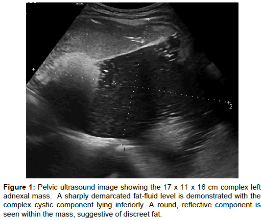
An ultrasound examination revealed the presence of a 17 × 11 × 16 cm complex left adnexal mass. Hyperechoic contents were seen layering above a cystic component, in keeping with a fat-fluid interface. The mass contained two 4 cm, discreet, hyperechoic components that were highly reflective, suggesting fat content (Figure 1). The appearances were felt to be most in keeping with a dermoid cyst.
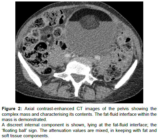
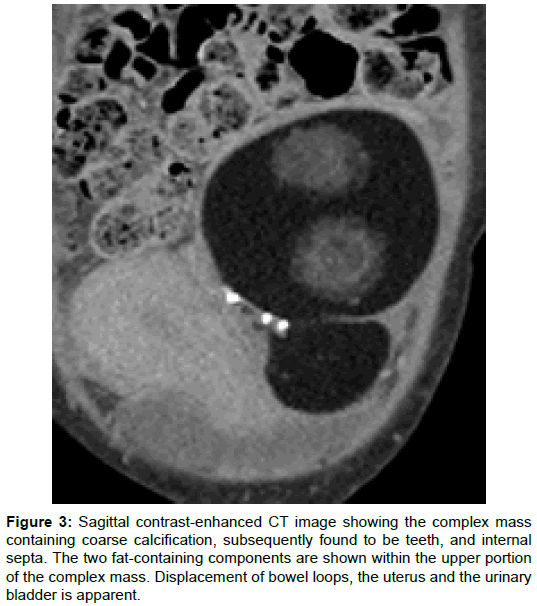
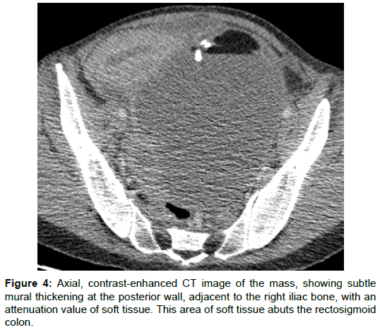
A CT scan was arranged and demonstrated that the large, complex mass was of left ovarian origin. Evaluation of the contents confirmed that the two 4 cm hyperechoic components, seen on the ultrasound study, were of attenuation in keeping with predominant fat content. These were floating at a fat-fluid interface within the mass (Figure 2). Internal foci of coarse calcification were also identified (Figure 3). A subtle area of thickened soft tissue was noted at the posterior wall of the cyst, abutting the adjacent rectosigmoid colon (Figure 4). There was a small volume of ascites, but no peritoneal disease or lymphadenopathy was seen. The lesion was reported as a left ovarian dermoid cyst. Blood tests were taken and showed a raised Ca125 value of 118 kU/L.
Figure 2: Axial contrast-enhanced CT images of the pelvis showing the complex mass and characterising its contents. The fat-fluid interface within the mass is demonstrated.
A discreet internal component is shown, lying at the fat-fluid interface; the ‘floating ball’ sign. The attenuation values are mixed, in keeping with fat and soft tissue components.
Figure 3: Sagittal contrast-enhanced CT image showing the complex mass containing coarse calcification, subsequently found to be teeth, and internal septa. The two fat-containing components are shown within the upper portion of the complex mass. Displacement of bowel loops, the uterus and the urinary bladder is apparent.
The patient underwent surgery where a left ovarian tumour was identified. This was focally adherent to the upper rectum, resulting in cyst rupture and leakage of sebaceous material during surgical excision. The omentum had a normal appearance and there was a small volume of ascites. A bilateral salpingo-oophorectomy and hysterectomy was performed and omental biopsies taken. There was no surgical resection of the bowel during this procedure.
The surgical specimens were sent for histological analysis, which revealed a 30 mm area of thickened wall within an enlarged, ruptured left ovarian cyst. Microscopically, this area of thickening was found to be moderate to poorly differentiated keratinising squamous cell carcinoma, arising from dysplastic squamous epithelium, lining a mature cystic teratoma. Incidentally, two teeth and hair were identified within the tumour. There was no evidence of lymphovascular invasion and omental biopsies were negative for neoplasia.
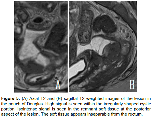
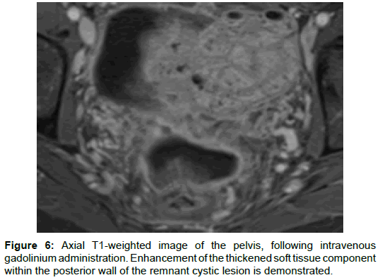
A post-operative MRI scan was performed and demonstrated a 5.3 cm × 2.7 cm × 5 cm remnant cystic lesions in the pouch of Douglas. A 3.4 cm × 2 cm × 2.1 cm focus of T2 isointense soft tissue was seen at the posterior portion of the cystic lesion. Furthermore, there was the suggestion of a focal implant at the anterior rectal wall, corresponding to the area of rectal adherence described in the operative note (Figure 5). This area of soft tissue demonstrated enhancement, following gadolinium administration, and restricted diffusion (Figures 6 and 7). Given this appearance, concern about local invasion was raised and the patient subsequently underwent further surgery where resection of the rectosigmoid colon and small bowel was performed.
Figure 5: (A) Axial T2 and (B) sagittal T2 weighted images of the lesion in the pouch of Douglas. High signal is seen within the irregularly shaped cystic portion. Isointense signal is seen in the remnant soft tissue at the posterior aspect of the lesion. The soft tissue appears inseparable from the rectum.
The features seen on the MRI were in keeping with subsequent histological results which showed two large deposits of moderately differentiated squamous cell carcinoma within the rectosigmoid colon and adherent small intestine. The findings were in keeping with metastatic squamous cell carcinoma, likely arising from the malignant tissue within the left ovarian dermoid cyst.
Discussion
Imaging adnexal masses
Imaging plays an important role in the detection and characterisation of an adnexal mass. Ultrasonography is typically the initial imaging investigation. Information concerning the location, morphology, composition and vascularity of the mass can be obtained.
A set of rules has been devised by the International Ovarian Tumour Analysis group (IOTA) to aid in the prediction of the risk of malignancy based on the sonographic appearances. The features that are typical for a benign tumour include a unilocular cyst, a smooth multilocular lesion measuring less than 10 cm in diameter, the presence of a posterior acoustic shadow and lack of internal blood flow. Additionally, if there is a solid component, this must measure less than 7 mm in diameter to be considered benign [1].
Conversely, the presence of an irregular solid tumour, a lesion with at least four papillary projections, an irregular multilocular lesion over 10 cm in diameter and the detection of marked internal blood flow are features of malignancy. Additional concerning findings include the presence of ascites and peritoneal nodularity, since these features indicate the presence of metastatic disease. Where there is a combination of benign and malignant features, the lesion is deemed indeterminate [1].
MRI can be used to determine the precise origin of an adnexal mass, to assess for local invasion and to better characterise the internal contents, owing to its high soft tissue resolution. Features that are suggestive of malignancy include vegetations within a cystic lesion, necrosis in a solid lesion, transmural extension of solid components, direct invasion of pelvic structures and the presence of peritoneal or serosal implants [2].
The administration of contrast medium accentuates differences in the relaxation characteristics of normal and pathological tissues, increasing the sensitivity of MRI in the detection of malignancy. For example, a contrast-enhanced sequence can be used to increase the conspicuity of nodules and septae within an ovarian mass and is helpful in identifying peritoneal disease [3]. Additionally, a dynamic profile of the passage of the contrast medium through a tissue can be obtained and has been used as a means for discriminating benign from malignant tissue [4,5].
Diffusion-weighted imaging (DWI) sequences are useful in assessing for the presence of malignant features in an adnexal mass. A diffusion-weighted image is derived from differences in the movement or diffusion of water within a tissue, which is related to its cellular contents [6]. A malignant lesion typically causes a restricted pattern of diffusion due to a combination of increased cellular density, restricted cellular permeability and a shift of water from extracellular to intracellular compartments [7]. However, caution in the interpretation of DWI is important in adnexal masses, given the potential overlap between malignant and benign causes of restricted diffusion. For example, a pattern of low diffusivity can be seen in benign cystic ovarian lesions, including abscesses and lesions with mucoid contents [6].
Contrast-enhanced CT demonstrates the presence of lymphadenopathy and metastases. This is important in both the initial staging of an ovarian malignancy and during the patient’s follow up, to determine whether there is recurrence or spread of disease.
Ovarian dermoid cysts
An ovarian dermoid cyst, also known as a mature cystic teratoma, is the most common ovarian germ cell tumour and accounts for 10- 20% of all ovarian tumours [8]. A rare complication, developing in 1-2% of cases, is malignant degeneration [9]. The annual incidence of malignant degeneration is reported to be in the range of 1.2-14.2 cases per 100,000 people [10]. The most common histological diagnosis in such cases is squamous cell carcinoma, representing transformation of ectodermal tissue elements [10].
Establishing a diagnosis of malignant transformation is challenging. Risk factors include increasing age, with most cases arising in those over 50. Findings that are associated with malignancy include high serum CA125 levels and tumour size greater than 10 cm [10]. The prognosis is determined by the stage of disease at diagnosis. Using the International Federation of Gynaecology and Obstetrics (FIGO) staging, 5-year survival for stage I disease was 76%, compared with 0% for stage IV disease, in a study of 220 cases [11]. Findings at surgery that are associated with a poor prognosis include the presence of transmural disease, vascular invasion, spillage of cyst contents and adhesions [12].
Ovarian dermoid cysts: imaging features
The radiological features of an ovarian dermoid cyst are wide ranging, reflecting the variable histological composition of the tumour. A sonographic feature which is highly suggestive of a dermoid is the appearance of hyperechoic contents within a cystic mass, in keeping with the presence of fat. In most cases, CT plays a limited role in the initial detection and characterisation of an adnexal mass. However, the diagnosis of a dermoid cyst can be made with CT imaging, if the presence of fat and calcification can be demonstrated [13].
MRI is useful in characterising the contents of a dermoid cyst and in determining whether there are features of malignant degeneration. As described above, DWI and contrast-enhanced sequences are particularly useful in assessing for the presence of malignant features. However, the interpretation of DWI may be particularly complex when applied to ovarian dermoid cysts, due to their heterogeneous tissue content. If, for example, there is a large cystic component and a small malignant focus, restricted diffusion may not be demonstrated on the DWI sequence, leading to a misdiagnosis of benign disease [6]. Therefore, interpretation of the appearances on a combination of MRI sequences is important in increasing the diagnostic accuracy.
In this case study, several radiological features that are pathognomonic for an ovarian dermoid were present on the different imaging modalities. These include a fat-fluid level within the lesion, the ‘floating ball’ sign and ‘tip of the iceberg’ sign [14]. The ‘floating ball’ sign describes focal fat within a complex mass sitting atop the lower density cystic contents (Figures 1 and 2). The sonographic appearance of fat within a mass may be likened to seeing the ‘tip of the iceberg’, describing an inability to visualise the contents beyond the fat, owing to its reflective properties. Finally, the areas of internal calcification described on the CT, which were found to represent teeth, are in keeping with a dermoid tumour (Figure 3).
Radiological features that are suggestive of a malignant process were also present in the case described. The tumour measured up to 17 cm in size, exceeding the threshold for benign disease [1]. An obtuse margin between the soft tissue component and wall of the cyst was apparent on the CT scan and is feature associated with malignancy (Figure 4) [12]. Additionally, MRI appearances that are concerning for malignancy were present, including enhancing soft tissue with restricted diffusion and transmural extension (Figures 5-7). In this case, these imaging features were consistent with the final histological diagnosis of squamous cell carcinoma within an ovarian dermoid cyst.
Conclusion
This case study has been used to provide a platform for discussing the clinical presentation, imaging modalities and radiological features encountered in the context of an adnexal mass. Some of the imaging appearances associated with ovarian dermoid cysts has been discussed, including clinical and radiological features that should raise concern about malignancy. Although malignant degeneration is a rare complication, it can carry a poor prognosis, depending on the stage at diagnosis. This mandates an awareness of both the clinical and radiological signs that should increase the clinician’s index of suspicion.
References
- Timmerman D, Testa AC, Bourne T, Amete L, Jurkovic D, et al. (2008) Simple ultrasound-based rules for the diagnosis of ovarian cancer. Ultrasound Obstet Gynecol 31: 681-690.
- Hubert J, Bergin D (2008) Imaging the female pelvis: When should MRI be considered? Appl Rad 37: 9.
- Sohaib SAA, Reznek RH (2007) MR imaging in ovarian cancer. Cancer Imag 7: S119-S129.
- Imaoka I, Wada A, Kaji Y, Hayashi T, Hayashi M, et al. (2006) Developing an MR imaging strategy for diagnosis of ovarian masses. RSNA Radiographics 26: 1431-1448.
- Thomassin-Naggara I, Darai E, Cuenod CA, Rouzier R, Callard P, et al. (2008) Dynamic contrast-enhanced magnetic resonance imaging: a useful tool for characterising ovarian epithelial tumours. J Magn Reson Imaging 28: 111-120.
- Whittaker CS, Coady A, Culver L, Rustin G, Padwick M, et al. (2009) Diffusion-weighted imaging of female pelvic tumours: a pictorial review. Radiographics 29: 759-774.
- Dhanda S, Thankur M, Kerkar R, Jagmohan P (2014) Diffusion-weighted imaging of gynaecologic tumors: diagnostic pearls and potential pitfalls. Radiographics 34: 1393-1416.
- Berek JS, Hacker NF (2005) Practical Gynecologic Oncology. Lippincott Williams & Wilkins, USA.
- Avci S, Selcukbiricik F, Bilici A, Özkan G, Özağarı AA, et al. (2012) Squamous cell carcinoma arising in a mature cystic teratoma. Case Rep Obs Gynaecol 2012: 314535.
- Hackethal A, Brueggmann D, Bohlmann MK, Franke FE, Tinneberg HR, et al. (2009) Squamous-cell carcinoma in mature cystic teratoma of the ovary: systematic review and analysis of published data. Lancet Oncol 9: 1173-1180.
- Chen RJ, Chen KY, Chang TC, Sheu BC, Chow SN, et al. Prognosis and treatment of squamous cell carcinoma from a mature cystic teratoma of the ovary. J Formos Med Assoc 107: 857-868.
- Goudeli C, Varytimiadi A, Koufopoulos N, Syrios J, Terzakis E (2017) An ovarian mature cystic teratoma evolving in squamous cell carcinoma: A case report and review of the literature. Gynecol Oncol Rep 19: 27-30.
- Foti PV, Attina G, Spadola S, Caltabiano R, Farina R, et al. (2016) MR imaging of ovarian masses: classification and differential diagnosis. Insights Imaging 7: 21-41.
- Hsiang CW, Liu WC, Huang GS, Hsu HH, Chang WC (2013) A floating ball: a pathognomonic sign of ovarian cystic teratoma. QJM 107: 319-320.
Citation: Crowe V, Wu Y (2018) Squamous Cell Carcinoma in a Newly Diagnosed Ovarian Dermoid Cyst. OMICS J Radiol 7:294. DOI: 10.4172/2167-7964.1000294
Copyright: © 2018 Crowe V, et al. This is an open-access article distributed under the terms of the Creative Commons Attribution License, which permits unrestricted use, distribution, and reproduction in any medium, provided the original author and source are credited.
Share This Article
Open Access Journals
Article Tools
Article Usage
- Total views: 7783
- [From(publication date): 0-2018 - Mar 29, 2025]
- Breakdown by view type
- HTML page views: 6921
- PDF downloads: 862

