Make the best use of Scientific Research and information from our 700+ peer reviewed, Open Access Journals that operates with the help of 50,000+ Editorial Board Members and esteemed reviewers and 1000+ Scientific associations in Medical, Clinical, Pharmaceutical, Engineering, Technology and Management Fields.
Meet Inspiring Speakers and Experts at our 3000+ Global Conferenceseries Events with over 600+ Conferences, 1200+ Symposiums and 1200+ Workshops on Medical, Pharma, Engineering, Science, Technology and Business
Case Report Open Access
Spontaneous Tricuspid Leaflet Partial Patch Closure of a Muscular Ventricular Septal Defect
| Ahmed N*, Dastidar AG, Lawton C and Bucciarelli-Ducci C | |
| Bristol Heart Institute, Bristol NIHR Cardiovascular Biomedical Research Unit (BRU), Bristol, United Kingdom | |
| Corresponding Author : | Ahmed N Bristol Heart Institute Bristol NIHR Cardiovascular Biomedical Research Unit (BRU) Bristol, United Kingdom Tel: +44 (0)1789 520304 E-mail: naumanzpk@yahoo.com |
| Received June 24, 2014; Accepted July 21, 2014; Published July 31, 2014 | |
| Citation: Ahmed N, Dastidar AG, Lawton C, Bucciarelli-Ducci C (2014) Spontaneous Tricuspid Leaflet Partial Patch Closure of a Muscular Ventricular Septal Defect. OMICS J Radiol 3:166 doi:10.4172/2167-7964.1000166 | |
| Copyright: © 2014 Ahmed N, et al. This is an open-access article distributed under the terms of the Creative Commons Attribution License, which permits unrestricted use, distribution, and reproduction in any medium, provided the original author and source are credited. | |
Visit for more related articles at OMICS Journal of Radiology
| Keywords |
| Ventricular septal defect; Muscular ventricular septal defect; Tricuspid valve |
| Introduction |
| Ventricular Septal Defect (VSD) is one of the commonest adult congenital cardiac lesions. Most of the small VSDs close spontaneously before the age of two years and they are unlikely to persist after the age of 10 years. Muscular VSDs usually undergo spontaneous closure as a result of muscular occlusion. Standard transthoracic echocardiography (TTE) is a non-invasive tool that mostly delineates the accurate morphology of VSDs and associated defects but sometime limitations in image quality of TTE prevent evaluation of VSD. We present a case of muscular VSD diagnosed on cardiovascular magnetic resonance (CMR) partially closed by septal leaflet of tricuspid valve. |
| Case Report |
| A 52 years old woman was referred to our hospital with few weeks’ history of central chest pain. There were no particular aggravating and relieving factors. Physical examination was normal. In view of her strong family history of coronary artery disease and being heavy smoking habits, she was investigated with a 12-lead ECG and single photon emission computed tomography (SPECT), both normal |
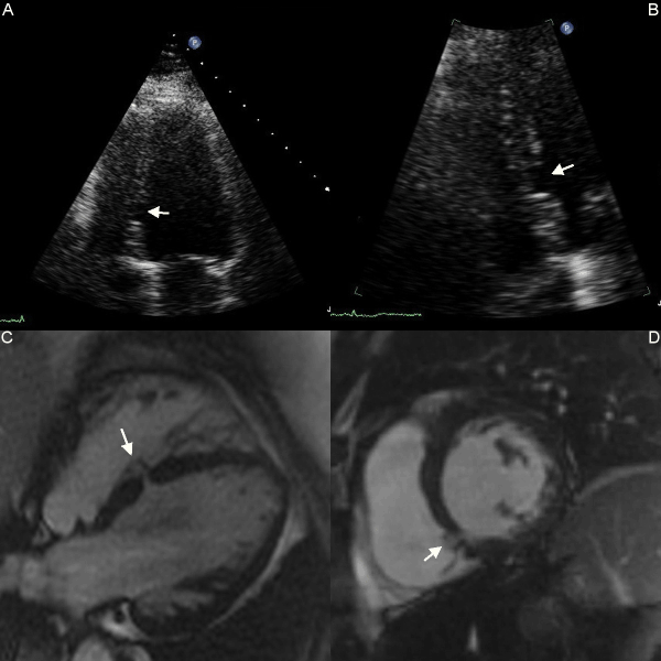
| Her symptoms were further investigated with a TTE which raised the possibility of a small muscular Ventricular Septal Defect (VSD) (Figure 1). A Cardiovascular Magnetic Resonance (CMR) scan was performed for further assessment as shown in Figure 1. The long-axis and short axis CMR cine images confirmed a small muscular VSD in the mid septum measuring 4×4 mm (area 0.4 cm2). The septal leaflet of tricuspid valve partially migrated to the septum providing a patch and partial closure of the VSD (Panels C and D, arrowheads of Figure 1-see also supplementary material online, Video). There was small residual left to right shunt (shunt volume=6 ml) with a normal QP/QS ratio. |
| Discussion |
| Ventricular septal defect is one the commonest adult congenital cardiac lesion encountered after bicuspid aortic valve, and reported up to 20% of congenital cardiac abnormalities as isolated abnormality [1]. Muscular VSDs are one of the most common subtypes of VSDs. Muscular VSDs usually undergo spontaneous closure as a result of muscular occlusion. A spontaneous closure of a muscular VSD’s is more common than that of perimembranous defects [2]. Partial or complete closure of perimembranous defects by the septal leaflet of the tricuspid valve has been described in the literature by progressive development of aneurysm formation by septal leaflet [3]. This is the first report to demonstrate that the septal tricuspid leaflet has migrated further towards the apex and partially patched a muscular VSD by CMR. CMR is an innovative non-invasive imaging technique that can identify and investigate cardiac abnormalities with high spatial resolution. |
References |
|
Figures at a glance
 |
| Figure 1 |
videos at a glance
| Video 1 |
Post your comment
Relevant Topics
- Abdominal Radiology
- Breast Imaging
- Cardiovascular Radiology
- Chest Radiology
- Clinical Radiology
- CT Imaging
- Diagnostic Radiology
- Emergency Radiology
- Fluoroscopy Radiology
- General Radiology
- Genitourinary Radiology
- Minimal Invasive surgery
- Musculoskeletal Radiology
- Neuroradiology
- Oral and Maxillofacial Radiology
- Radiography
- Radiology Imaging
- Surgical Radiology
- Tele Radiology
- Therapeutic Radiology
Recommended Journals
Article Tools
Article Usage
- Total views: 13175
- [From(publication date):
September-2014 - Jul 17, 2024] - Breakdown by view type
- HTML page views : 8851
- PDF downloads : 4324
