Spontaneous Transomental Hernia Combined with Incarcerated Inguinal Hernia: A Case Report
Received: 11-Oct-2017 / Accepted Date: 13-Oct-2017 / Published Date: 19-Oct-2017 DOI: 10.4172/2161-069X.1000532
Abstract
An 85-year-old woman was admitted to our hospital because of appetite loss and vomiting. She had no previous history of abdominal surgery, trauma, or intra-abdominal inflammation. Following the diagnosis of a small bowel obstruction due to an incarcerated left inguinal hernia, manual hernia repositioning was attempted, and proceeded smoothly. However, 42 hour after the procedure, she suddenly developed abdominal pain without recurrence of the incarcerated inguinal hernia. Abdominal computed tomography revealed signs of small bowel strangulation; therefore, emergency surgery was performed 48 hour after admission. On laparotomy, a defect was found in the greater omentum, and a part of the ileum was incarcerated by the defect. The operative diagnosis was spontaneous transomental hernia. Incision of the greater omentum was performed, and no ischemic changes were observed in the ileum; therefore, bowel resection was not performed, and the left inguinal hernia orifice was repaired. The postoperative course was uneventful. The transomental hernia in our case might have been induced because of the incarcerated inguinal hernia. For patients plagued with persistent small bowel obstruction after successful manual repositioning of anincarcerated inguinal hernia, concurrent internal hernia should be considered as a possible cause of the persistent small bowel obstruction.
Keywords: Transomental hernia; Incarcerated inguinal hernia; Internal hernia
6220Introduction
Transomental hernia (TOH) is a rare type of internal hernia, in which a loop of the small bowel herniates through an abnormal defect in the omentum, resulting in strangulation of the herniated bowel, with significant morbidity and mortality [1-6]. The incidence of TOH is rare, accounting for only 1-4% of total internal hernia cases [1], and most of the omental defects are acquired because of a previous intraabdominal event, such as abdominal surgery, trauma, or intraabdominal inflammation [2-6]. Here, we report a rare case of spontaneous TOH that presented 42 hour after successful manual repositioning of an incarcerated inguinal hernia. We also reviewed the relevant literature and discussed a possible mechanism of the presentation of TOH in our case.
Case Report
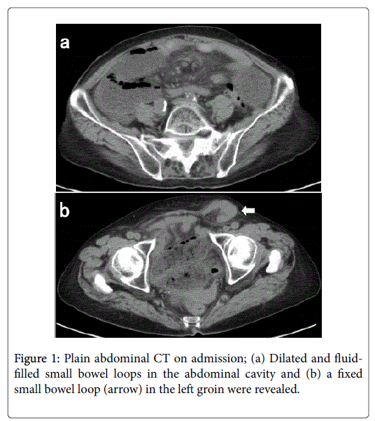
An 85-year-old woman with hypertension was admitted to our hospital because of appetite loss and vomiting that started approximately 24 hour prior to admission. She had no previous history of abdominal surgery, trauma, or intra-abdominal inflammation. On admission, her general condition was good, and vital signs were all within normal limits. On physical examination, her abdomen was mildly distended, but there was no tenderness, and a compressible hard mass, measuring approximately 50 mm in diameter, was palpated in the left groin. Plain abdominal computed tomography (CT) revealed dilated and fluid-filled small bowel loops in the abdominal cavity and a fixed small bowel loop in the left groin (Figure 1).
The patient was diagnosed with a small bowel obstruction (SBO) due to an incarcerated left inguinal hernia. Since there was no local redness or sign of peritoneal irritation, manual hernia repositioning was attempted, and proceeded smoothly. We hospitalized the patient after the procedure to observe her progress and considered elective definitive surgery for the inguinal hernia within several days. The following day, she remained stable, but nausea and abdominal distension persisted, and 42 hour after the manual hernia repositioning, she suddenly developed abdominal pain. Body temperature was 36.9°C; heart rate, 102 beats/min; respiratory rate, 20 breaths/min; and blood pressure, 118/83 mm Hg. On physical examination, her abdomen was severely distended with tenderness; however, there was no recurrence of the incarcerated inguinal hernia. Laboratory results revealed leukocytopenia (4,300/mm3) and elevated levels of blood urea nitrogen (48.7 mg/dL), creatinine (0.84 mg/dL) and C-reactive protein (19.8 mg/dL).
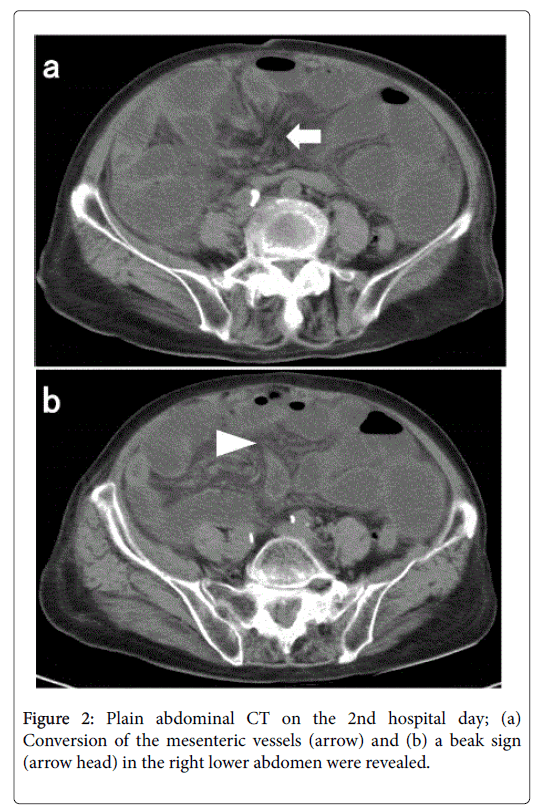
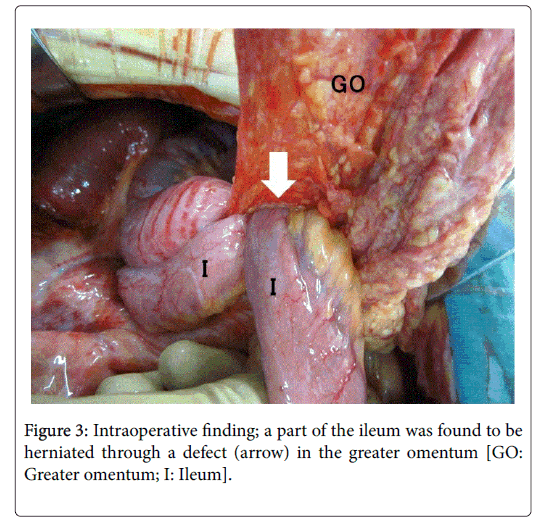
Plain abdominal CT revealed a cluster of dilated small bowel loops, a small amount of ascites, conversion of the mesenteric vessels, and a beak sign in the right lower abdomen (Figure 2). The patient was strongly suspected of developing a strangulated SBO, and emergency surgery was performed 48 hour after admission. On laparotomy, an omental defect without a hernia sac, measuring 40 mm in diameter, was found near the right edge of the greater omentum, and 50 cm of the ileum was incarcerated by the defect (Figure 3). The operative diagnosis was spontaneous TOH. To reduce the incarceration of the ileum, incision of the greater omentum from its right edge to the defect was performed, and no ischemic changes were observed in the ileum.
Therefore, bowel resection was not performed, and the left inguinal hernia orifice was repaired with mesh graft. The postoperative course was uneventful. Following rehabilitation for muscular atrophy of the lower legs, she was discharged 27 days later. At 1-year follow-up, she was doing well, with no sign of recurrence of the hernias. Retrospective reading of the initial CT on admission was performed. In the initial CT, conversion of the mesenteric vessels and a beak sign, which were revealed in the follow-up CT on the 2nd hospital day, were not observed at the same slice levels as in the follow-up CT; however, mesenteric edema was retrospectively identified at the same slice levels.
Discussion
Internal hernia is a characteristic condition of SBO, in which a loop of the small bowel herniates through a congenital or acquired peritoneal or mesenteric aperture within the abdominal cavity, resulting in strangulation of the herniated bowel [1,7-11]. Internal hernia is an unusual cause of SBO, accounting for only 0.6-5.8% of total SBO cases [7]. The types of internal hernia and frequency of each type are reported as follows: paraduodenal (53%), pericecal (13%), foramen of Winslow (8%), transmesenteric (8%), intersigmoid (6%), and transomental (1-4%) [1,8]. Thus, TOH is a rare entity among various causes of SBO. In children with TOH, the omental defect is usually a congenital anomaly caused by embryonic developmental failure [2,3]. In contrast, most of the omental defects in adult TOH cases are acquired because of a previous intraabdominal event [2-6]. In a few adult TOH cases, however, the defect might be created because of senile atrophy and/or weakness of the omentum without a predisposing history, as in our patient, and these cases are referred to as “spontaneous” TOH [2-5].
TOH is known to be more prone to develop strangulation of the herniated bowel than other types of internal hernia [2,4,7,9,10]. Therefore, for the treatment of TOH, emergency surgery must be attempted as soon as possible. However, the preoperative diagnosis of TOH is difficult because clinical manifestations are nonspecific, as compared with other causes or types of strangulated SBO [3-6,11]. As a result, precise diagnosis in most TOH cases, as in our case, is made during emergency surgery for strangulated SBO [3-6]. Recently, several studies have reported the usefulness of CT, especially multi-detector row CT, for the preoperative diagnosis of TOH [2,9,12-14]. In addition to CT signs of small bowel strangulation, (1) a cluster of dilated small bowel loops in the right paracolic gutter (2) medial and posterior displacement of the ascending colon and cecum by dilated loops and (3) absence of omental fat between the dilated loops and anterior abdominal wall, are suggested as characteristic CT findings of TOH [12,13].
In our case, conversion of the mesenteric vessels and a beak sign, which were revealed in the follow-up CT, were not observed in the initial CT; however, mesenteric edema, which may represent early ischemic change in the small bowel, i.e., an early CT sign of small bowel strangulation, was identified in the initial CT. Accordingly, we speculate a possible mechanism of the presentation of TOH in our case as follows. Initially, an abnormal defect was created in the greater omentum because of senile atrophy, which remained asymptomatic for some time. Under this condition, incarceration of an inguinal hernia incidentally occurred, leading to SBO. This SBO induced an increase in intra-abdominal pressure, which allowed the ileum to herniate through the formerly asymptomatic omental defect. Although the incarcerated inguinal hernia was successfully reduced with manual repositioning and the associated SBO was released, the transomental herniation of the ileum gradually progressed after the procedure, resulting in strangulated SBO due to TOH. Therefore, the TOH in our case might have been induced because of the incarcerated inguinal hernia through an interesting mechanism. To the best of our knowledge, there is no other case report of TOH combined with inguinal hernia in the literature.
SBO due to incarcerated inguinal hernia is a common gastrointestinal disease. Approximately 70% of incarcerated inguinal hernia cases are successfully reduced with manual repositioning [15], which may also facilitate the release of the associated SBO. If the associated SBO cannot be released and persists after successful manual hernia repositioning, the physician should consider recurrence of the incarcerated inguinal hernia or vascular compromise of the reduced small bowel [16], and further examination followed by emergency surgery must be performed. In our case, when the persistent SBO was recognized, we initially suspected stenosis of the reduced small bowel due to vascular compromise. However, the follow-up CT revealed signs of small bowel strangulation, as opposed to a stenotic small bowel segment. Further, on laparotomy, TOH proved to be the cause of the SBO. Therefore, we suggest that for patients plagued with persistent SBO after successful manual repositioning of an incarcerated inguinal hernia, concurrent internal hernia including TOH should be considered as a possible cause of the persistent SBO.
Conclusion
We experienced a rare case of spontaneous TOH combined with incarcerated inguinal hernia. The TOH in our case might have been induced because of the incarcerated inguinal hernia through an interesting mechanism. For patients plagued with persistent SBO after successful manual repositioning of an incarcerated inguinal hernia, concurrent internal hernia including TOH, although rare, should be considered by physicians.
References
- Ghahremani GG (1984) Internal abdominal hernias. SurgClin North Am 64: 393-406.
- Lee SH, Lee SH (2016) Spontaneous transomental hernia. Ann Coloproctol 32: 38-41.
- Camera L, De Gennaro A, Longobardi M, Masone S, Calabrese E, et al. (2014) A spontaneous strangulated transomental hernia: Prospective and retrospective multi-detector computed tomography findings. World J Radiol 6: 26-30.
- Yang DH, Chang WC, Kuo WH, Hsu WH, Teng CY, et al. (2009) Spontaneous internal herniation through the greater omentum. Abdom Imaging 34: 731-733.
- Tidjane A, Tabeti B, Serradj NB, Djellouli A, Benmaarouf N (2015) A spontaneous transomental hernia through the greater omentum. Pan Afr Med J 20: 384.
- Rathnakar SK, Muniyappa S, Vishnu VH, Kagali N (2016) Congenital defect in lesser omentum leading to internal hernia in adult: A rare case report. J Clin Diagn Res 10: PD08-09.
- Newsom BD, Kukora JS (1986) Congenital and acquired internal hernias: Unusual causes of small bowel obstruction. Am J Surg 152: 279-285.
- Martin LC, Merkle EM, Thompson WM (2006) Review of internal hernias: Radiographic and clinical findings. AJR Am J Roentgenol 186: 703-717.
- Blachar A, Federle MP (2002) Internal hernia: An increasingly common cause of small bowel obstruction. Semin Ultrasound CT MR 23: 174-183.
- Selcuk D, Kantarci F, Ogut G, Korman U (2005) Radiological evaluation of internal abdominal hernias. Turk J Gastroenterol 16: 57-64.
- Ghiassi S, Nguyen SQ, Divino CM, Byrn JC, Schlager A (2007) Internal hernias: Clinical findings, management, and outcomes in 49 nonbariatric cases. J GastrointestSurg 11: 291-295.
- Le Moigne F, Lamboley JL, de Charry C, Vitry T, Salamand P, et al. (2010) An exceptional case of internal transomental hernia: Correlation between CT and surgical findings. GastroenterolClinBiol 34: 562-564.
- Delabrousse E, Couvreur M, Saguet O, Heyd B, Brunelle S, et al. (2001) Strangulated transomental hernia: CT findings. Abdom Imaging 26: 86-88.
- Takagi Y, Yasuda K, Nakada T, Abe T, Matsuura H, et al. (1996) A case of strangulated transomental hernia diagnosed preoperatively. Am J Gastroenterol 91:1659-1660.
- Kauffman HM Jr, O’Brien DP (1970) Selective reduction of incarcerated inguinal hernia. Am J Surg 119: 660-673.
- Strauch ED, Voigt RW, Hill JL (2002) Gangrenous intestine in a hernia can be reduced. J Pediatr Surg 37: 919-920.
Citation: Ohtsuka Y, Takahara Y (2017) Spontaneous Transomental Hernia Combined with Incarcerated Inguinal Hernia: A Case Report. J Gastrointest Dig Syst 7: 532. DOI: 10.4172/2161-069X.1000532
Copyright: © 2017 Ohtsuka Y, et al. This is an open-access article distributed under the terms of the Creative Commons Attribution License, which permits unrestricted use, distribution, and reproduction in any medium, provided the original author and source are credited.
Share This Article
Recommended Journals
Open Access Journals
Article Tools
Article Usage
- Total views: 4903
- [From(publication date): 0-2017 - Dec 03, 2024]
- Breakdown by view type
- HTML page views: 4256
- PDF downloads: 647


