Make the best use of Scientific Research and information from our 700+ peer reviewed, Open Access Journals that operates with the help of 50,000+ Editorial Board Members and esteemed reviewers and 1000+ Scientific associations in Medical, Clinical, Pharmaceutical, Engineering, Technology and Management Fields.
Meet Inspiring Speakers and Experts at our 3000+ Global Conferenceseries Events with over 600+ Conferences, 1200+ Symposiums and 1200+ Workshops on Medical, Pharma, Engineering, Science, Technology and Business
Case Report Open Access
Spontaneous Regression of Large Cell Carcinoma of the Lung, a Case Report
| Margriet Kwint1*, Michel van den Heuvel2, Dominic Snijders3, Kim Monkhorst4 and Jose Belderbos1 | |
| 1Department of Radiation Oncology, The Netherlands Cancer Institute, Amsterdam, The Netherlands | |
| 2Department of Pulmonology, The Netherlands Cancer Institute, Amsterdam, The Netherlands | |
| 3Department of Pulmonology, Spaarne Hospital, Hoofddorp, The Netherlands | |
| 4Department of Pathology, The Netherlands Cancer Institute, Amsterdam, The Netherlands | |
| Corresponding Author : | Margriet Kwint Department of Radiation Oncology The Netherlands Cancer Institute Amsterdam, The Netherlands Tel: +0031205122125 E-mail: m.kwint@nki.nl |
| Received: September 21, 2015 Accepted: October 16, 2015 Published: October 19, 2015 | |
| Citation: Kwint M, van den Heuvel M, Snijders D, Monkhorst K, Belderbos J (2015) Spontaneous Regression of Large Cell Carcinoma of the Lung, a Case Report. OMICS J Radiol 4:206. doi:10.4172/2167-7964.1000206 | |
| Copyright: © 2015 Kwint M, et al. This is an open-access article distributed under the terms of the Creative Commons Attribution License, which permits unrestricted use, distribution, and reproduction in any medium, provided the original author and source are credited. | |
Visit for more related articles at Journal of Radiology
Abstract
Spontaneous regression of malignancies are a rare biological phenomenon, especially in lung cancer. Here, we present a case of an 80-year old man, with a cT2aN3M1b non-small cell lung cancer of the left upper lobe, whose tumor regressed spontaneously without any treatment. The cause of the spontaneous tumor remission remains unknown in this case. We describe this rare case and review the literature pertaining to spontaneous regression of lung cancer.
|
Abstract
Spontaneous regression of malignancies are a rare biological phenomenon, especially in lung cancer. Here, we present a case of an 80-year old man, with a cT2aN3M1b non-small cell lung cancer of the left upper lobe, whose tumor regressed spontaneously without any treatment. The cause of the spontaneous tumor remission remains unknown in this case. We describe this rare case and review the literature pertaining to spontaneous regression of lung cancer.
Introduction
Spontaneous regression of malignancies are a rare biological phenomenon, especially in lung cancer. It is defined as a complete or partial disappearance of the tumor in the absence of treatment or in the presence of therapy that is considered inadequate to exert a significant influence on malignant disease [1-3]. For several types of cancer spontaneous regression of cancer has been reported [4]. There are 76 cases of spontaneous regression of thoracic malignancies described between 1951 and 2008 [5]. Most of these cases concerned metastatic disease of malignancies that originated outside the thoracic cavity such as renal cell carcinoma (60%), hepatocellular carcinoma, endometrial stromal sarcoma, pleomorphic liposarcoma, esophageal cancer and leiomyosarcoma [5]. Only a few case reports described spontaneous regression of primary thoracic malignancies [5-16]. Here, we report on a case of spontaneous regression of pathologically proven non-small-cell lung cancer (NSCLC).
Case Report
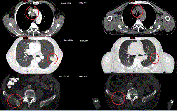
An 80-year-old male was referred to the chest physician in March 2014 with complaints of progressive dyspnea and anemia. He had lost 5 kgs of weight in the last months and also experienced pain in his right hip that radiated to his lower leg accompanied by muscle weakness. The patient had a smoking history of 65 pack years and a WHO performance score of 1. A CT-scan of the thorax/abdomen showed mediastinal and hilair lymphadenopathy, abdominal lymphadenopathy, a tumor in the left upper lobe and multiple pulmonary nodules in both lungs (Figure 1).
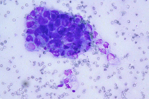
Furthermore, enlarged adrenal glands were seen on the CT-scan, and a small liver lesion in segment 4, suspect for metastasis. In the right ilium and 6th left costa, osteoblastic lesions suspect for bone metastases were visible (Figure 1). An MRI-scan of the lumbar-spine showed an abnormal signal in multiple corpora, suspect for metastases. Both fine needle aspirate of a pleural pulmonary nodule as well as a CT-guided biopsy of a lesion in the right ilium, were inconclusive. Endo-bronchial- ultra-sound (EBUS) fine needle aspiration of Naruke stations 4R and 7 showed atypical epitheloid cell groups highly suspicious of malignancy, compatible with a non-small cell lung carcinoma (Figure 2). There was not enough material however for immunocytochemical or molecular analysis. It was concluded that this patient had stage IV (cT2aN3M1b) NSCLC. Following the multidisciplinary tumor board discussion, a palliative plan was established consisting of irradiation of the bone metastasis in the pelvis to relieve the pain, thoracic irradiation for prevention of a vena cava superior syndrome, and chemotherapy. However, at the first visit of the radiotherapy department the patient reported an improvement in shortness of breath and condition. Besides this, he had gained 3 kg of weight. The pain in his right leg and right pelvis however was still present. A new CT-scan, planned to prepare for the irradiation and performed 2 months after the diagnostic CT, showed a spontaneous regression of the enlarged mediastinal lymph nodes (Figure 1). The vena cava was no longer compressed and the pulmonary lung nodules completely disappeared. Also the primary tumor had decreased in size. The femoral head showed a pathological fracture. Due to the spontaneous regression of the pulmonary lesions the pathology report was reassessed which confirmed the initial diagnosis of a NSCLC. Finally it was decided to irradiate the right femoral head and ilium, with a single fraction of 8 Gray. A Fluordeoxyglucose Positron Emission Tomography (FDG-PET)-CT- scan showed further regression with residual scarring in the left upper lobe without any FDG uptake. The mediastinal lymph nodes demonstrated low FDG uptake (SUV max 3.1). The irradiated location of the fracture in right femoral head showed an increase of sclerotic tissue and FDG uptake. In costa 6 sparse uptake was seen. A follow up CT-scan, 4 months later, showed further regression of the lesion in left upper lobe and mediastinal lymph nodes. The pulmonary nodules had totally disappeared. The patient was in good condition and there were no pulmonary complaints or weight loss. Discussion
Spontaneous regression (SR) of cancer is a rare phenomenon. The Everson and Cole review report 176 SR cases between 1900 and 1964 [17]. Challis and Stam updated that review to 489 cases in the period from 1900 to 1987 [4]. These case reports do not represent the true incidence of SR. Cole [18] estimated the incidence of SR to occur in 1 of 60.000 to 100.000 cancer individuals. However, most SR cases described, apparently occur in a few types of cancer: malignant melanoma, low-grade non-Hodgkin’s lymphoma, renal cell cancer, chronic lymphocytic leukemia and neuroblastoma in children [4,17]. SR seems even more uncommon in NSCLC [4,5,17]. To our knowledge only 15 cases of SR of NSCLC have been described in the literature [5-16]. The mechanisms for spontaneous regression are entirely hypothetical and the pathophysiology remains unclear. Associated mechanisms are immune response, tumor inhibition by growth factors and/or cytokines, apoptosis, differentiations, hormones, angiogenesis inhibition, telomerase inhibition and psychoneuroimmunological response [11]. In our case, the patient did not receive any treatment at the time of SR. Later on, radiotherapy was given for the bone metastasis, which might have further induced regression due to an abscopal effect [19] (A reaction produced following irradiation but occurring outside the zone of actual radiation absorption). A number of preclinical and clinical studies are ongoing to study the pro-immunogenic effects of local radiotherapy [20,21].
A limitation of this case report is that the diagnosis was based on a fine needle aspiration of mediastinal lymph nodes. There was no biopsy and not sufficient material for additional immunohistochemical or molecular analysis. We did not provide evidence of malignancy of the primary tumor in the left upper lung, nor the bone metastasis. However there was no doubt about the pathology diagnosis of NSCLC in the lymph nodes. The cause of the spontaneous tumor remission remains unknown in this case. Further studies are imperative to explain the underlying mechanism of SR. |
References
- Cole WH (1976) Spontaneous regression of cancer and the importance of finding its cause. Natl Cancer Inst Monogr 44: 5-9.
- EVERSON TC, COLE WH (1959) Spontaneous regression of malignant disease. J Am Med Assoc 169: 1758-1759.
- STEWART FW (1952) Experiences in spontaneous regression of neoplastic disease in man. Tex Rep Biol Med 10: 239-253.
- Challis GB, Stam HJ (1990) The spontaneous regression of cancer. A review of cases from 1900 to 1987. Acta Oncol 29: 545-550.
- Kumar T, Patel N, Talwar A (2010) Spontaneous regression of thoracic malignancies. Respir Med 104: 1543-1550.
- Cafferata MA, Chiaramondia M, Monetti F, Ardizzoni A (2004) Complete spontaneous remission of non-small-cell lung cancer: a case report. Lung Cancer 45: 263-266.
- Emerson GL, Emerson MS, Sherwood CE, Terry R (1968) Spontaneous regression of bronchogenic carcinoma. Twelve-year survival. J Thorac Cardiovasc Surg 55: 225-230.
- Haruki T, Nakamura H, Taniguchi Y, Miwa K, Adachi Y, et al. (2010) Spontaneous regression of lung adenocarcinoma: Report of a case. Surg Today 40: 1155-1158.
- Hwang ED, Kim YJ, Leem AY, Ji AY, Choi Y, et al. (2013) Spontaneous regression of non-small cell lung cancer in a patient with idiopathic pulmonary fibrosis: a case report. Tuberc Respir Dis (Seoul) 75: 214-217.
- Iwakami S, Fujii M, Ishiwata T, Iwakami N, Hara M, et al. (2013) Small-cell lung cancer exhibiting spontaneous regression. Intern Med 52: 2249-2252.
- Kappauf H, Gallmeier WM, Wunsch PH, Mittelmeier HO, Birkman J, et al. (1997) Complete spontaneous remission in a patient with metastatic non-small-cell lung cancer. Case report, review of the literature, and discussion of possible biological pathways involved. Annals of oncology 8:1031-1039.
- Kato M, Yoshitomo K, Mastushita T, Kato J, Hayashi Y, et al. (1986) [A case of spontaneous regression in lung cancer]. Nihon Kyobu Shikkan Gakkai Zasshi 24: 188-194.
- Leo F, Nicholson AG, Hansell DM, Corrin B, Pastorino U (1999) Spontaneous regression of large-cell carcinoma of the lung--a rare observation in clinical practice. Thorac Cardiovasc Surg 47: 53-55.
- Mizuno T, Usami N, Okasaka T, Kawaguchi K, Okagawa T, et al. (2011) Complete spontaneous regression of non-small cell lung cancer followed by adrenal relapse. Chest 140: 527-528.
- Saito H, Okuno M (1999) Spontaneous regression of a bulla with the development of adenocarcinoma of the lung. Intern Med 38: 439-441.
- Sperduto P, Vaezy A, Bridgman A, Wilkie L (1988) Spontaneous regression of squamous cell lung carcinoma with adrenal metastasis. Chest 94: 887-889.
- Everson TC (1967) Spontaneous regression of cancer. Prog Clin Cancer 3: 79-95.
- Cole WH (1981) Efforts to explain spontaneous regression of cancer. J Surg Oncol 17: 201-209.
- Siva S, MacManus MP, Martin RF, Martin OA (2015) Abscopal effects of radiation therapy: a clinical review for the radiobiologist. Cancer Lett 356: 82-90.
- Martin OA, Anderson RL, Russell PA, Cox RA, Ivashkevich A, et al. (2014) Mobilization of viable tumor cells into the circulation during radiation therapy. Int J Radiat Oncol Biol Phys 88: 395-403.
- van den Heuvel MM, Verheij M, Boshuizen R, Belderbos J, Dingemans AM, et al. (2015) Nhs-il2 combined with radiotherapy: Preclinical rationale and phase ib trial results in metastatic non-small cell lung cancer following first-line chemotherapy. J Transl Med 13: 32.
Figures at a glance
 |
 |
| Figure 1 | Figure 2 |
Post your comment
Relevant Topics
- Abdominal Radiology
- AI in Radiology
- Breast Imaging
- Cardiovascular Radiology
- Chest Radiology
- Clinical Radiology
- CT Imaging
- Diagnostic Radiology
- Emergency Radiology
- Fluoroscopy Radiology
- General Radiology
- Genitourinary Radiology
- Interventional Radiology Techniques
- Mammography
- Minimal Invasive surgery
- Musculoskeletal Radiology
- Neuroradiology
- Neuroradiology Advances
- Oral and Maxillofacial Radiology
- Radiography
- Radiology Imaging
- Surgical Radiology
- Tele Radiology
- Therapeutic Radiology
Recommended Journals
Article Tools
Article Usage
- Total views: 13804
- [From(publication date):
October-2015 - Mar 29, 2025] - Breakdown by view type
- HTML page views : 9261
- PDF downloads : 4543
