Solitary Fibrous Tumor in the Mediastinum: A Rare Entity with Wide-Spread Metastasis
Received: 02-Jan-2024 / Manuscript No. roa-24-126460 / Editor assigned: 05-Jan-2024 / PreQC No. roa-24-126460 / Reviewed: 19-Jan-2024 / QC No. roa-24-126460 / Revised: 24-Jan-2024 / Manuscript No. roa-24-126460 / Published Date: 31-Jan-2024
Abstract
Solitary fibrous tumor is a rare mesenchymal tumor, most frequently located in the pleura. Other locations have been recently described in the literature including the mediastinum. Solitary fibrous tumor of the mediastinum affects both men and women with a peak in frequency in the fifth and sixth decade. Most often asymptomatic and of incidental discovery, this tumor is evoked by a mediastinal opacity on thoracic radiography or a mediastinal tissue mass on CT scan and confirmed by histological examination through transcutaneous CT-guided aspiration biopsy or after complete
surgical removal.
Keywords
Aggressive mediastinal mass; Solitary fibrous tumor; Distant metastasis; CT
Introduction
Solitary fibrous tumors are uncommon tumors of non-mesothelial mesenchymal origin [1], initially described in the pleura as “benign local fibroma” and “submesothelial fibroma”[1,2]. Although the pleural location is the most frequent, the literature has recently described other sites, notably the peritoneal, mediastinal, bronchopulmonary, and orbital locations. The mediastinal location of these tumors is considered among the rarest and most aggressive [3,4]. We report a case of a solitary fibrous tumor of the mediastinum metastatic to the liver, peritoneal, and bone.
Case Report
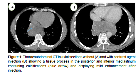
In the context of an altered general condition, a 48-year-old man without any prior medical or surgical history presented with symptoms including chest pain, coughing, dysphonia, respiratory discomfort, and a significant weight loss of 15 kg. The chest scan revealed a hypodense, heterogeneous tissue process in the posterior and inferior mediastinum. It showed weak enhancement after the injection of contrast product and contained calcifications. The tumor invaded the lower esophagus and the cardia. There was no pleural invasion or mediastinal adenopathy (Figure 1).
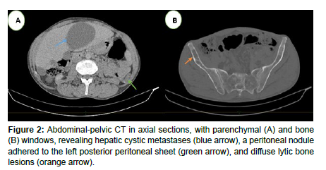
Abdominal CT was subsequently performed, demonstrating large, hypodense liver lesions and peritoneal nodules. Multiple lytic bone lesions were also discovered (Figure 2).
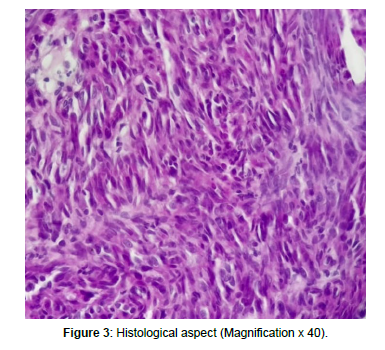
An ultrasound-guided biopsy of a liver lesion was performed (more easily accessible), and pathological examination showed an immunohistochemical profile in favour of a solitary fibrous tumour (Figure 3).
The patient underwent radiochemotherapy and died after 2 months of treatment
Discussion
The solitary fibrous tumor is a rare mesenchymal neoplasm initially characterized by the pleura [1]. While its primary occurrence in the pleural region is most prevalent, this tumor can manifest in various extra-pleural sites, including the mediastinum [5]. Notably, its presence in the mediastinum is exceptionally uncommon and recognized as the most aggressive form [3,4].
These tumors can affect both men and women, with a higher incidence observed during the fifth and sixth decades of life [5]. They typically remain asymptomatic and are often discovered incidentally through radiological examinations. However, in some cases, they may be accompanied by symptoms such as chest pain, cough, or dyspnea, as seen in our patient’s case.
The chest radiograph reveals a distinct mediastinal opacity characterized by well-defined outer boundaries that curve toward the lungs and seamlessly connect to the mediastinum. Additionally, internal boundaries are deeply embedded within the mediastinum. Performing a chest CT scan, both with and without contrast injection, is essential to confirm the tissue composition of the mediastinal mass and accurately determine its vascular connections with nearby structures [7]. In cases where vascular invasion is suspected, an MRI scan can provide valuable diagnostic information [8].
A histological study using transcutaneous scanno-guided aspiration biopsy confirms the diagnosis of this condition. It is important to note that the majority of these tumors are benign. Malignant progression with spread to distant sites is uncommon, occurring in only 13- 23% of cases [8,9]. In our patient’s case, extension work-up revealed liver, peritoneal and bone metastases. These tumors originate from CD34+ interstitial dendritic cells of non-mesothelial mesenchymal origin, which explains their occurrence in extrapleural sites [8]. An immunohistochemical study is crucial for accurate diagnosis, as it helps identify the expression of CD99, bcl2, and vimentin in these cells [10]. The recommended treatment for this condition entails a surgical intervention aimed at the complete resection of the mediastinal mass and any involved adjacent organs. Subsequently, the possibility of administering radiochemotherapy should be considered, especially in cases containing histological findings indicative of malignancy [8,9].
Conclusion
The solitary fibrous tumor is an uncommon and particularly aggressive tumor that primarily occurs in the mediastinum. Its clinical and radiological manifestations are nonspecific, making it challenging to diagnose definitively. Confirmation of the diagnosis is achieved through histological and immunohistochemical studies, either by performing a scanno-guided biopsy or through surgical removal. Although these mediastinal tumors are typically benign, there is a potential for malignant progression and the development of secondary distant metastases.
Conflict of Interest Statement
No conflict of interest.
References
- Bouchi J, Gharios E, Cortbawi E, Aftimos G (1993) Tumeur fibreuse solitaire de la plèvre avec coma et hypoglycémie. Rev Pneumol Clin 49: 243-246.
- Riss O, Mamlouk O, Troufléau P, Depardieu C, Stinés J (2000) Une tumeur fibreuse solitaire de la cuisse explorée par TDM et IRM. J Radiol 81: 1643-1646.
- Weidner N (1991) Solitary fibrous tumor of the mediastinum. Ultrastruct Pathol 15: 489-492.
- Vanderijn M, Lambard CM, Rousse RV (1994) Expression of CD34 by solitary fibrous tumor of the pleura, mediastinum, and lung. Am J Surg Pathol 18: 814-820.
- Suehisa H, Yamashita M, Komori E (2010) Solitary fibrous tumor of the mediastinum. Gen Thorac Cardiovasc Surg 58: 205-208.
- Baliga M, Flowers R, Heard K, Siddiqi A, Akhtar I (2007) Solitary fibrous tumor of the lung: a case report with a study of the aspiration biopsy, histopathology, immunohistochemistry, and autopsy findings. Diagn Cytopathol 35: 239-244.
- Fukushima K, Yamaguchi T, Take A, Ohara T, Hasegawa T, et al. (1992) A case report of so-called solitary fibrous tumor of the mediastinum. Nippon Kyobu Geka Gakkai Zasshi 40: 978-982.
- Jougon J, Minniti A, Begueret H, Dromer C, Delcamber F, Mac Bride T, et al. (2002) Tumeur fibreuse solitaire de la plèvre. Une tumeur rare d’évolution imprévisible. Rev Pneumol Clin 58: 135-138.
- Weidner N (1991) Solitary fibrous tumor of the mediastinum. Ultrastruct Pathol 15: 489-492.
- NEJJAR M, HIDRAOUI K, MIHRADI W (2004) Solitary fibrous mediastinal tumor. A case report. REV PNEUMOL CLIN 60: 235-238.
Indexed at, Google Scholar, Crossref
Indexed at, Google Scholar, Crossref
Indexed at, Google Scholar, Crossref
Indexed at, Google Scholar, Crossref
Indexed at, Google Scholar, Crossref
Citation: Zebbakh H, Bakkari A, Benbrahima F, Lemrabet A, Essaber H, et al.(2024) Solitary Fibrous Tumor in the Mediastinum: A Rare Entity with Wide-SpreadMetastasis. OMICS J Radiol 13: 528.
Copyright: © 2024 Zebbakh H, et al. This is an open-access article distributedunder the terms of the Creative Commons Attribution License, which permitsunrestricted use, distribution, and reproduction in any medium, provided theoriginal author and source are credited.
Share This Article
Open Access Journals
Article Usage
- Total views: 300
- [From(publication date): 0-0 - Aug 16, 2024]
- Breakdown by view type
- HTML page views: 270
- PDF downloads: 30


